
analyse_media
Reading File
Now let me search the medical library and web for these specific lesion types on the shins.
Finding Sources
Finding Sources
Searching the Web
violaceous atrophic nodules shins differential diagnosis pretibial lesions
Now let me search specifically for the most likely diagnoses visible here - the raised, dome-shaped, violaceous nodules on the pretibial area.
Finding Sources
Finding Sources
Finding Sources
Finding Sources
Finding Sources
Finding Sources
Reading File
Excellent - this is a very strong match. Let me fetch the textbook image for comparison.
Loading Image
The textbook image is a more advanced/severe case with more numerous and verrucous lesions, but the morphology (raised, discrete, dome-shaped, violaceous-to-grey-brown nodules on the anterior lower leg) is very similar. The user's photo shows an earlier/milder presentation. Now I have enough to give a thorough answer.
Clinical Assessment of the Leg Lesions
⚠️ Important disclaimer: This is not a medical diagnosis. Please consult a dermatologist for proper evaluation.
What the Image Shows
The photograph displays two discrete, well-demarcated, dome-shaped nodules on the anterior lower leg (pretibial area):
- Upper lesion: ~1.5-2 cm, raised, rounded, with a dusky violaceous-grey surface and surrounding hyperpigmentation halo
- Lower lesion: ~1 cm, similar morphology, slightly shiny surface
- Background skin has scattered flat brown macules (likely older, flatter lesions or post-inflammatory pigmentation)
- No active ulceration or crusting visible
Most Likely Diagnosis: Hypertrophic Lichen Planus (HLP)
The appearance is strongly consistent with hypertrophic lichen planus, a variant of lichen planus that almost exclusively affects the anterior lower legs/shins. Here is what the textbook says:
"Hypertrophic LP usually occurs on the shins but may occur anywhere. The anterior lower leg below the knee is the sole area of involvement in most patients. The typical lesions are verrucous plaques with variable amounts of scale... Lesions are of variable size but are frequently several centimeters in diameter and larger than the lesions of classic LP."
- Andrews' Diseases of the Skin, Clinical Dermatology (Hypertrophic Lichen Planus section)
The image in the textbook (Fig. 12.7, Hypertrophic Lichen Planus) shows an advanced case with multiple raised, discrete, violaceous-grey nodules on the lower leg - morphologically matching the user's photo but in a more advanced stage:
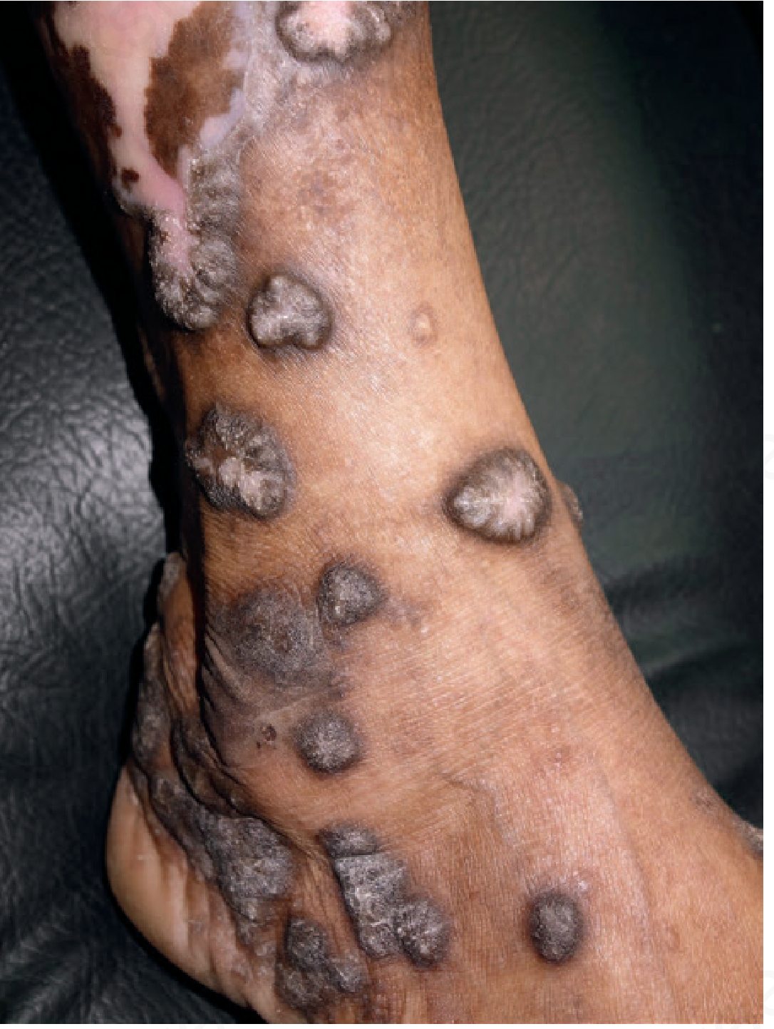
Key supporting features in the user's photo:
- Discrete, raised, dome-shaped nodules (vs. flat papules of classic LP)
- Dusky violaceous to grey-brown color - classic for LP in darker skin tones
- Shiny/atrophic surface
- Located specifically on the anterior lower leg/shin
- Surrounding post-inflammatory hyperpigmentation
Differential Diagnoses to Consider
| Condition | Supporting Features | Against |
|---|---|---|
| Hypertrophic Lichen Planus | Pretibial location, discrete dome-shaped violaceous nodules, shiny surface | Would expect more scaling/verrucous surface in later stages |
| Prurigo Nodularis | Discrete nodules, pretibial area, common with scratch-related trauma | Usually more excoriated/crusted tops |
| Lichen Amyloidosis | Hyperpigmented papules on shins, common in South/Southeast Asian patients | Usually smaller, more numerous ripple-pattern papules |
| Diabetic Dermopathy | Pretibial atrophic hyperpigmented spots | Usually flat macules/patches, not raised nodules |
| Necrobiosis Lipoidica | Pretibial location | Yellow-brown waxy appearance, not dome-shaped nodules |
| Erythema Nodosum | Pretibial nodules | Usually acute, tender, bilateral, resolves in weeks |
| Pretibial Myxedema | Pretibial nodules | Associated with thyroid disease, waxy/orange-peel texture |
What You Should Do
- See a dermatologist - A skin biopsy is often needed to confirm the diagnosis. Hypertrophic LP can mimic squamous cell carcinoma histologically, and long-standing HLP carries a small risk of malignant transformation.
- Tell the doctor:
- How long have these been present?
- Do they itch? (LP is often very pruritic)
- Any similar lesions in the mouth or nails? (LP commonly affects mucous membranes)
- Any medications recently started? (Drug-induced lichenoid reactions are common)
- Any known diabetes, thyroid disease, or hepatitis C? (HCV has a strong association with LP)
- Do not scratch - Trauma can worsen LP through the Koebner phenomenon (new lesions forming at sites of injury).
If Confirmed as Hypertrophic LP - Treatment Options
- First-line: Potent topical corticosteroids (clobetasol) under occlusion; intralesional triamcinolone injections
- Second-line: Topical tacrolimus/pimecrolimus, oral retinoids, phototherapy (PUVA/NB-UVB)
- Note: HLP is chronic and often refractory to topical therapy - early dermatology referral is advisable
Again, please see a dermatologist - this type of lesion genuinely requires in-person evaluation and likely a biopsy for definitive diagnosis.
This is a shared conversation. Sign in to Orris to start your own chat.