
write a detailed note on scapulohumeral rythym draw diagrams to explain and visualize glenohumeral rythym make an assignment for glenohumeral rythym
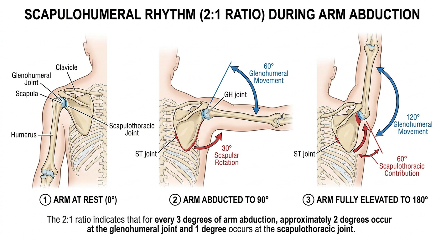
Medical anatomy diagram of scapulohumeral rhythm showing the 2:1 ratio during arm abduction. Three sequential positions illustrated side by side: (1) arm at rest 0 degrees, (2) arm abducted to 90 degrees showing 60 degrees glenohumeral movement and 30 degrees scapular rotation labeled separately, (3) arm fully elevated to 180 degrees showing 120 degrees glenohumeral and 60 degrees scapulothoracic contribution. Clear anatomical labels including glenohumeral joint, scapulothoracic joint, humerus, scapula, clavicle. Arrows showing direction of movement at each joint. Clean white background, medical textbook style illustration with colored annotations.
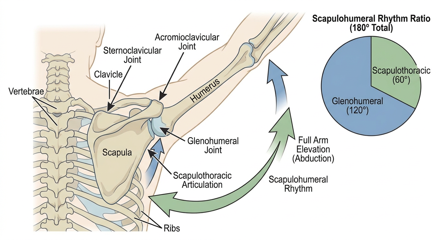
Medical diagram showing the joints involved in scapulohumeral rhythm: sternoclavicular joint, acromioclavicular joint, glenohumeral joint, and scapulothoracic articulation. Posterior view of shoulder girdle with labeled arrows showing contribution of each joint to full arm elevation. Include a pie chart or bar chart inset showing the 2:1 ratio (glenohumeral 120 degrees vs scapulothoracic 60 degrees out of 180 degrees total abduction). Anatomical landmarks clearly labeled. Clean educational medical illustration style.
Medical diagram showing abnormal vs normal scapulohumeral rhythm. Left side shows normal rhythm with smooth synchronized movement, right side shows disrupted rhythm with early scapular hiking due to rotator cuff pathology or frozen shoulder (adhesive capsulitis). Arrows and angle measurements. Labels: 'Normal 2:1 ratio' and 'Abnormal - early scapular elevation'. Clinical teaching illustration, clean white background, anatomical accuracy.
Scapulohumeral Rhythm - Detailed Clinical Note
Part I: Overview and Definition
Part II: The Shoulder Complex - Joints Involved
| Joint | Type | Contribution |
|---|---|---|
| Glenohumeral (GH) | Synovial ball-and-socket | Primary mover; contributes ~120° |
| Scapulothoracic (ST) | Functional articulation (not true synovial) | Upward rotation ~60° |
| Sternoclavicular (SC) | Saddle joint | Clavicular elevation ~30-35°, posterior rotation |
| Acromioclavicular (AC) | Plane synovial | Scapular tilting, rotation adjustment |
All four joints must contribute harmoniously. Restriction at any one joint alters the entire rhythm.
Part III: The 2:1 Ratio - The Core Rule
What it means:
- For every 3° of total arm abduction, the glenohumeral joint contributes 2° and the scapulothoracic articulation contributes 1°
- This ratio is maintained throughout most of the arc of abduction (after the initial set phase)
Breakdown over full 180° elevation:
| Total Arm Elevation | Glenohumeral Contribution | Scapulothoracic Contribution |
|---|---|---|
| 0° (resting) | 0° | 0° |
| 30° (initial setting phase) | ~30° | ~0° (scapula sets) |
| 90° | 60° | 30° |
| 120° | 80° | 40° |
| 180° (full elevation) | 120° | 60° |
Key Textbook Source:
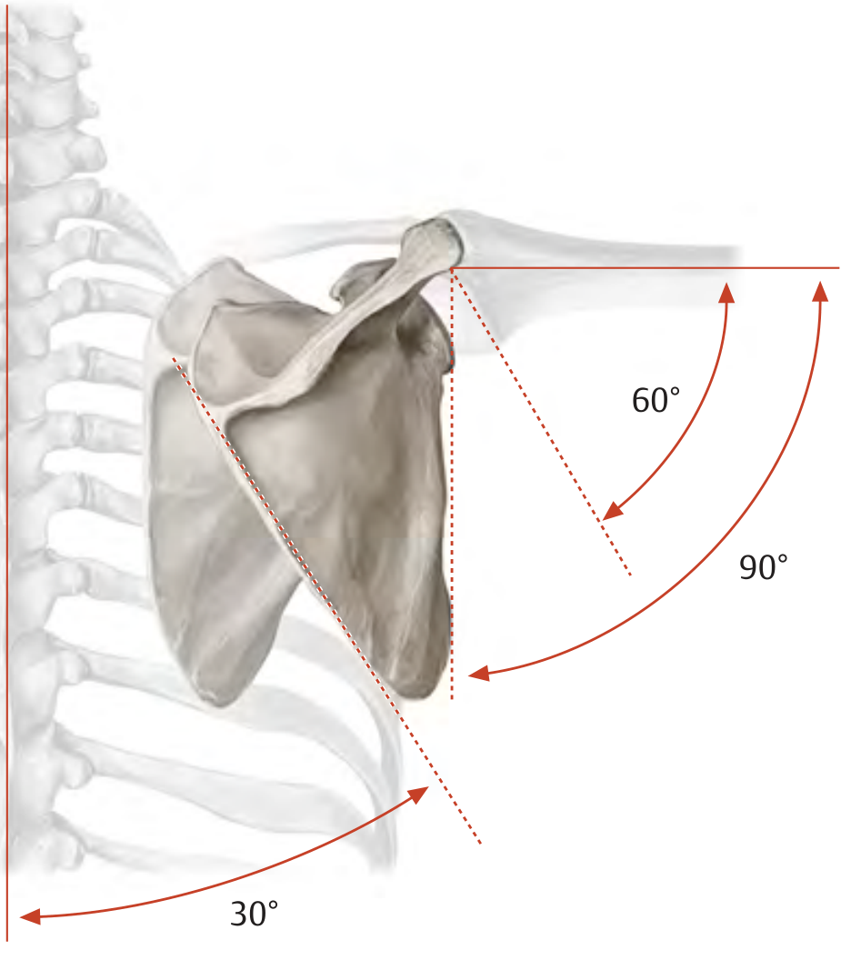
"The arm and scapula move in a 2:1 ratio during abduction. This means that when the arm is abducted 90°, for example, 60° of that movement occurs in the glenohumeral joint, while 30° is accomplished by concomitant movement of the shoulder girdle." - General Anatomy and Musculoskeletal System (THIEME Atlas of Anatomy)
Part IV: Diagram 1 - The 2:1 Ratio at Three Key Positions
Part V: Diagram 2 - Three Sequential Positions of Arm Abduction
Part VI: Diagram 3 - The Four Joints and Their Contributions
Part VII: Phase-by-Phase Analysis
Phase 1: The Setting Phase (0°- ~30°)
- The scapula "sets" its position against the thorax
- Most movement occurs at the glenohumeral joint
- The scapula may move minimally or variably
- Some sources describe a brief period of scapular retraction or protraction to find a stable position
Phase 2: Active Rhythm Phase (~30°-170°)
- The 2:1 ratio is established and maintained
- Glenohumeral abduction and scapular upward rotation occur simultaneously
- At 90° GH abduction, the greater tubercle would impinge against the acromion - this is prevented by automatic external rotation of the humerus
- The supraspinatus and deltoid are the primary GH abductors
- The serratus anterior and trapezius (upper and lower fibers) drive scapular upward rotation
Phase 3: Terminal Elevation (~170°-180°)
- Further elevation requires thoracic spine extension and lateral trunk flexion
- The spine and pelvis contribute to the final arc
- Contralateral trunk lean facilitates overhead reach
Part VIII: Muscles Driving Scapulohumeral Rhythm
Glenohumeral Abductors:
- Supraspinatus - initiates the first 15-30° of abduction; compresses humeral head into glenoid
- Deltoid (middle fibers) - primary abductor from 30° onward
- Infraspinatus and Teres Minor - laterally rotate the humerus, clearing the greater tubercle from the acromion
- Subscapularis - contributes to inferior translatory stability
Rotator Cuff as a Unit (Force Couple):
Scapular Upward Rotators (Scapulothoracic Force Couple):
| Muscle | Action | Role |
|---|---|---|
| Upper trapezius | Elevates and retracts scapula | Rotates scapula upward (superior angle) |
| Lower trapezius | Depresses and retracts scapula | Pulls inferior angle of spine, driving rotation |
| Serratus anterior | Protracts and upwardly rotates scapula | Pulls inferior angle forward and laterally |
Part IX: Diagram 4 - Normal vs. Abnormal Rhythm
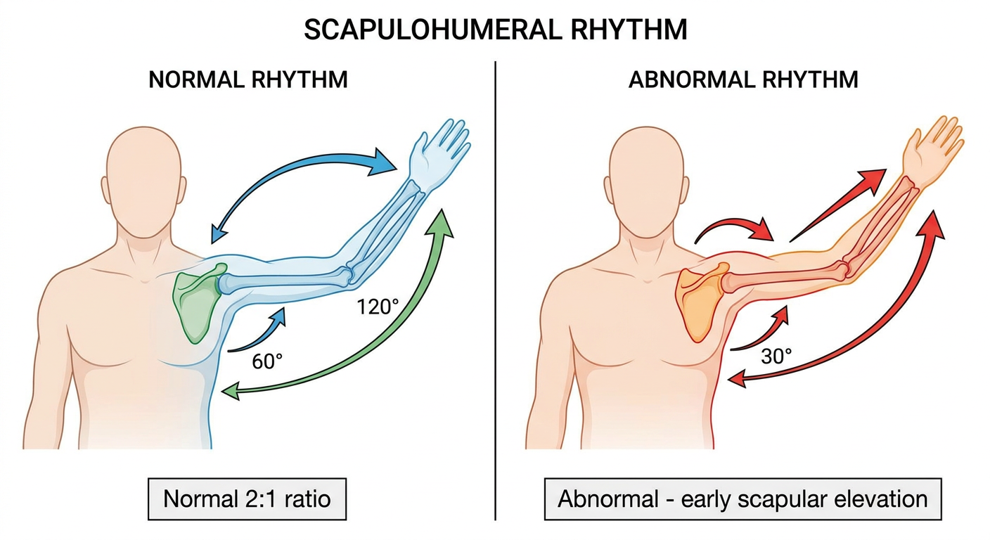
Part X: Role of the Clavicle and SC/AC Joints
- During full elevation, the clavicle elevates approximately 30-35°
- The clavicle also rotates posteriorly around its long axis by ~45° during elevation above 90°
- Without this posterior rotation, scapular upward rotation is limited
- Allows the scapula to rotate relative to the clavicle
- Contributes approximately 20° of scapular rotation independently
- AC joint pathology (arthrosis, dislocation) directly affects scapular kinematics
"For example, during humeral elevation in scapular plane, particularly beyond 90 degrees of elevation, the scapula rotates laterally, retracts, and tilts backwards, while the clavicle elevates, retracts, and rotates backward." - Imaging Anatomy (Bones, Joints, Vessels and Nerves)
Part XI: The Subacromial Space and Impingement
- External rotation of the humerus - rotates the greater tubercle posteriorly, away from the acromion
- Scapular upward rotation - elevates the acromion away from the rising humeral head
- Inferior translation of the humeral head - rotator cuff centering force keeps the head from riding superiorly
When the arm is abducted while internally rotated, the range of abduction is decreased to approximately 60° (as the greater tubercle cannot clear the acromion). - THIEME Atlas of Anatomy
Part XII: Clinical Significance
Assessment of Scapulohumeral Rhythm:
- Is the scapular rotation smooth or jerky?
- Does the scapula elevate excessively (early shrugging)?
- Is there asymmetry between sides?
- Is there a "hitch" or sudden increase in scapular movement at a specific angle?
Clinical Conditions Altering Rhythm:
| Condition | Alteration | Mechanism |
|---|---|---|
| Adhesive capsulitis (frozen shoulder) | Early, excessive scapular rotation | GH restriction forces compensation via ST joint |
| Rotator cuff tear | Loss of force couple; superior humeral migration | Deltoid unopposed; poor head centering |
| Subacromial impingement | Painful arc; scapular dyskinesis | Impingement alters muscle recruitment |
| GH ankylosis/arthrodesis | Pure scapulothoracic movement only | 40-60° abduction still possible via ST alone |
| Serratus anterior palsy (long thoracic nerve) | Winging + inability to elevate arm above 90° | Loss of scapular upward rotation force couple |
| Trapezius palsy (accessory nerve) | Scapular drop, winging, limited elevation | Loss of upper trapezius clavicular lever |
| AC joint dislocation | Disrupted clavicle-scapula coupling | Impaired posterior clavicular rotation |
"Diseases of the shoulder joint can alter this rhythm, often causing the scapula to begin its rotation considerably earlier. This is best illustrated by cases involving ankylosis or arthrodesis of the glenohumeral joint, in which movements in the shoulder girdle alone still permit the arm to be abducted 40 to 60° and still allow for one third the normal range of flexion/extension." - THIEME Atlas of Anatomy
Part XIII: Scapular Dyskinesis
- Type I: Inferior angle prominence (anterior tilt)
- Type II: Medial border prominence (excessive internal rotation/winging)
- Type III: Superior border elevation and dominant upper trapezius activity
Part XIV: Practical Note for Physiotherapy
- Retrain the serratus anterior (wall push-ups, protraction exercises) before overhead strengthening
- Activate lower trapezius (prone Y/T exercises) to restore rotatory force couple
- Avoid early overhead strengthening if scapular kinematics are abnormal
- Address thoracic spine mobility - thoracic kyphosis restricts scapular posterior tilting
ASSIGNMENT: Glenohumeral Rhythm
Section A: Short Answer Questions (2-3 marks each)
-
Define scapulohumeral rhythm and state the exact ratio that governs it during arm abduction. Why is this ratio clinically important?
-
Name the four articulations that make up the shoulder complex and briefly state the contribution of each to full arm elevation.
-
At 90° of total arm abduction, how many degrees of movement occur at (a) the glenohumeral joint and (b) the scapulothoracic articulation? Show your calculation.
-
What is the "setting phase" of scapulohumeral rhythm? Describe what happens during the first 30° of arm abduction.
-
Define a "force couple" and give one example of a force couple operating at the glenohumeral joint during abduction.
-
Why must the humerus externally rotate during abduction past 80-90°? Name the structures involved and the consequence if this does not occur.
-
What is the approximate range of abduction still possible in a shoulder with complete glenohumeral ankylosis? Explain the mechanism that allows this residual motion.
Section B: Structured Answer Questions (5-8 marks each)
| Total Arm Elevation | GH Contribution | ST Contribution |
|---|---|---|
| 30° | ||
| 60° | ||
| 90° | ||
| 150° | ||
| 180° |
Section C: Extended Response (10-15 marks each)
Section D: Clinical Application / Case Studies (10 marks each)
Section E: Diagram Labeling
Section F: True/False with Justification (2 marks each)
- During arm abduction, the scapula and humerus always move simultaneously from 0° onward.
- In a patient with complete glenohumeral fusion, no shoulder abduction is possible.
- The serratus anterior is essential for scapular upward rotation.
- The 2:1 ratio means that for every 1° of scapular rotation, the glenohumeral joint moves 2°.
- External rotation of the humerus is necessary to complete abduction beyond 90°.
- The clavicle rotates posteriorly around its long axis during elevation above 90°.
- A torn supraspinatus tendon does not affect scapulohumeral rhythm.
- The subacromial space normally measures approximately 9-10 mm.
Answer Key Highlights (for instructor use)
- Q3: GH = 60°, ST = 30° (2:1 ratio applied to 90°)
- Q7: 40-60° abduction is still possible through scapulothoracic motion alone
- Q8: Upper trapezius + Lower trapezius + Serratus anterior; upper trap pulls clavicular/acromial end up, lower trap and serratus anterior pull inferior angle outward - net effect is upward glenoid rotation
- Q10 answers: 30°→(20°/10°), 60°→(40°/20°), 90°→(60°/30°), 150°→(100°/50°), 180°→(120°/60°)
- Q13a: Long thoracic nerve - serratus anterior
- Section F answers: 1-F, 2-F, 3-T, 4-T, 5-T, 6-T, 7-F, 8-T
What Was Delivered
Detailed Clinical Note on Scapulohumeral Rhythm covers:
- Definition and historical context (Inman 1944)
- The four shoulder complex joints - GH, ST, SC, and AC - with their individual contributions
- The 2:1 ratio explained numerically across the full 0-180° arc, with a reference table
- Phase-by-phase analysis - setting phase (0-30°), active rhythm phase (30-170°), terminal elevation (170-180°)
- Muscles driving the rhythm - GH abductors and the two critical force couples (rotator cuff + deltoid; upper trap/lower trap/serratus)
- Role of SC and AC joints - clavicular elevation and posterior rotation
- Subacromial space mechanics - the three mechanisms that prevent impingement
- Clinical conditions - frozen shoulder, rotator cuff tears, nerve palsies, ankylosis, AC dislocation
- Scapular dyskinesis classification (Types I, II, III)
- Physiotherapy restoration principles
Four Diagrams:
- Textbook diagram from THIEME Atlas showing 60°/30° split at 90° elevation
- Generated diagram showing sequential positions (0°, 90°, 180°)
- Generated diagram showing all four joints and their contributions
- Normal vs. abnormal rhythm comparison
Assignment includes:
- Section A - 7 short answer questions
- Section B - 3 structured questions (including a clinical case)
- Section C - 2 extended essays (rotator cuff role; SC/AC joints)
- Section D - 2 case studies (long thoracic nerve palsy; post-arthroplasty rehab)
- Section E - Diagram labeling exercise
- Section F - 8 True/False with justification
- Answer Key for instructor use
make the assignment
ls /home/daytona/workspace/ 2>/dev/null && ls /home/daytona/skills/docx/
mkdir -p /home/daytona/workspace/glenohumeral-assignment
~/glenohumeral-assignment/build.js
const {
  Document, Packer, Paragraph, TextRun, Table, TableRow, TableCell,
  HeadingLevel, AlignmentType, BorderStyle, WidthType, ShadingType,
  VerticalAlign, PageNumber, PageBreak, Header, Footer, ImageRun,
  UnderlineType, TabStopType, TabStopPosition
} = require('docx');
const fs = require('fs');
const https = require('https');
const http = require('http');

// ── helpers ──────────────────────────────────────────────────────────────────

function heading1(text) {
  return new Paragraph({
    text,
    heading: HeadingLevel.HEADING_1,
    spacing: { before: 320, after: 120 },
  });
}

function heading2(text) {
  return new Paragraph({
    text,
    heading: HeadingLevel.HEADING_2,
    spacing: { before: 240, after: 80 },
  });
}

function heading3(text) {
  return new Paragraph({
    text,
    heading: HeadingLevel.HEADING_3,
    spacing: { before: 160, after: 60 },
  });
}

function para(text, opts = {}) {
  return new Paragraph({
    children: [new TextRun({ text, size: 24, font: 'Calibri', ...opts })],
    spacing: { after: 100 },
  });
}

function boldPara(text) {
  return new Paragraph({
    children: [new TextRun({ text, bold: true, size: 24, font: 'Calibri' })],
    spacing: { after: 100 },
  });
}

function bullet(text, bold_prefix = '') {
  const children = [];
  if (bold_prefix) {
    children.push(new TextRun({ text: bold_prefix, bold: true, size: 24, font: 'Calibri' }));
    children.push(new TextRun({ text, size: 24, font: 'Calibri' }));
  } else {
    children.push(new TextRun({ text, size: 24, font: 'Calibri' }));
  }
  return new Paragraph({
    children,
    bullet: { level: 0 },
    spacing: { after: 60 },
  });
}

function subbullet(text) {
  return new Paragraph({
    children: [new TextRun({ text, size: 22, font: 'Calibri' })],
    bullet: { level: 1 },
    spacing: { after: 50 },
  });
}

function numberedQ(num, text, marks = '') {
  return new Paragraph({
    children: [
      new TextRun({ text: `Q${num}. `, bold: true, size: 24, font: 'Calibri' }),
      new TextRun({ text, size: 24, font: 'Calibri' }),
      marks ? new TextRun({ text: `  (${marks})`, bold: true, italics: true, size: 22, font: 'Calibri' }) : new TextRun({ text: '' }),
    ],
    spacing: { before: 120, after: 100 },
  });
}

function subQ(letter, text, marks = '') {
  return new Paragraph({
    children: [
      new TextRun({ text: `   (${letter}) `, bold: true, size: 24, font: 'Calibri' }),
      new TextRun({ text, size: 24, font: 'Calibri' }),
      marks ? new TextRun({ text: `  [${marks}]`, bold: true, italics: true, size: 22, font: 'Calibri' }) : new TextRun({ text: '' }),
    ],
    spacing: { after: 80 },
    indent: { left: 360 },
  });
}

function answerLine(lines = 3) {
  const rows = [];
  for (let i = 0; i < lines; i++) {
    rows.push(new Paragraph({
      children: [new TextRun({ text: '', size: 24 })],
      border: { bottom: { style: BorderStyle.SINGLE, size: 4, color: 'AAAAAA' } },
      spacing: { after: 120 },
    }));
  }
  return rows;
}

function divider() {
  return new Paragraph({
    border: { bottom: { style: BorderStyle.SINGLE, size: 6, color: '2E74B5' } },
    spacing: { before: 200, after: 200 },
  });
}

function pageBreak() {
  return new Paragraph({ children: [new PageBreak()] });
}

function noteBox(text) {
  return new Table({
    width: { size: 100, type: WidthType.PERCENTAGE },
    rows: [
      new TableRow({
        children: [
          new TableCell({
            shading: { type: ShadingType.CLEAR, fill: 'EBF3FB' },
            borders: {
              top: { style: BorderStyle.SINGLE, size: 6, color: '2E74B5' },
              bottom: { style: BorderStyle.SINGLE, size: 6, color: '2E74B5' },
              left: { style: BorderStyle.SINGLE, size: 12, color: '2E74B5' },
              right: { style: BorderStyle.NONE, size: 0 },
            },
            children: [new Paragraph({
              children: [new TextRun({ text, size: 22, font: 'Calibri', italics: true })],
              spacing: { before: 80, after: 80 },
              indent: { left: 160 },
            })],
          }),
        ],
      }),
    ],
    spacing: { after: 160 },
  });
}

function makeTable(headers, rows, shadeHeader = true) {
  const headerRow = new TableRow({
    children: headers.map(h =>
      new TableCell({
        shading: shadeHeader ? { type: ShadingType.CLEAR, fill: '2E74B5' } : {},
        children: [new Paragraph({
          children: [new TextRun({ text: h, bold: true, color: shadeHeader ? 'FFFFFF' : '000000', size: 22, font: 'Calibri' })],
          alignment: AlignmentType.CENTER,
        })],
        verticalAlign: VerticalAlign.CENTER,
      })
    ),
  });

  const dataRows = rows.map((row, ri) =>
    new TableRow({
      children: row.map(cell =>
        new TableCell({
          shading: ri % 2 === 1 ? { type: ShadingType.CLEAR, fill: 'F0F7FF' } : {},
          children: [new Paragraph({
            children: [new TextRun({ text: cell, size: 22, font: 'Calibri' })],
            alignment: AlignmentType.CENTER,
          })],
          verticalAlign: VerticalAlign.CENTER,
        })
      ),
    })
  );

  return new Table({
    width: { size: 100, type: WidthType.PERCENTAGE },
    rows: [headerRow, ...dataRows],
  });
}

function sectionBox(letter, title, marks) {
  return new Table({
    width: { size: 100, type: WidthType.PERCENTAGE },
    rows: [
      new TableRow({
        children: [
          new TableCell({
            shading: { type: ShadingType.CLEAR, fill: '1F3864' },
            children: [new Paragraph({
              children: [
                new TextRun({ text: `SECTION ${letter}: `, bold: true, color: 'FFFFFF', size: 28, font: 'Calibri' }),
                new TextRun({ text: title, bold: true, color: 'FFD700', size: 28, font: 'Calibri' }),
                new TextRun({ text: `   [${marks}]`, color: 'FFFFFF', size: 24, font: 'Calibri' }),
              ],
              alignment: AlignmentType.CENTER,
              spacing: { before: 100, after: 100 },
            })],
          }),
        ],
      }),
    ],
  });
}

// ── cover page ────────────────────────────────────────────────────────────────

function coverPage() {
  return [
    new Paragraph({ spacing: { after: 800 } }),
    new Paragraph({
      children: [new TextRun({ text: 'MUSCULOSKELETAL ANATOMY & BIOMECHANICS', bold: true, size: 36, font: 'Calibri', color: '1F3864' })],
      alignment: AlignmentType.CENTER,
      spacing: { after: 120 },
    }),
    new Paragraph({
      children: [new TextRun({ text: 'ASSIGNMENT', bold: true, size: 56, font: 'Calibri', color: '2E74B5' })],
      alignment: AlignmentType.CENTER,
      spacing: { after: 200 },
    }),
    new Table({
      width: { size: 80, type: WidthType.PERCENTAGE },
      rows: [
        new TableRow({ children: [
          new TableCell({
            shading: { type: ShadingType.CLEAR, fill: '2E74B5' },
            children: [new Paragraph({
              children: [new TextRun({ text: 'Glenohumeral & Scapulohumeral Rhythm', bold: true, color: 'FFFFFF', size: 32, font: 'Calibri' })],
              alignment: AlignmentType.CENTER,
              spacing: { before: 120, after: 120 },
            })],
          }),
        ]}),
      ],
    }),
    new Paragraph({ spacing: { after: 400 } }),
    new Table({
      width: { size: 80, type: WidthType.PERCENTAGE },
      rows: [
        ['Topic', 'Shoulder Complex Biomechanics – Scapulohumeral Rhythm'],
        ['Level', 'Undergraduate / Graduate Health Sciences'],
        ['Total Marks', '100 marks'],
        ['Sections', '6 Sections (A–F)'],
        ['Time Allowed', '2 Hours'],
        ['Instructions', 'Attempt ALL questions. Write legibly. Show all calculations.'],
      ].map((row, i) => new TableRow({
        children: [
          new TableCell({
            shading: { type: ShadingType.CLEAR, fill: i % 2 === 0 ? 'EBF3FB' : 'FFFFFF' },
            children: [new Paragraph({
              children: [new TextRun({ text: row[0], bold: true, size: 22, font: 'Calibri' })],
              indent: { left: 120 },
            })],
            width: { size: 30, type: WidthType.PERCENTAGE },
          }),
          new TableCell({
            shading: { type: ShadingType.CLEAR, fill: i % 2 === 0 ? 'EBF3FB' : 'FFFFFF' },
            children: [new Paragraph({
              children: [new TextRun({ text: row[1], size: 22, font: 'Calibri' })],
              indent: { left: 120 },
            })],
          }),
        ],
      })),
    }),
    new Paragraph({ spacing: { after: 300 } }),
    new Table({
      width: { size: 80, type: WidthType.PERCENTAGE },
      rows: [
        new TableRow({ children: [
          new TableCell({
            shading: { type: ShadingType.CLEAR, fill: 'F0F7FF' },
            children: [
              new Paragraph({ children: [new TextRun({ text: 'Student Name: ', bold: true, size: 24, font: 'Calibri' })], spacing: { after: 40 } }),
              new Paragraph({ border: { bottom: { style: BorderStyle.SINGLE, size: 4, color: '2E74B5' } }, spacing: { after: 160 } }),
              new Paragraph({ children: [new TextRun({ text: 'Student ID: ', bold: true, size: 24, font: 'Calibri' })], spacing: { after: 40 } }),
              new Paragraph({ border: { bottom: { style: BorderStyle.SINGLE, size: 4, color: '2E74B5' } }, spacing: { after: 160 } }),
              new Paragraph({ children: [new TextRun({ text: 'Date: ', bold: true, size: 24, font: 'Calibri' })], spacing: { after: 40 } }),
              new Paragraph({ border: { bottom: { style: BorderStyle.SINGLE, size: 4, color: '2E74B5' } }, spacing: { after: 100 } }),
            ],
            margins: { top: 200, bottom: 200, left: 200, right: 200 },
          }),
        ]}),
      ],
    }),
    pageBreak(),
  ];
}

// ── mark scheme summary table ─────────────────────────────────────────────────

function markScheme() {
  return [
    heading2('Mark Scheme Summary'),
    new Paragraph({ spacing: { after: 100 } }),
    makeTable(
      ['Section', 'Topic', 'Marks'],
      [
        ['A', 'Short Answer Questions', '14 marks'],
        ['B', 'Structured Questions', '21 marks'],
        ['C', 'Extended Response / Essays', '25 marks'],
        ['D', 'Clinical Case Studies', '20 marks'],
        ['E', 'Diagram Labeling', '10 marks'],
        ['F', 'True / False with Justification', '16 marks'],
        ['', 'TOTAL', '106 marks'],
      ]
    ),
    new Paragraph({ spacing: { after: 200 } }),
    divider(),
    pageBreak(),
  ];
}

// ── main document build ───────────────────────────────────────────────────────

async function buildDoc() {

  const children = [
    ...coverPage(),
    ...markScheme(),

    // ── SECTION A ──
    sectionBox('A', 'SHORT ANSWER QUESTIONS', '14 marks – 2 marks each'),
    new Paragraph({ spacing: { after: 120 } }),
    noteBox('Answer each question clearly and concisely. Full sentences are expected.'),

    numberedQ(1, 'Define scapulohumeral rhythm. State the exact numerical ratio that governs it during arm abduction and explain why this ratio has clinical importance.', '2 marks'),
    ...answerLine(4),

    numberedQ(2, 'Name the four articulations that make up the shoulder complex. For each joint, state whether it is a true synovial joint or a functional articulation.', '2 marks'),
    ...answerLine(4),

    numberedQ(3, 'At 90° of total arm abduction, calculate how many degrees of movement occur at (a) the glenohumeral joint and (b) the scapulothoracic articulation. Show your working.', '2 marks'),
    ...answerLine(4),

    numberedQ(4, 'Describe the "setting phase" of scapulohumeral rhythm. What happens during the first 30° of arm abduction and why is this phase important?', '2 marks'),
    ...answerLine(4),

    numberedQ(5, 'Define a "force couple" in the context of shoulder biomechanics. Provide one example of a force couple operating at the glenohumeral joint during abduction and name the muscles involved.', '2 marks'),
    ...answerLine(4),

    numberedQ(6, 'Why must the humerus externally rotate during abduction past 80–90°? Name the structure at risk if this rotation does not occur and explain the resulting clinical syndrome.', '2 marks'),
    ...answerLine(4),

    numberedQ(7, 'What is the approximate range of abduction still possible in a shoulder with complete glenohumeral ankylosis? Explain the biomechanical mechanism that permits this residual motion.', '2 marks'),
    ...answerLine(4),

    divider(),
    pageBreak(),

    // ── SECTION B ──
    sectionBox('B', 'STRUCTURED QUESTIONS', '21 marks'),
    new Paragraph({ spacing: { after: 120 } }),

    numberedQ(8, 'Describe the muscular force couple responsible for scapular upward rotation.', '8 marks'),
    new Paragraph({
      children: [new TextRun({ text: 'Your answer must include:', italics: true, size: 22, font: 'Calibri' })],
      spacing: { after: 60 }, indent: { left: 360 },
    }),
    subbullet('The names of the three muscles involved'),
    subbullet('The individual mechanical action of each muscle'),
    subbullet('How their combined actions produce upward rotation of the glenoid fossa'),
    subbullet('The clinical consequence if any one of these three muscles is paralysed'),
    ...answerLine(8),

    numberedQ(9, 'Using the 2:1 ratio, complete the table below showing glenohumeral (GH) and scapulothoracic (ST) contributions at each degree of total arm elevation. Show your calculations in the space provided.', '5 marks'),
    new Paragraph({ spacing: { after: 100 } }),
    makeTable(
      ['Total Arm Elevation', 'GH Contribution (°)', 'ST Contribution (°)', 'Calculation'],
      [
        ['30°', '', '', ''],
        ['60°', '', '', ''],
        ['90°', '', '', ''],
        ['150°', '', '', ''],
        ['180°', '', '', ''],
      ]
    ),
    new Paragraph({ spacing: { after: 200 } }),

    numberedQ(10, 'A 55-year-old woman presents with right shoulder pain. On observation, her right scapula elevates markedly very early during arm abduction, well before the arm reaches 60°. She has limited glenohumeral abduction.', '8 marks'),
    subQ('a', 'What clinical term describes the abnormal scapular movement pattern you observe?', '1 mark'),
    ...answerLine(2),
    subQ('b', 'Name two shoulder pathologies that most commonly produce this finding.', '2 marks'),
    ...answerLine(2),
    subQ('c', 'Explain the biomechanical reason why the scapula compensates in this way. Refer to the 2:1 ratio in your answer.', '3 marks'),
    ...answerLine(4),
    subQ('d', 'How does this altered rhythm affect the subacromial space and what clinical syndrome may result?', '2 marks'),
    ...answerLine(3),

    divider(),
    pageBreak(),

    // ── SECTION C ──
    sectionBox('C', 'EXTENDED RESPONSE', '25 marks'),
    new Paragraph({ spacing: { after: 120 } }),
    noteBox('Write in continuous prose. Use subheadings where appropriate. Diagrams are encouraged and will be credited.'),

    numberedQ(11, 'Write a detailed essay on the role of the rotator cuff in maintaining glenohumeral rhythm during shoulder abduction.', '15 marks'),
    new Paragraph({
      children: [new TextRun({ text: 'Your essay must address all of the following:', italics: true, size: 22, font: 'Calibri' })],
      spacing: { after: 60 }, indent: { left: 360 },
    }),
    subbullet('Anatomy of the rotator cuff – the four muscles, their origins, insertions, and primary actions'),
    subbullet('The concept of concavity-compression and how it centres the humeral head in the glenoid'),
    subbullet('The inferior force couple between the deltoid and the rotator cuff – why this is essential to abduction'),
    subbullet('How supraspinatus initiates abduction and its relationship to the first 15–30°'),
    subbullet('The role of infraspinatus/teres minor in clearing the greater tubercle from the acromion'),
    subbullet('Consequences of a full-thickness supraspinatus tear on scapulohumeral rhythm (superior humeral migration, impingement arc, loss of rhythm)'),
    subbullet('Clinical and physiotherapy management strategies to restore rhythm in rotator cuff pathology'),
    new Paragraph({ spacing: { after: 120 } }),
    ...answerLine(16),
    pageBreak(),

    numberedQ(12, 'Compare the movement contributions of the sternoclavicular (SC) and acromioclavicular (AC) joints during full arm elevation to 180°.', '10 marks'),
    new Paragraph({
      children: [new TextRun({ text: 'Your answer must cover:', italics: true, size: 22, font: 'Calibri' })],
      spacing: { after: 60 }, indent: { left: 360 },
    }),
    subbullet('Type of each joint and its axes of movement'),
    subbullet('The degrees of clavicular elevation and posterior rotation at the SC joint during full elevation'),
    subbullet('Why posterior rotation of the clavicle at the SC joint is essential for scapular upward rotation beyond 90°'),
    subbullet('The contribution of the AC joint to independent scapular rotation (~20°)'),
    subbullet('What happens to scapulohumeral rhythm when the AC joint is disrupted (e.g., Grade III AC dislocation) – discuss both mechanical and muscular consequences'),
    new Paragraph({ spacing: { after: 120 } }),
    ...answerLine(16),

    divider(),
    pageBreak(),

    // ── SECTION D ──
    sectionBox('D', 'CLINICAL CASE STUDIES', '20 marks – 10 marks each'),
    new Paragraph({ spacing: { after: 120 } }),

    heading2('Case Study 1'),
    noteBox('A 40-year-old cricket bowler presents unable to lift his right arm above shoulder height. On examination there is winging of the right scapula – the medial border lifts away from the thorax on forward flexion against resistance. He has no pain. Passive range of motion at the glenohumeral joint is completely full and pain-free.'),

    numberedQ(13, '', ''),
    subQ('a', 'Which nerve, if damaged, would produce this clinical picture? Name the primary muscle it supplies and describe where this nerve is vulnerable to injury.', '2 marks'),
    ...answerLine(3),
    subQ('b', 'Explain in detail how denervation of this muscle disrupts scapulohumeral rhythm. Refer specifically to the scapular force couple and the 2:1 ratio.', '4 marks'),
    ...answerLine(5),
    subQ('c', 'Why is full passive range of motion preserved despite the patient being unable to actively elevate the arm fully? What does this tell you about where the lesion is?', '2 marks'),
    ...answerLine(3),
    subQ('d', 'Outline a physiotherapy rehabilitation programme for this patient. State the goals and key exercises in the correct sequence.', '2 marks'),
    ...answerLine(4),

    new Paragraph({ spacing: { after: 200 } }),
    heading2('Case Study 2'),
    noteBox('A physiotherapist is treating a 62-year-old patient post-total shoulder arthroplasty. At 8 weeks post-op, the patient can only actively abduct to 70°. Observation reveals the scapula is elevating (shrugging) instead of rotating upward. Pain is minimal. There is marked atrophy of the posterior shoulder musculature.'),

    numberedQ(14, '', ''),
    subQ('a', 'Based on the 2:1 ratio, what is the expected glenohumeral contribution and scapulothoracic contribution at 70° total elevation? Show your calculation.', '2 marks'),
    ...answerLine(3),
    subQ('b', 'The patient is substituting with upper trapezius dominance instead of proper scapular upward rotation. Name the THREE muscles that form the correct scapular upward rotation force couple, and describe the specific exercise for each.', '4 marks'),
    ...answerLine(6),
    subQ('c', 'List four muscles the therapist should prioritise assessing and strengthening to restore normal scapulohumeral rhythm. For each muscle, state the test used to assess it clinically.', '4 marks'),
    makeTable(
      ['Muscle', 'Clinical Assessment Test', 'Significance to Rhythm'],
      [
        ['1.', '', ''],
        ['2.', '', ''],
        ['3.', '', ''],
        ['4.', '', ''],
      ]
    ),
    new Paragraph({ spacing: { after: 200 } }),

    divider(),
    pageBreak(),

    // ── SECTION E ──
    sectionBox('E', 'DIAGRAM LABELING', '10 marks – 2 marks each label'),
    new Paragraph({ spacing: { after: 120 } }),
    para('Study the diagram of scapulohumeral rhythm below and answer the questions that follow. The diagram shows the right shoulder at 90° of total arm abduction.'),
    new Paragraph({ spacing: { after: 160 } }),

    // Embed textbook image
    new Paragraph({
      children: [
        new ImageRun({
          data: fs.readFileSync('/home/daytona/workspace/glenohumeral-assignment/rhythm_diagram.png'),
          transformation: { width: 400, height: 450 },
        }),
      ],
      alignment: AlignmentType.CENTER,
      spacing: { after: 60 },
    }),
    new Paragraph({
      children: [new TextRun({ text: 'Figure: Scapulohumeral rhythm at 90° arm abduction. (Source: THIEME Atlas of General Anatomy and Musculoskeletal System)', italics: true, size: 20, font: 'Calibri', color: '595959' })],
      alignment: AlignmentType.CENTER,
      spacing: { after: 200 },
    }),

    numberedQ(15, 'Using the diagram above, answer the following:'),
    subQ('a', 'Identify and label the angle of glenohumeral abduction shown in the diagram. What is this value?', '2 marks'),
    ...answerLine(2),
    subQ('b', 'Identify and label the angle of scapular rotation shown. What is this value and how does it relate to the GH movement?', '2 marks'),
    ...answerLine(2),
    subQ('c', 'Draw an arrow on the diagram (or describe in words) indicating the direction of scapular upward rotation. Which muscles produce this movement?', '2 marks'),
    ...answerLine(2),
    subQ('d', 'The diagram represents 90° of total elevation. If the arm were raised to 120° total, predict the new GH and ST values using the 2:1 ratio.', '2 marks'),
    ...answerLine(2),
    subQ('e', 'Explain what would happen to the diagram if the patient had adhesive capsulitis (frozen shoulder). How would the angles change?', '2 marks'),
    ...answerLine(3),

    divider(),
    pageBreak(),

    // ── SECTION F ──
    sectionBox('F', 'TRUE / FALSE WITH JUSTIFICATION', '16 marks – 2 marks each'),
    new Paragraph({ spacing: { after: 120 } }),
    noteBox('Circle TRUE or FALSE. A correct T/F answer earns 1 mark. A correct one-sentence justification earns the second mark. No justification = maximum 1 mark.'),
    new Paragraph({ spacing: { after: 100 } }),

    ...[
      ['During arm abduction, the scapula and humerus begin moving simultaneously from 0° onward, with no initial setting phase.', 'FALSE'],
      ['In a patient with complete glenohumeral fusion (ankylosis), no shoulder abduction whatsoever is possible.', 'FALSE'],
      ['The serratus anterior is essential for scapular upward rotation and loss of its function produces medial scapular winging.', 'TRUE'],
      ['The 2:1 ratio means that for every 1° of scapular rotation, the glenohumeral joint moves 2°.', 'TRUE'],
      ['External rotation of the humerus during abduction beyond 80° is necessary to prevent impingement of the greater tubercle against the acromion.', 'TRUE'],
      ['The clavicle rotates posteriorly around its long axis during arm elevation above 90°, and this motion is essential to allow full scapular upward rotation.', 'TRUE'],
      ['A full-thickness supraspinatus tear does not affect scapulohumeral rhythm because the deltoid can fully compensate for supraspinatus function.', 'FALSE'],
      ['The subacromial space normally measures approximately 9–10 mm at rest and must be actively maintained during abduction by the rotator cuff and scapular rotators.', 'TRUE'],
    ].flatMap(([statement, answer], i) => [
      new Paragraph({
        children: [
          new TextRun({ text: `${i + 1}. `, bold: true, size: 24, font: 'Calibri' }),
          new TextRun({ text: statement, size: 24, font: 'Calibri' }),
        ],
        spacing: { before: 140, after: 60 },
      }),
      new Paragraph({
        children: [
          new TextRun({ text: '    Circle one:   ', size: 22, font: 'Calibri' }),
          new TextRun({ text: '  TRUE  ', bold: true, size: 22, font: 'Calibri', highlight: answer === 'TRUE' ? undefined : undefined }),
          new TextRun({ text: '          ', size: 22, font: 'Calibri' }),
          new TextRun({ text: '  FALSE  ', bold: true, size: 22, font: 'Calibri' }),
        ],
        spacing: { after: 40 },
        indent: { left: 360 },
      }),
      new Paragraph({
        children: [new TextRun({ text: 'Justification: ', bold: true, size: 22, font: 'Calibri' })],
        spacing: { after: 20 }, indent: { left: 360 },
      }),
      new Paragraph({
        border: { bottom: { style: BorderStyle.SINGLE, size: 4, color: 'AAAAAA' } },
        spacing: { after: 120 },
        indent: { left: 360 },
      }),
    ]),

    divider(),
    pageBreak(),

    // ── ANSWER KEY ──
    sectionBox('', 'ANSWER KEY (Instructor Use Only)', ''),
    new Paragraph({ spacing: { after: 120 } }),
    noteBox('This section is for instructor / examiner use only. Do not distribute to students before the assessment.'),

    heading2('Section A – Model Answers'),
    boldPara('Q1.'), para('Scapulohumeral rhythm is the coordinated, synchronised movement between the glenohumeral joint and the scapulothoracic articulation during arm elevation. The ratio is 2:1 (GH:ST). Clinically important because disruption of this ratio indicates shoulder pathology and can cause pain, impingement, or loss of function.'),
    boldPara('Q2.'), para('Glenohumeral (synovial ball-and-socket), Scapulothoracic (functional articulation – not true synovial), Sternoclavicular (synovial saddle joint), Acromioclavicular (synovial plane joint).'),
    boldPara('Q3.'), para('At 90° total: GH = 60°, ST = 30°. Using 2:1 ratio: 90° × (2/3) = 60° GH; 90° × (1/3) = 30° ST.'),
    boldPara('Q4.'), para('The setting phase (0–30°) is when the scapula finds its stable position against the thorax. During this phase, most movement occurs at the GH joint and the scapula moves variably or minimally. This phase is important as it establishes a stable base for subsequent scapular rotation.'),
    boldPara('Q5.'), para('A force couple is when two equal and opposite forces act on an object to produce rotation without translation. Example: The rotator cuff (inferior cuff – infraspinatus, teres minor, subscapularis) pulls the humeral head inferiorly and centrally while the deltoid pulls it superiorly – together they produce pure abduction without superior humeral migration.'),
    boldPara('Q6.'), para('External rotation swings the greater tubercle posteriorly, clearing it from the coracoacromial arch. Muscles: infraspinatus, teres minor. Without this rotation, the greater tubercle impinges on the acromion – causing subacromial impingement syndrome (painful arc).'),
    boldPara('Q7.'), para('40–60° of abduction is still possible via scapulothoracic motion alone. The scapula rotates upward via the trapezius and serratus anterior, elevating the glenoid fossa and producing apparent arm elevation even with a fused GH joint.'),

    heading2('Section B – Key Points'),
    boldPara('Q8 (8 marks):'), para('Three muscles: Upper trapezius (elevates/retracts clavicular end), Lower trapezius (depresses/retracts inferior scapular spine), Serratus anterior (protracts and pulls inferior angle forward). Combined: UT and LT/SA form a rotatory couple. Loss of serratus → winging, failure of overhead elevation. Loss of trapezius → scapular drop and tilt.'),
    boldPara('Q9 Table:'),
    makeTable(
      ['Total Elevation', 'GH (°)', 'ST (°)', 'Calculation'],
      [
        ['30°', '20°', '10°', '30 × 2/3 = 20; 30 × 1/3 = 10'],
        ['60°', '40°', '20°', '60 × 2/3 = 40; 60 × 1/3 = 20'],
        ['90°', '60°', '30°', '90 × 2/3 = 60; 90 × 1/3 = 30'],
        ['150°', '100°', '50°', '150 × 2/3 = 100; 150 × 1/3 = 50'],
        ['180°', '120°', '60°', '180 × 2/3 = 120; 180 × 1/3 = 60'],
      ]
    ),
    new Paragraph({ spacing: { after: 120 } }),
    boldPara('Q10: (a)'), para('Scapular dyskinesis (excessive early scapular elevation / abnormal scapulohumeral rhythm).'),
    boldPara('Q10: (b)'), para('Adhesive capsulitis (frozen shoulder) / rotator cuff pathology.'),
    boldPara('Q10: (c)'), para('With restricted GH joint, the 2:1 ratio is lost. To achieve any arm elevation, the scapula is forced to rotate earlier and more extensively than normal, substituting for the lost GH range.'),
    boldPara('Q10: (d)'), para('The acromion does not elevate adequately relative to the humeral head; the subacromial space narrows dynamically, predisposing to subacromial impingement.'),

    heading2('Section F – True / False Answers'),
    makeTable(
      ['Statement', 'Answer', 'Key Justification'],
      [
        ['1', 'FALSE', 'A setting phase (0–30°) exists where the scapula settles before the 2:1 rhythm begins.'],
        ['2', 'FALSE', '40–60° abduction is still possible via scapulothoracic motion alone.'],
        ['3', 'TRUE', 'Serratus anterior is a key component of the scapular upward rotation force couple.'],
        ['4', 'TRUE', 'For every 1° ST rotation, there are 2° GH movement – correct expression of 2:1.'],
        ['5', 'TRUE', 'External rotation swings greater tubercle away from the coracoacromial arch.'],
        ['6', 'TRUE', 'Posterior clavicular rotation at SC joint is essential for full scapular upward rotation.'],
        ['7', 'FALSE', 'Supraspinatus tear → loss of inferior force couple → superior humeral migration, impingement, loss of rhythm.'],
        ['8', 'TRUE', 'Normal subacromial space is 9–10 mm; maintained by rotator cuff centering and scapular rotation.'],
      ]
    ),
    new Paragraph({ spacing: { after: 200 } }),

    // footer note
    new Paragraph({
      children: [new TextRun({ text: 'Sources: THIEME Atlas of General Anatomy and Musculoskeletal System; Imaging Anatomy Text and Atlas Vol. 3 (Bones, Joints, Vessels and Nerves); Campbell\'s Operative Orthopaedics 15th Ed 2026.', size: 18, font: 'Calibri', italics: true, color: '595959' })],
      alignment: AlignmentType.CENTER,
      spacing: { before: 200 },
    }),
  ];

  const doc = new Document({
    styles: {
      default: {
        document: { run: { font: 'Calibri', size: 24 } },
      },
      paragraphStyles: [
        {
          id: 'Heading1', name: 'Heading 1', basedOn: 'Normal', next: 'Normal',
          run: { bold: true, size: 32, color: '1F3864', font: 'Calibri' },
          paragraph: { spacing: { before: 320, after: 120 } },
        },
        {
          id: 'Heading2', name: 'Heading 2', basedOn: 'Normal', next: 'Normal',
          run: { bold: true, size: 28, color: '2E74B5', font: 'Calibri' },
          paragraph: { spacing: { before: 240, after: 80 } },
        },
        {
          id: 'Heading3', name: 'Heading 3', basedOn: 'Normal', next: 'Normal',
          run: { bold: true, size: 26, color: '2F5597', font: 'Calibri' },
          paragraph: { spacing: { before: 160, after: 60 } },
        },
      ],
    },
    sections: [{
      properties: {
        page: {
          margin: { top: 1080, bottom: 1080, left: 1080, right: 1080 },
        },
      },
      headers: {
        default: new Header({
          children: [
            new Paragraph({
              children: [
                new TextRun({ text: 'Glenohumeral & Scapulohumeral Rhythm – Assignment', size: 18, font: 'Calibri', color: '595959' }),
                new TextRun({ text: '\t\t', size: 18 }),
              ],
              border: { bottom: { style: BorderStyle.SINGLE, size: 4, color: '2E74B5' } },
            }),
          ],
        }),
      },
      footers: {
        default: new Footer({
          children: [
            new Paragraph({
              children: [
                new TextRun({ text: 'Musculoskeletal Anatomy & Biomechanics  |  Scapulohumeral Rhythm  |  Page ', size: 18, font: 'Calibri', color: '595959' }),
                new PageNumber(),
              ],
              alignment: AlignmentType.CENTER,
              border: { top: { style: BorderStyle.SINGLE, size: 4, color: '2E74B5' } },
            }),
          ],
        }),
      },
      children,
    }],
  });

  const buffer = await Packer.toBuffer(doc);
  fs.writeFileSync('/home/daytona/workspace/glenohumeral-assignment/Glenohumeral_Rhythm_Assignment.docx', buffer);
  console.log('Done.');
}

buildDoc().catch(e => { console.error(e); process.exit(1); });
cd /home/daytona/workspace/glenohumeral-assignment && curl -s "https://cdn.orris.care/cdss_images/ecbb1a5a67f02a3cbe818179df0d53ba333ec5606139f50a2d5c860608e9a276.png" -o rhythm_diagram.png && ls -lh rhythm_diagram.png
cd /home/daytona/workspace/glenohumeral-assignment && node build.js 2>&1
node -e "const d = require('docx'); console.log(Object.keys(d).filter(k => k.toLowerCase().includes('page')))"
node -e "const {PageNumber, PageNumberElement} = require('docx'); console.log(typeof PageNumber, typeof PageNumberElement)"
PageNumber is an object (not a constructor), PageNumberElement is the correct class. Let me fix that and also check ImageRun usage:node -e "const {ImageRun} = require('docx'); const fs = require('fs'); const buf = fs.readFileSync('/home/daytona/workspace/glenohumeral-assignment/rhythm_diagram.png'); const img = new ImageRun({data: buf, transformation:{width:400,height:450}}); console.log('ImageRun OK', typeof img);"
~/glenohumeral-assignment/build.js
Error: String not found in file: ' PageNumber, PageBreak, Header, Footer, ImageRun,'
~/glenohumeral-assignment/build.js
Successfully replaced 1 instance(s) of the string in '/home/daytona/workspace/glenohumeral-assignment/build.js'
new PageNumber() call in the footer:"new PageNumber" in ~/glenohumeral-assignment
~/glenohumeral-assignment/build.js
Successfully replaced 1 instance(s) of the string in '/home/daytona/workspace/glenohumeral-assignment/build.js'
cd /home/daytona/workspace/glenohumeral-assignment && node build.js 2>&1
python3 /home/daytona/skills/docx/scripts/office/validate.py /home/daytona/workspace/glenohumeral-assignment/Glenohumeral_Rhythm_Assignment.docx 2>&1 && ls -lh /home/daytona/workspace/glenohumeral-assignment/Glenohumeral_Rhythm_Assignment.docx
ls -lh /home/daytona/workspace/glenohumeral-assignment/Glenohumeral_Rhythm_Assignment.docx && python3 -c "import zipfile; z=zipfile.ZipFile('/home/daytona/workspace/glenohumeral-assignment/Glenohumeral_Rhythm_Assignment.docx'); print('Valid ZIP, files:', len(z.namelist())); print(z.namelist()[:8])"
Glenohumeral Rhythm Assignment
Word Document · DOCX
- Q8 (8 marks): Scapular upward rotation force couple essay
- Q9 (5 marks): Fill-in table for GH/ST contributions at 30°, 60°, 90°, 150°, 180°
- Q10 (8 marks): 4-part structured clinical scenario (scapular dyskinesis case)
- Q11 (15 marks): Full essay on rotator cuff role in maintaining rhythm
- Q12 (10 marks): Comparative essay on SC vs AC joint contributions
- Case 1: Long thoracic nerve palsy / serratus anterior winging
- Case 2: Post-total shoulder arthroplasty rehab with disrupted rhythm
- The embedded THIEME Atlas textbook diagram of the 2:1 rhythm with 5 sub-questions
- 8 statements with T/F circles and justification lines