

Elaborate and explain
genetic toxicity testing assays genotoxicity pharmacology drug development explained
Genetic Toxicity Testing (Genotoxicity Testing)
What Is It?
- Gene mutations - permanent changes in the DNA base sequence of a single gene
- Chromosomal aberrations - structural damage to chromosomes (deletions, inversions, translocations)
- DNA sequence alterations - broader changes in the arrangement or integrity of DNA
Why It Is Done
- Regulatory requirement - All new drugs must pass genotoxicity tests before human clinical trials (with the notable exception for anticancer drugs explained below).
- Safety prediction - A genotoxic signal can predict carcinogenic potential long before expensive, time-consuming long-term carcinogenicity studies.
- Dose guidance - Helps set safe exposure limits in early human trials.
Test Systems Used
| System | Examples |
|---|---|
| Microorganisms | Bacteria (e.g., Salmonella in the Ames test) |
| Plants | Tradescantia micronucleus assay |
| Mammalian cells (in vitro) | CHO cells, lymphocytes in culture |
| Whole animals (in vivo) | Rodents - mice and rats (preferred) |
Specific Assays Described in the Text
1. Rodent Chromosome Assay (Chromosomal Aberration Test)
- Cells from rodents (typically bone marrow or peripheral blood) are examined under a microscope after drug exposure.
- Looks for broken or rearranged chromosomes.
- Detects clastogenic agents (those that break chromosomes).
2. Dominant Lethal Assay
- A dominant lethal mutation is a genetic change that occurs inside a germ cell (sperm or egg).
- Crucially, the mutation does NOT kill the germ cell itself - the sperm/egg still functions and can fertilize.
- However, after fertilization, the resulting embryo inherits the mutated DNA and dies early (pre-implantation or post-implantation death).
- How it works in practice: Male rodents are exposed to the test drug, then mated with unexposed females. The females are later examined for the number of dead implants, early fetal deaths, and reduced litter sizes - all indicators of dominant lethal mutations in the male's sperm.
- What it detects: DNA strand breaks, chromosomal damage in germ cells - especially relevant for assessing risk of heritable mutations.
3. Mouse Specific Locus Test
- Exposes mice to the test compound and then looks for mutations at specific, easily visible gene loci (e.g., coat color genes).
- If mutations arise, the offspring show observable phenotypic changes (like a different coat color).
- A direct, in vivo measure of gene mutation frequency in germ cells.
4. Micronucleus Test
- One of the most widely used and internationally recognized genotoxicity tests.
- After cell division, chromosomal fragments or whole chromosomes that were not incorporated into the main nucleus form small separate nuclei called micronuclei.
- Increased micronuclei in bone marrow erythrocytes (immature red blood cells) of rodents = evidence of chromosomal damage or spindle disruption.
- Simple, fast, and highly reliable.
5. Heritable Translocation Assay
- Checks whether a drug causes chromosomal translocations (pieces of chromosomes swapping places) that are passed on to offspring.
- Male mice are exposed to the drug, mated, and the resulting offspring (F1 generation) are examined for reduced fertility, which signals a balanced translocation inherited from the father.
- Detects transmissible chromosomal structural damage.
6. Sister Chromatid Exchange (SCE) Assay
- During DNA replication, the two identical copies of a chromosome (sister chromatids) normally do not exchange segments.
- Genotoxic agents cause abnormal swapping of DNA segments between sister chromatids.
- Detected using special staining techniques (differential staining after BrdU incorporation).
- A very sensitive indicator of DNA strand breakage and repair activity - even low-level genotoxic exposure can elevate SCE frequency.
Special Rule for Anticancer Drugs
"Genotoxic data of an anticancer drug is not required until it passes Phase 2 trial."
- Many anticancer drugs are intentionally genotoxic - they work by damaging the DNA of rapidly dividing cancer cells (e.g., alkylating agents, topoisomerase inhibitors).
- Requiring full genotoxicity data before Phase 1/2 would prevent potentially life-saving drugs from being tested in terminal patients.
- The risk-benefit calculation shifts significantly when the patient has cancer vs. a healthy volunteer or a patient with a minor condition.
- Once Phase 2 demonstrates efficacy, full genotoxicity profiling is then required before broader Phase 3 trials.
Summary Table of Assays
| Assay | What It Detects | In vivo / In vitro |
|---|---|---|
| Rodent Chromosome Assay | Chromosomal breaks/rearrangements | In vivo |
| Dominant Lethal Assay | Germ cell DNA damage causing embryo death | In vivo |
| Mouse Specific Locus Test | Gene point mutations in germ cells | In vivo |
| Micronucleus Test | Chromosomal breakage / spindle damage | In vivo (bone marrow) |
| Heritable Translocation Assay | Transmissible chromosomal translocations | In vivo |
| Sister Chromatid Exchange (SCE) | DNA strand breaks / repair | In vitro or In vivo |
How do these assays differ from in vitro vs in vivo testing?

Provisional diagnosis
in vitro vs in vivo genotoxicity assays differences advantages limitations ICH S2
leukocytosis neutrophilia 74 year old male differential diagnosis bacterial infection
Part 1: In Vitro vs In Vivo Genotoxicity Assays
The Core Distinction
| Feature | In Vitro | In Vivo |
|---|---|---|
| Setting | Test tube / cell culture / bacterial plate | Living animal (usually rodent) |
| System | Isolated cells, bacteria, or cell lines | Whole organism with intact physiology |
| Metabolism | Often absent or artificially added (S9 mix) | Full metabolic processing by liver, gut, etc. |
| Cost | Low | High |
| Speed | Days | Weeks to months |
| Regulatory weight | Screening level | Confirmatory / definitive |
In Vitro Assays - Explained
Ames Test (Bacterial Reverse Mutation Assay)
- Uses specially engineered strains of Salmonella typhimurium and E. coli that cannot grow without histidine.
- If the drug causes a gene mutation that "reverses" this defect, the bacteria grow - counted as colonies.
- Fast, cheap, and the gold standard first-line screen for point mutations.
- Limitation: Bacteria lack human metabolism. A "pro-mutagen" (a drug that becomes toxic only after liver metabolism) may be missed. To address this, a rat liver extract called S9 mix is added to mimic hepatic metabolism.
In Vitro Chromosomal Aberration Test
- Human or Chinese hamster ovary (CHO) cells are grown in culture and exposed to the drug.
- Cells are arrested in metaphase and chromosomes are examined under microscope for breaks, deletions, or rearrangements.
- Detects clastogens (chromosome-breaking agents).
In Vitro Micronucleus Test
- Cells are exposed to the drug and examined after division.
- Micronuclei (tiny fragments of broken chromosomes or entire lagging chromosomes) are counted.
- Detects both clastogens (chromosome breakers) and aneugens (agents that disrupt the spindle, causing whole chromosomes to be lost).
Mouse Lymphoma tk Assay
- Uses mouse lymphoma cells with one functional copy of the thymidine kinase (tk) gene.
- Mutations in this gene allow cells to survive in a selective medium - colonies indicate mutagenic activity.
- Sensitive to a wide range of mutation types.
In Vivo Assays - Explained
Why In Vivo Matters
- Metabolism is real - the liver converts the drug into active or inactive metabolites naturally.
- DNA repair mechanisms are intact and functional.
- Pharmacokinetics (absorption, distribution) affect how much drug actually reaches target cells.
- A drug that looks genotoxic in vitro may be harmless in vivo because it is rapidly detoxified, cannot penetrate cells, or is repaired efficiently.
Rodent Bone Marrow Micronucleus Test (In Vivo)
- Rodents are given the drug, and bone marrow cells (erythrocytes) are examined for micronuclei.
- The bone marrow is used because it divides rapidly, and immature red blood cells (polychromatic erythrocytes) lack a nucleus - making micronuclei easy to spot.
- The most widely accepted in vivo confirmatory test.
Comet Assay (Single Cell Gel Electrophoresis)
- Individual cells from any tissue are embedded in gel and subjected to electrophoresis.
- Damaged (broken) DNA strands migrate away from the nucleus, forming a "comet tail."
- The longer the tail, the more DNA strand breakage.
- Can be performed on any tissue (liver, stomach, lung) - useful when testing a drug that concentrates in a specific organ.
- Often paired with the micronucleus test in vivo to satisfy ICH S2(R1) requirements.
Dominant Lethal, Heritable Translocation, Specific Locus Test
- As described in your textbook - these target germ cells specifically, assessing whether mutations can be passed to future generations.
- Critical for reproductive toxicology.
ICH S2(R1) Standard Battery (Regulatory Requirement)
- Ames test (bacterial mutation, in vitro)
- In vitro chromosomal aberration OR micronucleus test (mammalian cells)
- One in vivo test (usually bone marrow micronucleus)
- Ames test (in vitro)
- Two in vivo tests in two different tissues (e.g., micronucleus + comet in liver)
Summary: Why Both Are Needed
In Vitro  →  Fast, cheap, sensitive screening
              BUT: high false positives, no real metabolism, no repair

In Vivo   →  Slower, costly, but reflects true biological reality
              Confirms or refutes in vitro signals
              Considers metabolism, repair, pharmacokinetics
Part 2: CBC Report - Provisional Diagnosis
Abnormal Findings at a Glance
| Parameter | Result | Reference | Status |
|---|---|---|---|
| Total WBC | 11,800 /cumm | 4000-11000 | HIGH |
| Neutrophils % | 86% | 50-70% | HIGH |
| Lymphocytes % | 10% | 25-50% | LOW |
| Absolute Neutrophil Count | 10,148 /cumm | 2000-7000 | HIGH |
| Haemoglobin | 12.2 g/dL | 12-17 | Low-normal |
| Platelets | 1.58 lakh/cumm | 1.50-4.10 | Low-normal |
| RBC indices (MCV, MCH, MCHC, RDW) | All normal | - | Normal |
Interpretation of the Pattern
1. Leukocytosis with Marked Neutrophilia
- WBC of 11,800 with 86% neutrophils = absolute neutrophil count of 10,148 - nearly 1.5x the upper limit of normal.
- This pattern - elevated total WBC driven almost entirely by neutrophils - is the classic response to acute bacterial infection or significant tissue inflammation/stress.
- The neutrophilia is not extreme enough to suggest a primary haematological malignancy (e.g., CML typically causes WBC >50,000 with a "left shift" showing immature forms like bands, metamyelocytes).
2. Relative Lymphopenia (10%)
- Lymphocytes are suppressed to 10% (absolute count 1,180, which is actually low-normal).
- Relative lymphopenia in the setting of neutrophilia is expected - it does NOT indicate a primary lymphocyte problem. The neutrophils are "crowding out" the percentage.
- This pattern is typical of: acute stress response, bacterial sepsis, corticosteroid use, or acute severe illness.
3. Haemoglobin 12.2 g/dL (Low-Normal for a 74-year-old Male)
- While technically within the male reference range (12-17), a Hb of 12.2 in a 74-year-old man is concerning.
- WHO defines anaemia in adult males as Hb <13 g/dL. By this standard, this patient has mild anaemia.
- The RBC indices are normocytic normochromic (MCV 82.7, MCH 27, MCHC 32.6, RDW 13.9) - pointing toward anaemia of chronic disease or early iron deficiency rather than B12/folate deficiency.
4. Platelets 1.58 lakh/cumm (Low-Normal)
- Within range but at the lower end. Should be monitored. Not thrombocytopenic.
Provisional Differential Diagnosis
- Acute Bacterial Infection - pneumonia, urinary tract infection, cholangitis, or bacteraemia - is the most probable cause of leukocytosis + neutrophilia in a 74-year-old. Elderly patients often have atypical presentations (no fever, minimal symptoms).
- Systemic Inflammatory Response - post-procedure, post-fall injury, or tissue necrosis (e.g., myocardial infarction, bowel ischemia).
Recommended Next Steps
- Clinical correlation is essential - what symptoms does the patient have? (Fever, cough, dysuria, abdominal pain, chest pain?)
- Peripheral blood smear - to look for toxic granulation, Dohle bodies (infection), or immature myeloid cells (leukaemia).
- CRP / Procalcitonin - elevated in bacterial infection; helps differentiate from non-infectious causes.
- Blood cultures if bacteraemia is suspected.
- Urine routine and culture - UTI is the most common infection in elderly males causing these findings.
- Chest X-ray - to rule out pneumonia.
- LFTs, serum ferritin, iron studies - to characterize the mild anaemia.
Important note: This analysis is for educational purposes only. Clinical correlation, full history, examination findings, and further investigations are required before any diagnosis or treatment decision.

Explain it
Cre loxP tissue specific knockout mice mechanism explained floxed gene
Tissue-Specific Knockout Mice: The Cre/loxP System
The Problem It Solves
- If the gene is essential for early development, the mouse dies as an embryo - you never get to study the gene's role in adults.
- You cannot determine which tissue the gene's effects come from. For example, if you knock out an insulin receptor gene everywhere, you cannot tell whether the effects are from muscle, liver, or brain.
The Two Components: Cre and loxP
loxP Sites
- loxP stands for "locus of X-over of P1" - named after bacteriophage P1, where this system was first discovered.
- A loxP site is a short, specific 34 base-pair DNA sequence that does NOT naturally exist in mouse or human genomes - it must be artificially inserted.
- Two loxP sites are inserted flanking (one on each side of) the gene exon you want to delete. A gene sandwiched between two loxP sites is called a "floxed" gene (flanked by loxP).
- The floxed gene is fully functional - the loxP sites do not disrupt its expression on their own.
Cre Recombinase
- Cre (Cyclization Recombinase) is an enzyme originally produced by bacteriophage P1 (a virus that infects E. coli).
- Cre is a site-specific recombinase - it recognizes loxP sequences specifically and cuts the DNA at those sites.
- When Cre encounters two loxP sites on the same DNA molecule, it cuts both sites and loops out and excises the DNA between them - permanently deleting the gene sandwiched in between.
- The result: the target exon is precisely and irreversibly removed, silencing that gene.
Before Cre acts:
——[loxP]——[TARGET GENE EXON]——[loxP]——
           ↓ Cre cuts both loxP sites
After Cre acts:
——[loxP]——   (gene is gone, only one loxP remains)
How Tissue Specificity Is Achieved
- A promoter is the "switch" region of DNA that controls when and where a gene is turned on.
- If you attach Cre to a promoter that only activates in, say, liver cells, then Cre will only be produced in liver cells.
- Cre only acts where it is expressed - so the floxed gene is only deleted in that one tissue.
The Two-Mouse Breeding Strategy
Mouse Line 1: The "Floxed Mouse" (Cre-Deletor Mouse)
- A mouse engineered to carry the target gene flanked by loxP sites on both chromosomes.
- This mouse looks and behaves completely normally because loxP sites do not affect gene function.
- Written as: Exon^fl/fl (homozygous floxed)
Mouse Line 2: The "Cre Mouse" (Cre-Deleter Mouse)
- A mouse that expresses Cre recombinase from a tissue-specific promoter.
- The Cre gene is present and active only in the desired tissue (e.g., neurons, cartilage, heart muscle, etc.).
- This mouse also looks normal because it does not have any floxed gene to delete.
The Breeding Cross
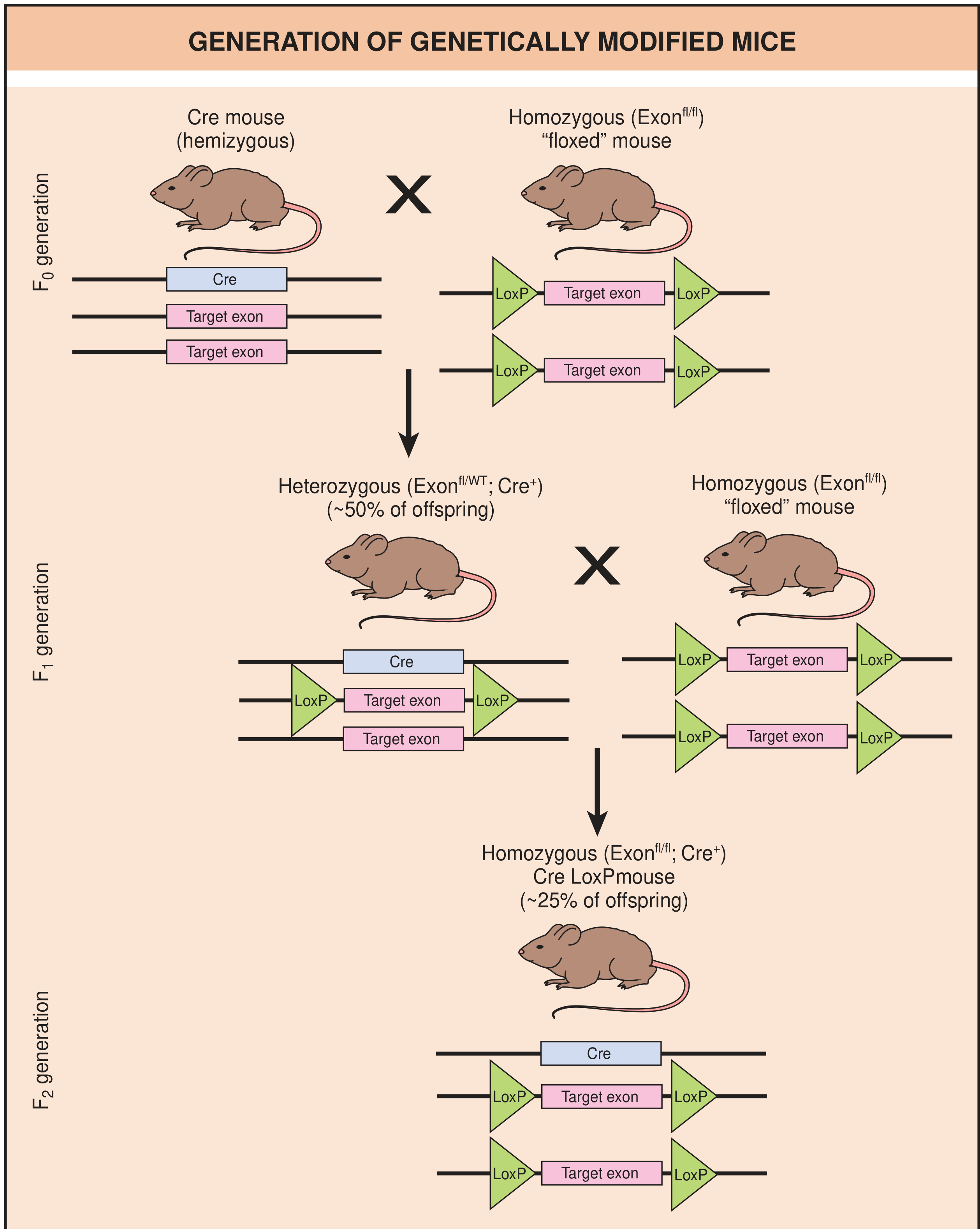
- Cre mouse (hemizygous) × Floxed mouse (homozygous, Exon^fl/fl)
- All offspring carry one floxed allele and ~50% also carry the Cre gene.
- F1 mice that are Exon^fl/WT; Cre+ are bred back to homozygous floxed mice (Exon^fl/fl)
- This produces mixed litters.
- ~25% of F2 offspring are homozygous floxed AND carry Cre (Exon^fl/fl; Cre+)
- In these mice: wherever the tissue-specific promoter activates Cre → Cre enzyme cuts the loxP sites → target exon is looped out and deleted → that tissue has no functional copy of the gene.
- Every other tissue: gene intact and working normally.
The "Knock-in" Technology Mention
Why This Matters: Applications
| Application | Example |
|---|---|
| Study gene function in one organ | Delete insulin receptor only in brain to study diabetes and cognition separately |
| Avoid embryonic lethality | Many genes essential for development can be studied in adult tissue if deleted only later |
| Disease modeling | Delete cartilage-specific genes in joints to study osteoarthritis |
| Cancer research | Delete tumor suppressor genes only in specific tissues to generate organ-specific cancer models |
| Neuroscience | Delete synaptic genes in specific brain regions (e.g., hippocampus) to study memory |
Summary in Simple Steps
- Identify the gene you want to knock out in a specific tissue.
- Flank it with loxP sites using embryonic stem cell engineering → create the "floxed mouse."
- Generate a Cre mouse that expresses Cre only in your target tissue using a tissue-specific promoter.
- Breed the two lines together.
- Offspring that carry both the floxed gene AND the Cre gene will have their target gene silenced only in that specific tissue - a conditional tissue-specific knockout.
Gene knock in vs knock out
gene knock-in vs knock-out difference mechanism comparison molecular biology
Gene Knock-Out vs. Gene Knock-In
The One-Line Difference
| What happens to the gene | |
|---|---|
| Knock-Out (KO) | A gene is disabled / deleted - it no longer produces a functional protein |
| Knock-In (KI) | A new or modified gene sequence is inserted at a precise, defined location |
Shared Foundation: Homologous Recombination
- Contains sequences identical to the regions flanking the gene of interest (called "homology arms")
- Carries whatever modification they want to make (deletion, insertion, point mutation)
Gene Knock-Out (KO) - Loss of Function
Goal
Mechanism
-
A targeting vector is built containing:
- Left homology arm (matches DNA upstream of target gene)
- A neomycin resistance gene (neo) or other selectable marker inserted in the middle - this physically disrupts the target gene's coding sequence
- Right homology arm (matches DNA downstream of target gene)
- A thymidine kinase (tk) gene at the outer end - used for negative selection
-
The vector is transfected into mouse embryonic stem (ES) cells.
-
Double selection is applied:
- Neomycin kills cells where no insertion occurred at all
- Ganciclovir kills cells where random (non-targeted) insertion occurred - because only randomly inserted DNA retains the tk gene, which converts ganciclovir to a toxic product
- Only cells where homologous recombination occurred survive both - they have neo but NOT tk
-
Surviving ES cells have one disrupted copy of the target gene (heterozygous KO).
-
These ES cells are injected into a mouse blastocyst and implanted into a pseudopregnant female.
-
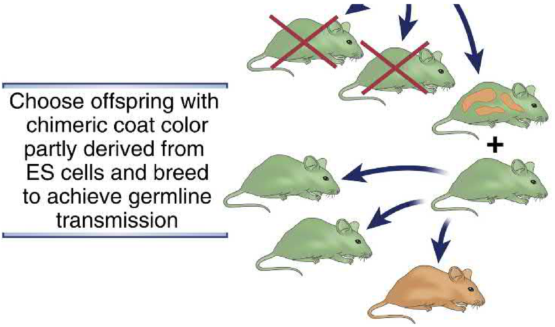
A chimeric mouse is born - some tissues come from normal blastocyst cells, others from the engineered ES cells. Chimeric mice can be identified by their mixed coat color (see diagram below).
-
Chimeric mice are bred further until offspring that are homozygous for the knocked-out gene are obtained (both copies disrupted = complete knockout).
Result
Limitations (from Cellular & Molecular Immunology textbook)
- Developmental compensation - other genes may take over the function of the deleted gene during embryonic development, masking the phenotype.
- Embryonic lethality - if the knocked-out gene is essential for development, the embryo dies before you can study it in adults.
- Cannot isolate tissue-specific effects - if a gene matters in both liver and brain, a global KO cannot tell you which tissue is responsible (→ this limitation is solved by the Cre/loxP conditional KO system covered previously).
- Selectable marker interference - the inserted neo gene remains in the genome and may unexpectedly affect nearby gene expression.
Gene Knock-In (KI) - Gain or Replacement of Function
Goal
Mechanism
- Instead of a disruptive selectable marker, it carries a desired exogenous gene or a point-mutated version of the endogenous gene.
- The homology arms still guide the vector to the correct chromosomal location.
- Homologous recombination swaps in the new sequence in place of the original.
Types of Knock-In Models
| Type | What Is Inserted | Purpose |
|---|---|---|
| Point mutation KI | One or a few altered base pairs | Model a human disease mutation (e.g., sickle cell anemia point mutation) |
| Reporter KI | Fluorescent protein (GFP, tdTomato) | Track when/where a gene is expressed in real time |
| Humanized KI | Human gene replaces the mouse gene | Study human proteins in a living animal (e.g., human HLA genes) |
| Disease model KI | Pathogenic mutation from a patient | Recreate a genetic disease exactly as it occurs in humans |
| Defined-site transgene | Any exogenous gene at a specific locus | Ensure transgene is under control of a known endogenous promoter/enhancer |
Key Advantage Over Random Transgenic Mice
Side-by-Side Comparison Table
| Feature | Knock-Out | Knock-In |
|---|---|---|
| Core action | Gene disruption / deletion | Gene insertion / replacement |
| Effect on protein | No protein produced (loss of function) | New or modified protein produced |
| Mechanism | Homologous recombination with disruptive vector | Homologous recombination with replacement vector |
| Uses selectable markers | Yes (neo, tk) | Yes, but different design |
| Question it answers | "What does this gene do?" | "What happens with this specific mutation or gene?" |
| Disease models | Models loss-of-function diseases (e.g., SCID, CF) | Models gain-of-function or point-mutation diseases (e.g., sickle cell, cancer mutations) |
| Can introduce reporter genes? | No | Yes (GFP knock-ins track gene expression) |
| Risk of embryonic lethality | High (if gene is essential) | Lower (gene may still be partially functional) |
| Modern tool | CRISPR indel (creates frameshift = KO) | CRISPR + HDR template (precise insertion = KI) |
The Modern Approach: CRISPR/Cas9
-
Knockout with CRISPR: Cas9 cuts the DNA at the target gene. The cell repairs it via NHEJ (error-prone repair) → random insertions/deletions (indels) → frameshift mutation → non-functional protein. Fast, cheap, and highly efficient.
-
Knock-in with CRISPR: Cas9 cuts the DNA, but a repair template (HDR template) containing the desired sequence is also provided. The cell uses HDR (homology-directed repair) to incorporate the new sequence precisely. More difficult than KO but increasingly routine.
Practical Examples
| Disease/Research Goal | Technique Used |
|---|---|
| Study what the BRCA1 gene does | BRCA1 knockout mouse |
| Model sickle cell disease in mice | Knock-in of the human HbS point mutation |
| Track B-cell development visually | GFP knocked into a B-cell specific gene locus |
| Study Alzheimer's disease | Knock-in of human presenilin mutations (PS1, PS2) |
| Understand immune deficiency | RAG1/RAG2 knockout = severe combined immunodeficiency (SCID) model |
| Study a cancer driver mutation | KRAS^G12D knock-in = pancreatic cancer mouse model |
In Summary
- Knock-out = take something away → reveals what a gene does through its absence
- Knock-in = put something new in at a precise location → models specific mutations, introduces reporters, or replaces a gene with a human version
- Both exploit homologous recombination (or CRISPR) to make precise, heritable changes to the genome
- Both require ES cells, blastocyst injection, chimeric mice, and selective breeding to establish stable lines
Transgenic animal
transgenic animals definition methods applications examples biology
Transgenic Animals
Definition
- Heritable - passed on to offspring
- Expressed - produces a protein in the relevant tissues
- Stably integrated - becomes a permanent part of that animal's chromosomes
The Transgene Construct
[ PROMOTER / ENHANCER ] ——→ [ GENE OF INTEREST (coding sequence) ]
        ↑                              ↑
  Controls when/where           The actual gene to be expressed
  the gene is turned on
-
Promoter: Acts as a switch. Can be:
- Constitutive (always on, in all tissues) - e.g., CMV promoter
- Tissue-specific - e.g., rat insulin promoter → expression only in pancreatic β-cells
- Inducible - turned on/off by a drug (e.g., tetracycline-inducible system)
-
Gene of interest: Can encode any protein - a human disease gene, a reporter (like GFP), a hormone, an oncogene, etc.
Methods of Creating Transgenic Animals
Method 1: Pronuclear Microinjection (Classic Method - Most Common)
- Superovulation - A female mouse is given hormones to produce many eggs at once.
- Fertilization - Eggs are fertilized (in vitro or by mating).
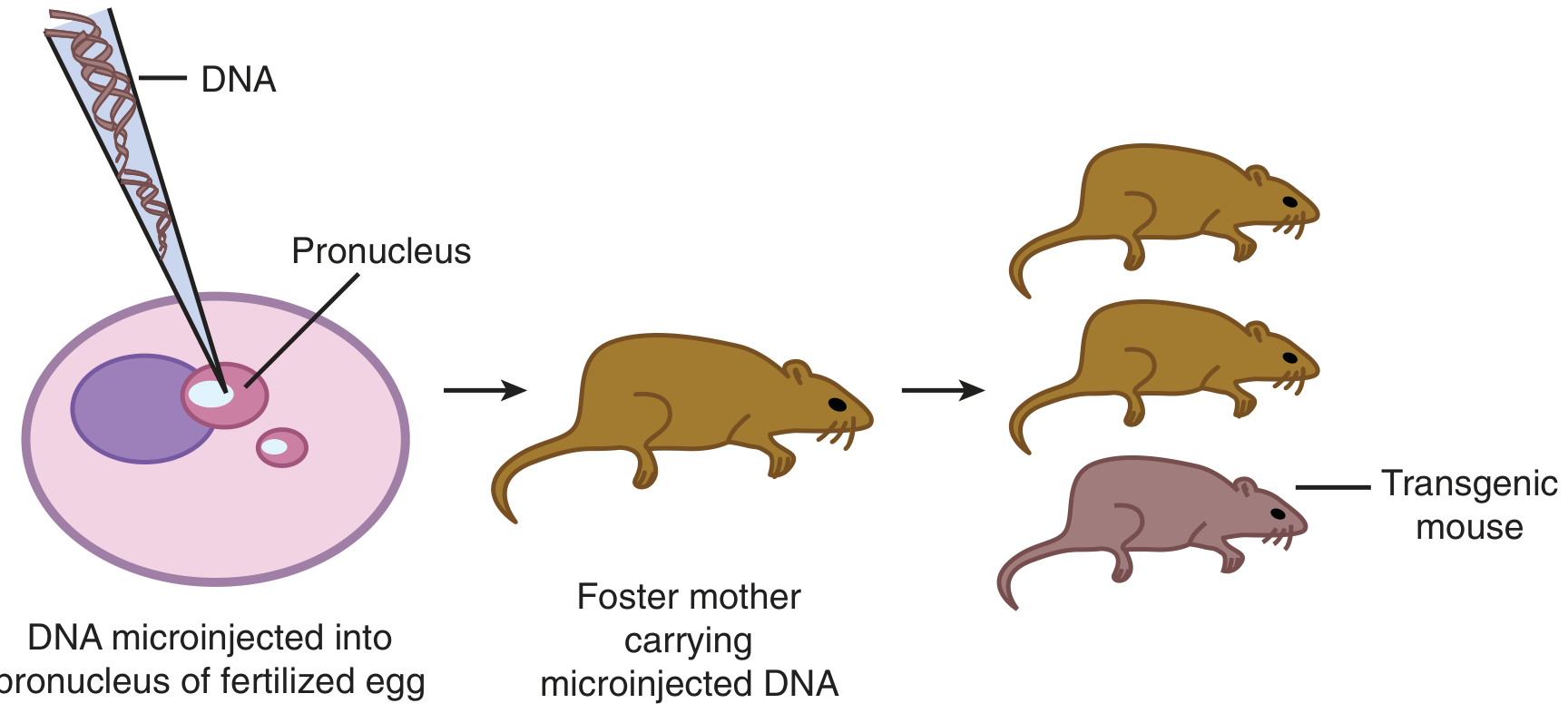
- Microinjection - A very fine glass needle injects hundreds of copies of the linearized transgene DNA directly into the pronucleus of the fertilized egg (the pronucleus is the nucleus of the sperm or egg just after fertilization, before they fuse).
- Implantation - The injected egg is implanted into the uterus of a pseudopregnant foster mother (a female prepared hormonally to accept embryos).
- Birth of Founder Mouse - Offspring that were born carry the transgene integrated into one chromosome = heterozygous founder mouse.
- Genotyping - A small piece of tail tissue is taken and DNA is extracted. PCR or Southern blot analysis confirms which pups incorporated the transgene.
- Breeding - Founder mice are bred with normal mice. Per Mendelian genetics, ~50% of offspring will be heterozygous transgenic. Two heterozygous mice are then bred to produce homozygous transgenics (transgene on both chromosomes = double dose).
Method 2: Embryonic Stem (ES) Cell-Mediated Transgenesis
- Transgene is introduced into pluripotent ES cells in culture via electroporation or lipofection.
- Engineered ES cells are injected into a blastocyst.
- A chimeric mouse is born; selected by coat color and bred to transmit the transgene through the germline.
- This method allows more controlled integration (can combine with homologous recombination for knock-in).
Method 3: Retrovirus-Mediated Gene Transfer
- A retrovirus carrying the transgene is used to infect early embryos.
- The viral machinery integrates the transgene into the host genome.
- Limitation: transgene size is restricted to ~8 kb; there is a risk of insertional mutagenesis.
Method 4: Sperm-Mediated Gene Transfer (SMGT)
- Sperm cells are incubated with foreign DNA, which they can absorb.
- These sperm are then used to fertilize eggs.
- Simpler but less reliable - efficiency is variable.
Method 5: Somatic Cell Nuclear Transfer (SCNT) / Cloning
- A somatic (body) cell is engineered to carry the transgene in the lab.
- Its nucleus is transferred into an enucleated egg cell.
- The egg develops into an embryo - all cells carry the transgene.
- Used to produce large transgenic farm animals (e.g., Dolly the sheep).
Modern Method: CRISPR/Cas9
- Increasingly replacing older methods.
- Can inject Cas9 protein + guide RNA + donor template directly into the fertilized egg.
- Allows precise, targeted integration - not random like pronuclear injection.
- Much faster and cheaper.
Breeding to Establish a Stable Line
Founder (heterozygous, one chromosome has transgene)
            ↓ × normal mouse
    50% transgenic offspring (heterozygous)
            ↓ × another heterozygous transgenic
    25% homozygous transgenic (two copies of transgene)
Applications of Transgenic Animals
1. Basic Research - Understanding Gene Function
- Overexpress a growth factor or oncogene to see what happens.
- Express dominant-negative mutants that block endogenous protein function.
- Use tissue-specific promoters to study a gene's role in just one organ.
- Example: Transgenic mice overexpressing HER2/neu oncogene in the mammary gland develop breast tumors → model for breast cancer research.
2. Disease Modeling
- Introduce human disease genes into mice to recreate the disease.
- Examples:
- Alzheimer's disease: Mice expressing mutant human amyloid precursor protein (APP) develop amyloid plaques.
- Sickle cell disease: Mice with human HbS gene develop sickling.
- Cystic fibrosis: Mice with CFTR mutation.
- HLA-B27 transgenic rats → develop spontaneous spondyloarthropathy (used in rheumatology research). - Rheumatology 2022, Elsevier
- TNF transgenic mice → spontaneously develop inflammatory arthritis.
3. Reporter Lines
- A reporter gene (GFP - green fluorescent protein, luciferase, β-galactosidase) is fused to a promoter.
- Whenever that promoter is active, cells glow or produce a detectable signal.
- Used to track exactly when and where a gene is normally expressed during development.
4. Drug Testing & Therapeutic Development
- Transgenic disease models allow preclinical testing of new drugs before human trials.
- Example: Alzheimer's transgenic mice are used to test amyloid-clearing therapies.
5. Biopharmaceutical Production ("Pharming")
- Human proteins that are difficult or expensive to produce in bacteria can be expressed in transgenic animal milk (mammary gland-specific promoter).
- Examples:
- Transgenic goats producing human antithrombin in milk (approved by FDA/EMA)
- Cows producing human lactoferrin or insulin
- Pigs producing human growth hormone
6. Xenotransplantation
- Pigs are genetically engineered to express human surface proteins (e.g., human CD46, CD55, CD59) to reduce immune rejection when their organs are transplanted into humans.
- Also, human genes like human leukocyte antigens are knocked in to make pig organs "look more human" to the immune system.
7. Immunology Research
- TCR transgenic mice: All T cells express the same T-cell receptor of known antigen specificity → ideal tool to study antigen-specific immune responses with large, pure populations. - Cellular & Molecular Immunology, Abbas et al., 10th Ed.
Limitations
| Limitation | Explanation |
|---|---|
| Random integration | Transgene lands anywhere - may disrupt another gene or be silenced by nearby chromatin |
| Variable expression | Different founder lines have different expression levels depending on insertion site |
| Insertional mutagenesis | Integration may accidentally knock out an endogenous gene |
| Species differences | A human transgene expressed in mice may behave differently than in humans |
| Only adds, doesn't remove | Classic transgenics only add a gene; you cannot study gene deletion with this alone (→ need knockout) |
| Timing control is difficult | Constitutive expression from birth may cause developmental compensation |
Transgenic vs. Knockout - Quick Reminder
| Transgenic | Knockout | |
|---|---|---|
| Action | Adds a new gene | Removes/disables an existing gene |
| Question | "What does this gene DO when present/overexpressed?" | "What happens when this gene is ABSENT?" |
| Integration | Random (pronuclear injection) or targeted (ES cells) | Targeted (homologous recombination) |
| Loss or gain? | Usually gain-of-function | Loss-of-function |
Nudes mouses
nude mice athymic immunodeficient mouse model research uses explained
https://www.jax.org/news-and-insights/jax-blog/2019/december…
Nude Mice (Athymic Mice)
What Are They?
- No functional thymus (athymic) → no mature T lymphocytes → severely compromised immune system
- Hairless (nude) appearance → sparse, abnormal whiskers and fur that break as they grow
The Genetic Cause: Foxn1 Mutation
The Gene
- The nude phenotype is caused by a recessive mutation in the Foxn1 gene (Forkhead Box N1), located on mouse chromosome 11.
- Written as nu/nu in homozygous form (both copies mutated = full nude phenotype).
- Heterozygous mice (nu/+) have one functional copy → normal thymus, normal fur → they look and behave like wild-type mice.
What Foxn1 Normally Does
- The thymic stroma - shapes the thymic microenvironment needed for T-cell maturation
- The skin - drives normal hair follicle and nail development
What Happens When Foxn1 Is Mutated
Foxn1 mutated (nu/nu)
        ↓
Thymic epithelial cells fail to differentiate properly
        ↓
No functional thymic microenvironment
        ↓
T-cell progenitors arrive from bone marrow but CANNOT undergo
positive or negative selection → no mature T cells are released
        ↓
Near-total absence of CD4+ helper T cells AND CD8+ cytotoxic T cells
in blood, spleen, and lymph nodes
Immune Profile of Nude Mice
| Cell Type | Status | Consequence |
|---|---|---|
| T cells (CD4+ and CD8+) | Absent | Cannot reject foreign tissue; cannot fight viruses |
| B cells | Present but reduced/dysfunctional | Without T-cell help, B cells produce minimal antibodies |
| NK cells | Present and active | Key limitation - they can still kill tumor cells |
| Macrophages | Present | Some innate immunity retained |
| Neutrophils | Present | Innate responses intact |
Why This Makes Them Invaluable in Research
- Human cells, tissues, or tumors transplanted into a nude mouse will survive and grow - something that would be instantly rejected in a normal mouse.
- They are essentially a "living test tube" for foreign biological material.
Key Research Applications
1. Xenograft Models (Cancer Research - Most Important Use)
- Human cancer cell lines (e.g., MCF-7 breast cancer, A549 lung cancer, HCT116 colon cancer) are injected subcutaneously into the flank of a nude mouse.
- The tumor grows predictably.
- New anticancer drugs are tested on these tumors to assess:
- Does the drug shrink the tumor?
- Is there toxicity to the mouse?
- What is the effective dose?
- A patient's actual tumor biopsy (not a cell line) is implanted directly into nude mice.
- Preserves the tumor's original heterogeneity and microenvironment more accurately than cell lines.
- Used for personalized medicine - testing which drug works best for a specific patient's tumor.
2. Metastasis Models
- Human cancer cells can be injected into the tail vein or cardiac circulation.
- They travel through the bloodstream and form metastatic deposits in lungs, liver, bone.
- Used to study the biology and treatment of cancer metastasis.
3. Immunology Research
- Adoptive transfer experiments: Human immune cells can be engrafted into nude mice to partially reconstitute a human immune system - used to study human immune responses in vivo.
- Study what happens in the complete absence of T-cell mediated immunity.
- Can transfer specific T-cell populations to study their individual roles.
4. Tissue Transplantation Research
- Organ or tissue grafts from other species survive in nude mice, allowing study of tissue function, vascularization, and rejection mechanisms without interference from T cells.
5. Stem Cell Research
- Human pluripotent stem cells can be injected into nude mice - they form teratomas (benign tumors containing multiple tissue types), which is a standard test to confirm pluripotency.
Limitations of Nude Mice
| Limitation | Why It Matters |
|---|---|
| NK cells are intact | NK cells can still kill human tumor cells → some xenografts fail. For NK-resistant tumors this is less of a problem. |
| Some residual T cells | A tiny number of atypical T cells (called "leaky" T cells) can develop via extrathymic pathways. |
| No T-cell biology possible | Cannot study T-cell dependent immune responses. |
| Not suitable for humanized immune studies | NK cells and macrophages can eliminate injected human immune cells. Better models (NSG, NRG) exist for humanization. |
| Susceptible to infections | Must be housed in strict specific pathogen-free (SPF) conditions, often in individually ventilated cages (IVCs) or isolators. |
Comparison with Other Immunodeficient Mouse Models
| Model | T cells | B cells | NK cells | Best Use |
|---|---|---|---|---|
| Nude (nu/nu) | Absent | Present (reduced) | Present | CDX tumor models, basic xenografts |
| SCID | Absent | Absent | Present | Better for B-cell studies |
| NOD-SCID | Absent | Absent | Reduced | Humanized mice, improved xenografts |
| NSG / NRG / NOG | Absent | Absent | Absent | Best human cell engraftment, humanized immune system models |
Housing Requirements
- Housed in laminar flow hoods, isolators, or individually ventilated cages (IVCs).
- Strictly germ-free or SPF conditions.
- All bedding, food, and water must be autoclaved/sterilized.
- Handlers must wear full protective gear.
- Antibiotic supplementation in water is sometimes used prophylactically.
Summary
| Feature | Detail |
|---|---|
| Mutation | Foxn1 (nu/nu) - recessive |
| Thymus | Absent / non-functional |
| T cells | Absent |
| Appearance | Hairless, whiskerless |
| Key property | Cannot reject foreign tissue |
| Primary use | Xenograft tumor models, cancer drug testing |
| Limitation | NK cells still present |
| Alternative | NSG mice (no T, B, or NK cells) |
Cpcsea guidlines
CPCSEA guidelines India animal experiments Committee Purpose Control Supervision 2018
https://jssaherdatalake.blob.core.windows.net/web-jssaher/CP…
https://wjpsronline.com/images/d192e24f99b2f3ba65b43234055ef…
CPCSEA Guidelines
What is CPCSEA?
- A statutory committee established under Section 15 of the Prevention of Cruelty to Animals (PCA) Act, 1960 by the Government of India.
- Functions under the Ministry of Environment, Forest and Climate Change (MoEFCC).
- Has been operational since 1991.
- The current consolidated reference is the Compendium of CPCSEA 2018.
- Enforces compliance with the Breeding of and Experiments on Animals (Control and Supervision) Rules, 1998 and its amendments.
Core Mandate
"To ensure that animals are not subjected to unnecessary pain or suffering before, during, or after the performance of experiments on them."
Why CPCSEA Exists
- Prevent unnecessary cruelty and suffering to animals
- Promote humane and ethical science
- Ensure reproducibility and scientific validity of animal experiments
- Bring Indian standards in line with international norms (like the 3Rs, OECD guidelines)
The 3Rs Principle - Foundation of CPCSEA Ethics
| R | Meaning | What It Requires |
|---|---|---|
| Replace | Avoid using animals wherever possible | Use cell cultures, computer simulations, in vitro models, or lower organisms first |
| Reduce | Use the minimum number of animals necessary | Statistical justification for sample size; avoid redundancy |
| Refine | Minimize pain, distress, and suffering | Better analgesia, anaesthesia, humane endpoints, improved housing |
CPCSEA's Key Functions
- Registration of institutions and establishments that conduct animal experiments or breed animals for research purposes.
- Appointment of nominees to the Institutional Animal Ethics Committees (IAECs) of registered establishments.
- Approval of animal house facilities based on inspection reports.
- Permission for conducting experiments on large animals (canines, primates, large livestock).
- Training programs for IAEC nominees and animal house personnel.
- Inspection and monitoring of registered facilities (routine/annual inspections).
- Revocation of registration if norms are violated.
The IAEC: Institutional Animal Ethics Committee
Composition (Mandatory - 8 Members)
| # | Member |
|---|---|
| 1 | One biological scientist |
| 2 | Two scientists from different biological disciplines |
| 3 | One veterinarian involved in animal care |
| 4 | One scientist in-charge of the animal facility |
| # | Member |
|---|---|
| 6 | One CPCSEA nominee (external expert) |
| 7 | One scientist from a different institution |
| 8 | One socially aware community member / lay person (to represent public interest) |
Quorum
- Minimum members must be present for a valid meeting; the CPCSEA nominee's presence is mandatory.
- If meetings are not held for 6 consecutive months, the nominee must notify CPCSEA.
Powers of the IAEC
- Reviews and approves all research protocols involving animals before the study begins.
- Can approve experiments using small animals (rats, mice, guinea pigs, rabbits) independently.
- For large animals (dogs, primates, cattle, sheep): IAEC can only recommend - final approval comes from CPCSEA directly.
- Monitors ongoing studies and receives periodic reports.
- Conducts visits to the animal house and laboratory.
Ethical Principles for Animal Experiments (CPCSEA Principles)
Principle 1 - Permitted Purposes
- Advancement of new physiological knowledge
- Knowledge expected to save or prolong human life or alleviate suffering
- Significant gains in the well-being of people
- Combating any disease (human, animal, or plant)
Principle 2 - Phylogenetic Scale
- Prefer bacteria → invertebrates → fish → rodents → over primates or dogs
- Use animals with the least capacity to perceive pain and suffering.
Principle 3 - Minimum Numbers
Principle 4 - Avoid Unnecessary Procedures
Principle 5 - Analgesia and Anaesthesia
- Experiments involving pain must be performed under appropriate anaesthesia.
- Post-operative analgesia must be provided.
- Animals must not be allowed to recover from surgery only to experience severe pain.
Principle 6 - Humane Endpoints
Principle 7 - Qualified Personnel
- Experiments shall be performed only by or under the supervision of qualified persons (degree/diploma in Veterinary Science, Medicine, or Laboratory Animal Science).
- All personnel handling animals must be trained.
Animal House Facility Guidelines
Location and Infrastructure
- Must be housed in an isolated building, separated from offices, public areas, and human habitation.
- Located away from dust, smoke, noise, wild rodents, insects, and birds.
- Avoid sharp fluctuations in temperature, humidity, light, sound, and ventilation.
- Must have dedicated rooms for each species (species should not be housed together).
- Separate areas for: quarantine, surgery, post-operative recovery, necropsy, storage.
Environmental Controls
| Parameter | Requirement |
|---|---|
| Temperature | 21-23°C (rodents); species-specific |
| Humidity | 50-70% relative humidity |
| Light cycle | 12 hours light / 12 hours dark (consistent) |
| Ventilation | 10-15 air changes per hour |
| Noise | Minimal; avoid sudden loud sounds |
Housing and Caging
- Cages must allow natural behavior - social housing where species-appropriate.
- Environmental enrichment (nesting material, tunnels, toys) is encouraged to promote mental well-being.
- Adequate space per animal as per species-specific norms.
- Bedding must be clean, non-toxic, absorbent.
Feed and Water
- Nutritionally adequate, standardized diet.
- Fresh, uncontaminated water at all times (autoclaved for immunodeficient animals).
- Feed and water containers cleaned and sterilized regularly.
Record-Keeping (Mandatory)
- Species, strain, source, and number of animals
- Date of arrival, acclimatization, and experiment
- Health records and veterinary interventions
- Breeding records
- Experimental protocols and outcomes
- Records of euthanasia
Registration and Approval Process
Who Needs to Register?
- Any institution, laboratory, pharmaceutical company, or educational establishment that uses or breeds animals for scientific purposes must be registered with CPCSEA.
- Over 665 laboratories are registered nationwide.
Registration Application Requires:
- Name and address of the organization
- Purpose of registration (research, education, breeding, commercial, etc.)
- List of species to be used
- Composition of IAEC (with qualifications)
- Minutes of the last IAEC meeting
- Details of animal house facility (location, dimensions, environmental controls)
- Annual renewal with inspection reports
Inspection
- CPCSEA conducts routine/annual inspections of all registered facilities.
- Inspection report covers: species maintained, cage conditions, environmental controls, documentation, compliance with 3Rs, staff training, and IAEC functioning.
Protocol Approval (Form B)
- Title of the project
- Species, strain, sex, age, and number of animals to be used
- Scientific justification for using animals (why in vitro/alternatives are insufficient)
- Justification for species and number chosen (3Rs compliance)
- Experimental procedures in detail
- Pain and distress assessment category
- Anaesthesia and analgesia plan
- Proposed humane endpoints
- Fate of animals after experiment (continued use, re-homing, euthanasia)
Special Provisions
Non-Human Primates (NHPs)
- Highly restricted use.
- Must be approved directly by CPCSEA (IAEC alone cannot authorize).
- Only specific institutions with full facilities and expertise may house and use NHPs.
- Strong preference for non-primate alternatives.
Import of Animals
- No animal may be imported without CPCSEA permission.
- Exception: genetically defined laboratory rats and mice (e.g., inbred strains) may be imported by registered breeders.
Immune Biologicals from Equines (Horses)
- Production of antiserum (e.g., anti-snake venom, anti-tetanus) from horses has a specific protocol ratified by the Supreme Court of India through CPCSEA oversight.
Breeding Establishments
- Facilities that breed animals purely for supply to research institutes must also be registered with CPCSEA.
- Must maintain genetic purity and health records.
Penalties for Violation
- Conducting experiments without CPCSEA registration = criminal offense.
- CPCSEA can cancel/suspend registration.
- CPCSEA nominees can recommend halting of ongoing experiments if cruelty or non-compliance is found.
- Fines and imprisonment possible under PCA Act provisions.
Summary Table
| Aspect | Details |
|---|---|
| Governing Act | Prevention of Cruelty to Animals Act, 1960 |
| Rules | Breeding of and Experiments on Animals Rules, 1998 (amended) |
| Ministry | MoEFCC, Government of India |
| Foundation principle | 3Rs (Replace, Reduce, Refine) |
| On-ground body | IAEC (Institutional Animal Ethics Committee) |
| IAEC composition | 8 members (5 internal + 3 external including CPCSEA nominee) |
| Small animal approval | IAEC alone can approve (rats, mice, guinea pigs, rabbits) |
| Large animal approval | CPCSEA directly (dogs, primates, cattle) |
| Documentation | Form B (standard) / Form C (non-human primates) |
| Facility requirements | Isolated building, controlled environment, enrichment, records |
| Current reference | CPCSEA Compendium 2018 |
Pcr technique
PCR polymerase chain reaction steps mechanism types applications explained
Polymerase Chain Reaction (PCR)
What Is PCR?
- Invented by Kary Mullis in 1983 → Nobel Prize in Chemistry, 1993
- With 30 cycles, PCR achieves approximately 10⁹-fold (1 billion times) amplification of the target sequence.
- The amplified product is called an amplicon.
Essential Components (What Goes Into the Tube)
| Component | Role |
|---|---|
| Template DNA | The source DNA containing the sequence you want to copy |
| Forward Primer | Short, single-stranded DNA (~20 bases) that binds to one strand of the template at the start of the target region |
| Reverse Primer | Binds to the opposite strand at the other end of the target region |
| Taq DNA Polymerase | Thermostable enzyme (from Thermus aquaticus bacteria) that synthesizes new DNA strands |
| dNTPs | Deoxyribonucleotide triphosphates (dATP, dCTP, dGTP, dTTP) - the building blocks for new DNA |
| MgCl₂ buffer | Magnesium acts as a cofactor for Taq polymerase; the buffer maintains optimal pH and ionic conditions |
Why Taq Polymerase Specifically?
The Three Steps of One PCR Cycle
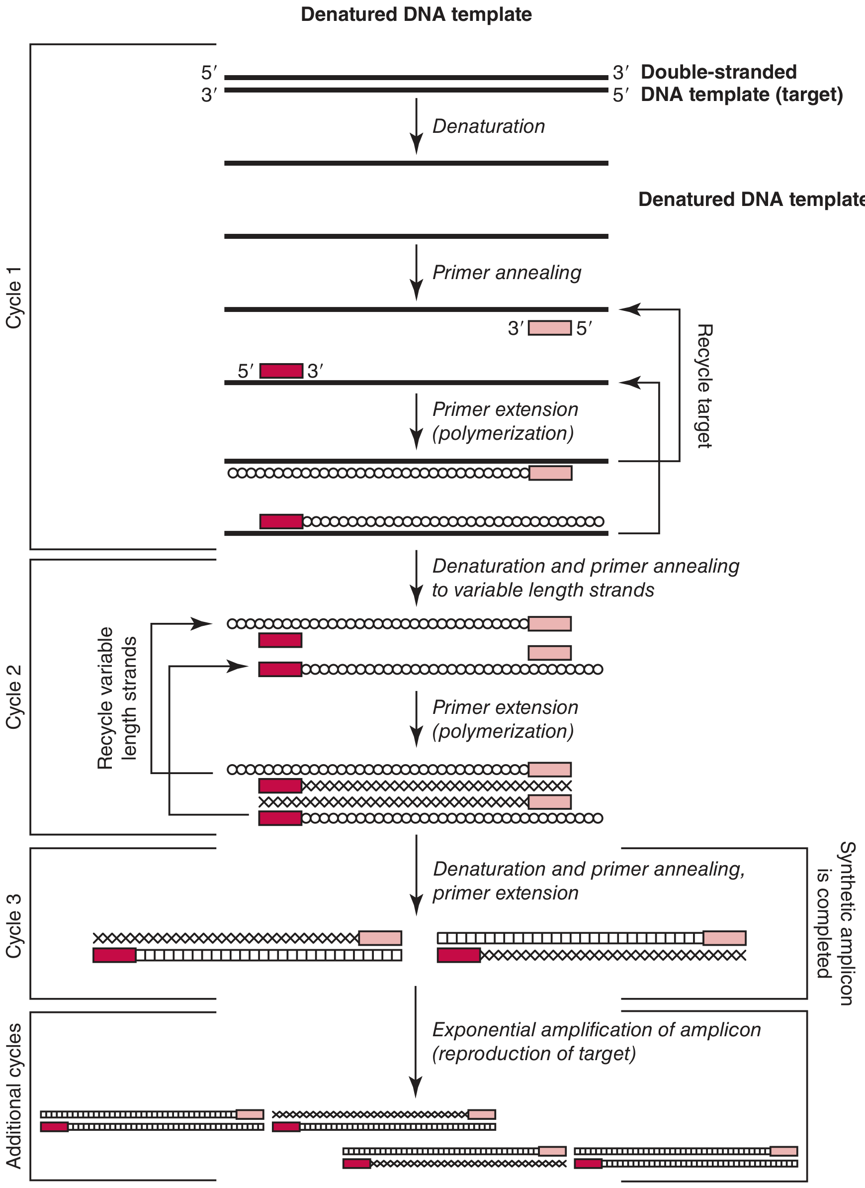
Step 1: Denaturation - 94-95°C (30-60 seconds)
- The reaction tube is heated to ~94-95°C.
- The hydrogen bonds between the two strands of the double-stranded DNA template are broken.
- The double helix "unzips" into two single-stranded templates.
- Both single strands are now available as templates for copying.
Double-stranded DNA  →(heat to 94°C)→  Two single strands
5'——————————————3'                     5'——————————3'
3'——————————————5'                     3'——————————5'
Step 2: Annealing (Primer Hybridization) - 50-65°C (30-60 seconds)
- The temperature is lowered to allow the primers to bind.
- The forward primer binds (anneals) to one single strand.
- The reverse primer binds to the complementary strand.
- Primers bind by Watson-Crick base pairing (A-T, G-C) to their complementary sequences.
- The annealing temperature is carefully chosen - too low → non-specific binding; too high → primers fall off.
Step 3: Extension (Elongation) - 72°C (1 minute per 1 kb of product)
- Temperature is raised to 72°C - the optimal temperature for Taq polymerase activity.
- Taq polymerase binds to the primer-template junction and begins reading the template in the 3' direction.
- It adds complementary dNTPs one by one, synthesizing a new DNA strand.
- Extension proceeds until the end of the template (or the time allotted).
- Result: two new double-stranded DNA molecules where there was one.
Exponential Amplification - The Mathematics
| Cycle | Number of DNA copies |
|---|---|
| 0 (start) | 1 |
| 1 | 2 |
| 2 | 4 |
| 3 | 8 |
| 10 | 1,024 |
| 20 | 1,048,576 (>1 million) |
| 30 | >1 billion |
| 35 | ~34 billion |
Initial and Final Steps
- Initial denaturation: 94-95°C for 2-5 minutes at the start - fully denatures all template DNA before cycling begins.
- Final extension: 72°C for 5-10 minutes at the end - ensures all partially extended strands are completed.
- Hold: 4°C to store the completed PCR product.
Detection of PCR Products
Agarose Gel Electrophoresis (Classic)
- PCR product is loaded onto an agarose gel.
- Electric current separates DNA by size (smaller fragments travel faster).
- Gel is stained with ethidium bromide or SYBR Safe.
- A band at the expected size = positive result.
- A DNA ladder (size marker) in the adjacent lane confirms the correct product size.
Real-Time PCR (see below) - detects fluorescence during the reaction itself.
Types of PCR
1. Standard / Conventional PCR
- Basic amplification; product detected after the reaction by gel electrophoresis.
- Used for cloning, genotyping, detecting presence/absence of a gene.
2. Real-Time PCR (qPCR - Quantitative PCR)
- Fluorescence is measured at every cycle as the reaction progresses.
- Amount of product is monitored in real time.
- The Ct value (Cycle Threshold) = the cycle number at which fluorescence crosses a set threshold.
- Lower Ct = more starting template (earlier detection)
- Higher Ct = less starting template
- Can precisely quantify how much DNA/RNA was present in the original sample.
- Detection methods:
- SYBR Green - fluorescent dye that binds to any double-stranded DNA; simple but less specific.
- TaqMan probes (hydrolysis probes) - probe with a fluorophore and quencher; Taq polymerase degrades the probe during extension, releasing fluorescence. Highly specific.
- Molecular beacons / FRET probes - other probe-based methods for even greater specificity.
3. RT-PCR (Reverse Transcription PCR)
- Used when the starting material is RNA (not DNA).
- RNA cannot be directly amplified by Taq polymerase.
- Step 1: Reverse transcriptase enzyme converts RNA → cDNA (complementary DNA)
- Step 2: Standard PCR amplifies the cDNA.
- Key uses:
- Measuring gene expression (mRNA levels) - called RT-qPCR when quantitative
- Detecting RNA viruses (e.g., COVID-19, Influenza, HIV, Hepatitis C)
- "A reverse transcription reaction step is required before the PCR amplification of RNA targets (RT-PCR) that relies on enzymes required for the replication of RNA viruses (reverse transcriptase)." - Tietz Textbook of Laboratory Medicine, 7th Ed.
4. Multiplex PCR
- Multiple primer pairs used in a single reaction.
- Amplifies several different target sequences simultaneously.
- Used for detecting multiple pathogens at once, or multiple mutations in one run.
5. Nested PCR
- Two rounds of PCR with two sets of primers.
- First PCR amplifies a large region; second PCR uses primers nested inside that product.
- Greatly increases specificity and sensitivity.
- Used for detecting very low amounts of target (e.g., TB DNA in CSF).
6. Hot-Start PCR
- Taq polymerase is kept inactive at low temperatures (either by antibody or wax barrier).
- Only activates at 94-95°C during the first denaturation.
- Prevents non-specific amplification that can occur when primers bind non-specifically at room temperature.
7. Digital PCR (dPCR)
- The sample is partitioned into thousands of tiny individual reactions.
- Each partition either contains the target or does not.
- After amplification, each partition is read as positive (1) or negative (0) - like digital bits.
- Provides absolute quantification without needing a standard curve.
- More accurate than qPCR for rare targets (e.g., circulating tumor DNA). - Tietz Textbook of Laboratory Medicine, 7th Ed.
8. LAMP (Loop-Mediated Isothermal Amplification)
- A PCR-related technique that amplifies DNA at a constant temperature (no thermocycler needed).
- Used in rapid point-of-care diagnostics in resource-limited settings.
Applications of PCR
1. Medical Diagnostics
- Infectious diseases: Detect bacterial, viral, fungal, parasitic DNA/RNA - COVID-19, TB, HIV, Hepatitis, STIs, sepsis workup
- Genetic disorders: Detect mutations causing cystic fibrosis, sickle cell disease, hemophilia
- Cancer: Detect oncogene mutations (KRAS, BRAF, EGFR), chromosomal translocations (BCR-ABL in CML), minimal residual disease
- Prenatal diagnosis: Detect genetic disorders from fetal DNA (amniocentesis, chorionic villus sampling)
- Tissue typing: HLA typing for organ transplantation
2. Forensic Science
- DNA fingerprinting from blood, saliva, hair, or semen at crime scenes.
- PCR amplifies microsatellite repeats (STRs) for individual identification.
- Paternity testing.
3. Research
- Gene cloning - amplify a gene before inserting into a vector.
- Site-directed mutagenesis - introduce specific mutations.
- Genotyping transgenic or knockout mice.
- Gene expression analysis (RT-qPCR).
- Sequencing preparation (amplify the region first, then sequence).
4. Agriculture and Food Safety
- Detect GMO crops.
- Identify animal species in food products (e.g., horse meat scandal).
- Detect plant pathogens.
Limitations of PCR
| Limitation | Explanation |
|---|---|
| Primer design is critical | Primers must be highly specific; poor design causes non-specific products or no amplification |
| Contamination risk | Extremely sensitive - even trace DNA from another source gives false positives |
| No viability information | Detects DNA even from dead organisms - cannot distinguish live from dead bacteria |
| Short amplicons | Standard PCR amplifies fragments up to ~3-5 kb; long-range PCR needed for larger targets |
| Inhibitors | Substances in clinical samples (blood, stool, tissue) can inhibit Taq polymerase |
| Cannot detect unknown sequences | Requires prior knowledge of the sequence to design primers |
Summary
Template DNA
     ↓
[94°C] DENATURATION → strands separate
     ↓
[50-65°C] ANNEALING → primers bind
     ↓
[72°C] EXTENSION → Taq builds new strands
     ↓
Repeat 30-40 times
     ↓
2ⁿ copies of the target sequence
     ↓
Detect by gel electrophoresis / real-time fluorescence