
Dentinogenesis imperfecta
dentinogenesis imperfecta teeth clinical appearance
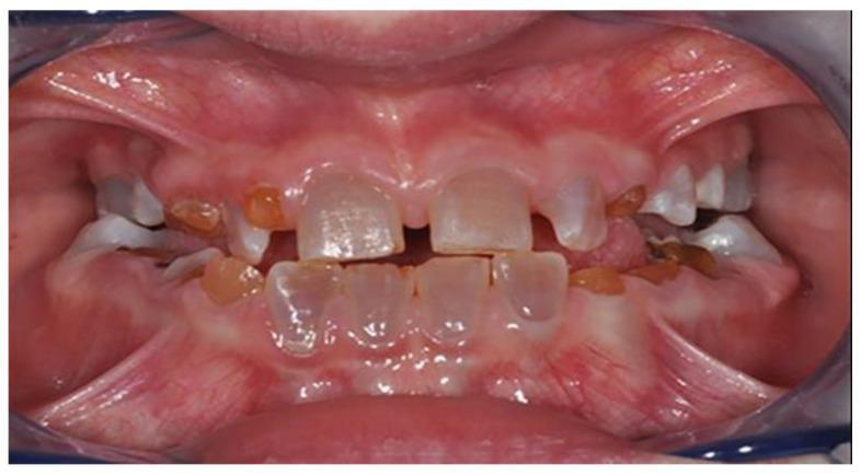
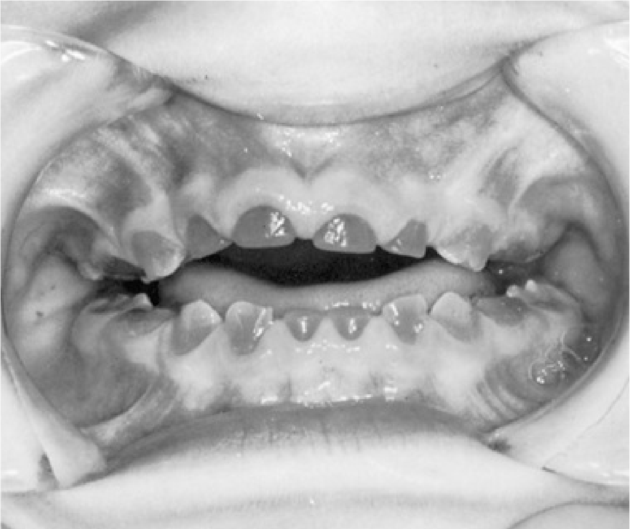
This intraoral clinical photograph showcases the dental manifestations of dentinogenesis imperfecta (DI) in a patient with Osteogenesis Imperfecta (OI). The primary findings include significant dental dyschromia, with the dentition exhibiting a characteristic opalescent, blue-grey to yellowish-brown translucent appearance. This discoloration results from the underlying defective dentin structure visible through the enamel. Structurally, the teeth show evidence of attrition and wear on the incisal and occlusal surfaces. The photograph also demonstrates clinical signs of increased enamel fragility, with apparent chipping and fractures. Malocclusion and irregular spacing are evident across both the maxillary and mandibular arches. This image serves as an educational example of how systemic connective tissue disorders, specifically OI, can impact dental development and lead to the clinical diagnosis of hereditary dentin defects.
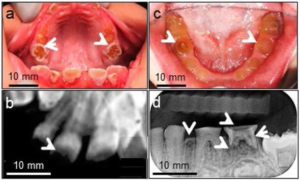
A multi-panel figure displaying clinical and radiographic findings of Dentinogenesis Imperfecta Type II (DGI-II). (a, c) Intraoral clinical photographs of the primary and early mixed dentition show hallmark features including opalescent, yellow-brown discoloration and a translucent appearance of the teeth. Severe occlusal attrition (white arrowheads) is visible, with significant loss of crown height down to the gingival level in some areas. (b, d) Dental radiographs demonstrate the structural internal consequences of the disease. Key findings include significantly reduced dental mineral density, thin dentine layers, and rapid enamel loss. The pulp chambers and root canals appear constricted or obliterated (decreased pulp space). White arrowheads in panel (d) highlight periapical radiolucencies consistent with periapical abscesses, a common complication arising from early pulp exposure due to rapid wear. These images illustrate the phenotypic manifestations of DSPP gene mutations affecting dentinogenesis.
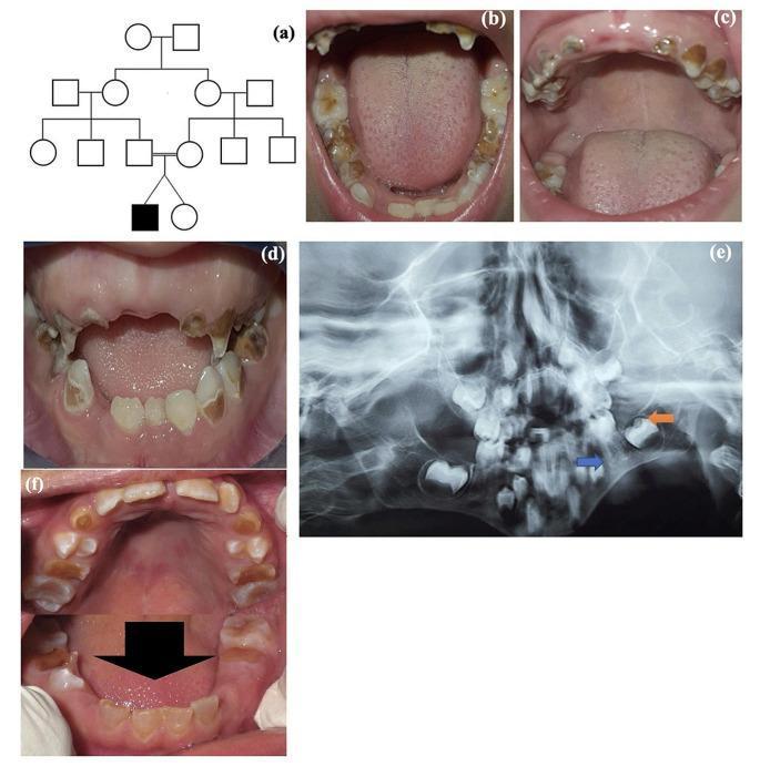
This composite image illustrates clinical and radiographic features of Dentinogenesis Imperfecta (DI) in a pediatric patient. Panel (a) shows a pedigree indicating a familial inheritance pattern. Panels (b, c, d, f) are intraoral clinical photographs showing both primary and permanent dentition. The teeth exhibit characteristic amber-to-opalescent discoloration, significant enamel attrition, and bulbous crown morphology. Severe destruction of the clinical crowns is visible, particularly in the deciduous teeth. Panel (e) is a panoramic radiograph demonstrating hallmark diagnostic features of DI, including bulbous molar crowns with cervical constriction, thin or shortened roots (blue arrow), and enlarged pulp chambers (orange arrow), indicating a failure of normal dentin deposition. The radiographic appearance also shows a lack of clear demarcation between the enamel and dentin layers. This collection of images serves as a clinical reference for the diagnosis of hereditary dentin defects, specifically DI Type II, commonly associated with DSPP gene mutations.
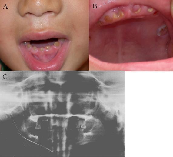
This composite medical image displays clinical and radiographic findings characteristic of Dentinogenesis Imperfecta (DGI) Type II in a pediatric patient. Panels A and B are clinical photographs of the oral cavity showing the primary dentition. The teeth exhibit a distinct amber to brownish discoloration and a translucent appearance. There is evidence of severe attrition, with the enamel layer appearing significantly thinned or absent, leading to the rapid wear of the underlying dentin and a flattened crown morphology. Panel C is a panoramic radiograph (Panorex) of the same individual, providing a comprehensive view of the maxillary and mandibular dental arches. The radiograph reveals typical features of DGI, which often include bulbous crowns, cervical constriction (neck-like appearance at the cementoenamel junction), and partial or complete obliteration of the pulp chambers and root canals due to excessive dentin deposition. The image serves as a clinical reference for hereditary dentin defects associated with DSPP gene mutations, highlighting the diagnostic transition from visual clinical signs to radiographic confirmation of structural anomalies.
dentinogenesis imperfecta
Dentinogenesis Imperfecta (DI)
Definition and Genetics
- Gene locus: chromosome 4q (non-syndromic form)
- Causative gene: DSPP (Dentin Sialophosphoprotein) - encodes dentin sialoprotein and dentin phosphoprotein
- Prevalence: relatively common in white children; estimated 1 in 6,000-8,000 individuals
Classification (Shields Classification)
| Type | Description | Association |
|---|---|---|
| Type I | DI occurring in patients with Osteogenesis Imperfecta (OI) | OI types I, III, IV |
| Type II | Isolated ("hereditary opalescent dentin") - no systemic disease | DSPP mutation |
| Type III | "Brandywine type" - seen in a Maryland triracial isolate; shell teeth with large pulps | Rare, distinct DSPP variant |
Pathogenesis
-
Poor bonding between enamel and dentin
-
Enamel chips away, exposing the soft underlying dentin
-
Progressive and rapid wear of exposed dentin
-
The Developing Human (Moore & Persaud), p. 1207
Clinical Features
- Teeth have an opalescent sheen - translucent gray to yellow-brown or amber discoloration
- Affects both primary and permanent dentitions (primary teeth often more severely affected)
- Bulbous crown morphology with cervical constriction (bell-shaped crowns)
- Enamel fractures and chips easily off the crown surface
- Severe attrition and wear, sometimes down to gingival level
- Malocclusion and irregular spacing
Radiographic Features
| Feature | Description |
|---|---|
| Obliterated pulp chambers | Progressive dentin deposition occludes pulp spaces early |
| Bulbous crowns | Bell-shaped appearance with cervical constriction |
| Thin, shortened roots | Narrow root canals |
| Periapical radiolucencies | From pulp necrosis through dentinal tubules without frank caries |
| Reduced mineral density | On radiograph, dentin appears less opaque |
Association with Osteogenesis Imperfecta (OI)
| OI Type | DI Presence |
|---|---|
| Type I | Uncommon |
| Type II | Not present (lethal perinatally) |
| Type III | Common |
| Type IV | Common |
| Type V-IX | Absent |
- Goldman-Cecil Medicine, p. 2751
- Robbins Pathologic Basis of Disease, p. 1085
Complications
- Pulp necrosis and periapical abscesses - bacterial penetration through dentinal tubules even without caries
- Rapid loss of vertical dimension (bite collapse)
- Severe functional and aesthetic impairment
- Dentinal hypersensitivity
Diagnosis
- Clinical: characteristic translucent discoloration + rapid attrition
- Radiographic: obliterated pulp chambers, bulbous crowns, thin roots
- Genetic testing: DSPP mutation confirmation
- Family history (autosomal dominant - 50% of offspring affected)
Management
- Stainless steel crowns to protect remaining tooth structure and maintain vertical dimension
- Early intervention to prevent further attrition
- Full coverage crowns (porcelain-fused-to-metal or zirconia)
- Overdentures if teeth are severely compromised
- Dental implants (caution in OI-associated DI due to underlying bone fragility)
- Orthodontic treatment as needed
Differential Diagnosis
| Condition | Key Distinction |
|---|---|
| Amelogenesis imperfecta | Enamel defect only (normal dentin); no pulp obliteration on X-ray |
| Tetracycline staining | History of drug exposure; no structural weakness; no pulp obliteration |
| Dentin dysplasia | Radiographically shows nearly absent pulp chambers even in minimally worn teeth |
| Erythroblastosis fetalis | Blue-black staining; no structural fragility |
Key Takeaways
- Autosomal dominant; DSPP gene (chromosome 4q) for isolated form
- Both dentitions affected; primary teeth often worse
- Opalescent gray-brown teeth + rapid attrition + obliterated pulp chambers on X-ray
- Type I DI = part of OI; Types II/III = isolated
- Treatment is restorative/preventive; early stainless steel crowns in children
Myasthenia gravis
myasthenia gravis ptosis clinical neuromuscular junction
This clinical photograph is a side-by-side comparison illustrating a positive Tensilon (edrophonium) test in a patient with Myasthenia Gravis (MG). Panel A shows the patient's orbital region at baseline, demonstrating significant bilateral asymmetrical ptosis (drooping of the upper eyelids). The left eyelid is more severely affected, covering a substantial portion of the visual axis. Panel B shows the same patient after the administration of edrophonium, an acetylcholinesterase inhibitor. There is a marked improvement in the bilateral ptosis, characterized by an increased palpebral fissure width and elevation of the upper lid margins, exposing more of the iris and pupil. This visual change is a classic diagnostic indicator for disorders of the neuromuscular junction, specifically MG associated with anti-acetylcholine receptor (AchR) antibodies. The image serves as a clinical teaching tool for neurology and ophthalmology to demonstrate the transient reversal of fatiguable muscle weakness through pharmacological intervention.
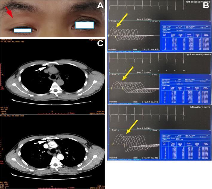
A composite medical image illustrating diagnostic manifestations of myasthenia gravis across three modalities. Panel A is a clinical photograph of the periorbital region showing asymmetrical ophthalmoparesis; a red arrow indicates significant ptosis of the right upper eyelid, which obscures the upper half of the pupil compared to the normal left eyelid position. Panel B displays results from a repetitive nerve stimulation (RNS) test of the bilateral accessory and left axillary nerves; yellow arrows highlight a decremental response in compound muscle action potential (CMAP) amplitudes, characteristic of neuromuscular junction dysfunction. Panel C contains two axial computed tomography (CT) images of the chest; white arrows point to an enlarged soft tissue mass in the anterior mediastinum, consistent with thymic hyperplasia. Together, these panels demonstrate the classic clinical triad of ocular symptoms (ptosis), electrophysiological evidence of fatigue, and associated anatomical abnormalities (thymus) often found in autoimmune myasthenia gravis.
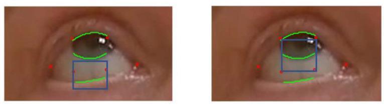
This clinical image consists of two side-by-side close-up photographs of a human eye, demonstrating computer vision and machine learning techniques for ophthalmologic assessment. The images utilize annotated overlays to track key anatomical landmarks related to Myasthenia Gravis (MG) evaluation, specifically for ptosis (eyelid droop). Red dots represent machine learning-derived landmarks at the eye corners and lid margins. Green curved lines delineate the upper eyelid contour, the lower eyelid margin, and the limbus (the interface between the iris and sclera). Blue bounding boxes define 'Regions of Interest' (ROI); in the left frame, the box targets the lower eyelid-sclera junction, while in the right frame, it frames the inferior iris-sclera boundary. This methodology is designed for automated, quantitative monitoring of eyelid distance and eye area during sustained upward gaze, providing objective data for telemedicine-based clinical examinations of neuromuscular junction disorders.
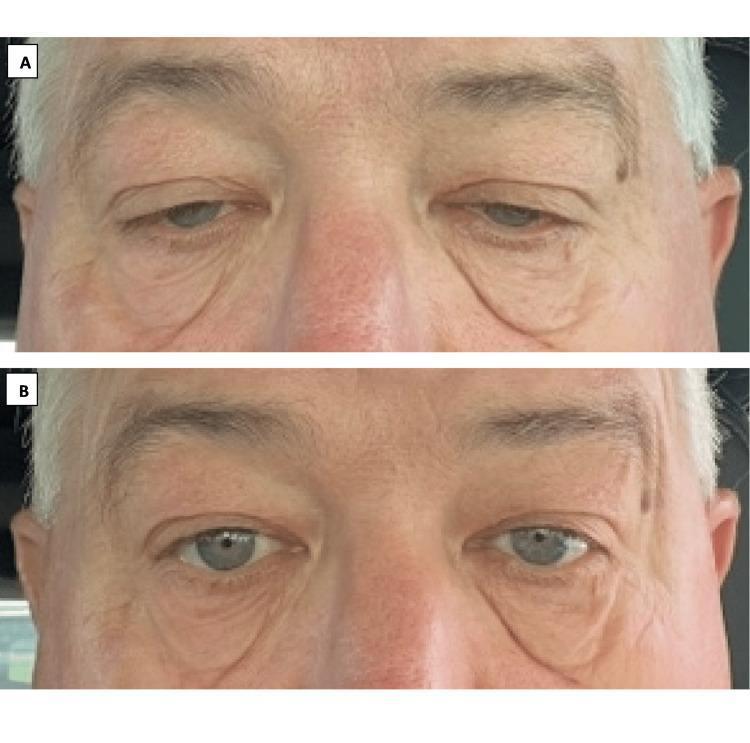
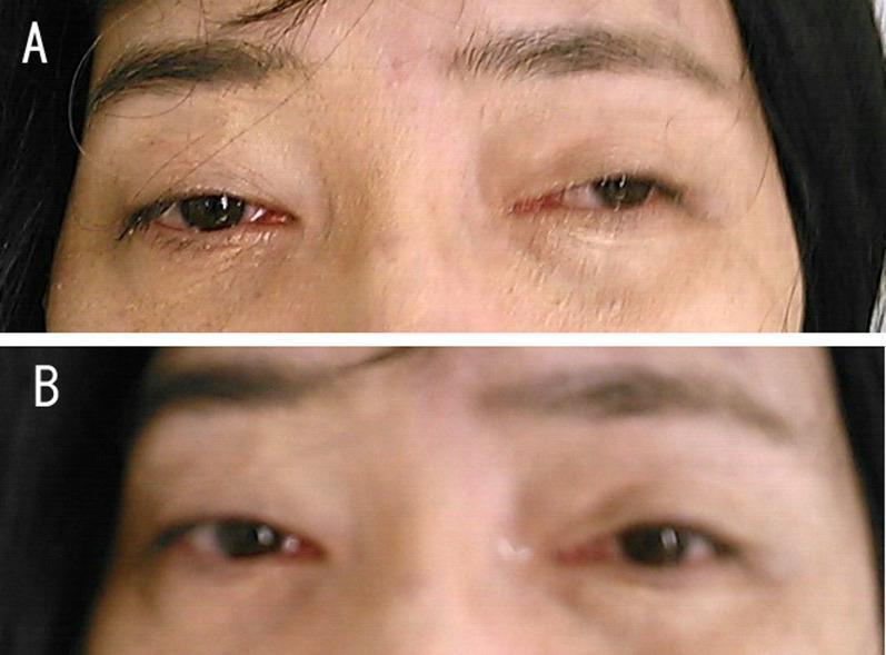
This clinical photograph consists of a side-by-side comparison (labeled A and B) demonstrating the pharmacological management of bilateral eyelid ptosis in a patient with Myasthenia Gravis (MG). Panel A (Baseline): Displays severe bilateral upper eyelid drooping, which obscures approximately 30-40% of the superior iris and limits the palpebral fissure width. The periorbital skin shows significant laxity and skin folds characteristic of chronic ptosis and aged skin. Panel B (Post-Treatment): Shows the same patient 30 minutes after the administration of oxymetazoline hydrochloride 0.1% ophthalmic solution. There is a notable elevation of both upper eyelids, resulting in increased exposure of the iris and sclera, with only 5-10% of the iris remaining covered. Educational Focus: The images illustrate the clinical efficacy of alpha-adrenergic agonists in stimulating Müller’s muscle (sympathetic smooth muscle) to improve eyelid elevation. This comparison highlights a non-surgical management option for symptomatic ptosis in patients with neuromuscular junction disorders or refractory weakness.
myasthenia gravis
Myasthenia Gravis (MG)
Definition
-
Prevalence: 150-200 per 1,000,000
-
Bimodal age distribution: young women (peak 28-35 years) and older men (peak 42-49 years); female-to-male ratio 2:1 in young adults, male predominance in older adults
-
Robbins Pathologic Basis of Disease, p. 2573; Harrison's Principles of Internal Medicine 22E
Pathogenesis
The Neuromuscular Junction in MG
Antibody Mechanisms
| Antibody | Prevalence | Mechanism | Features |
|---|---|---|---|
| Anti-AChR (IgG) | ~85% generalized MG; ~50% ocular MG | Complement activation, receptor cross-linking and degradation, postsynaptic membrane damage | Classic MG |
| Anti-MuSK | ~40% of AChR-negative generalized MG | Interferes with AChR trafficking and clustering (no complement fixation) | More prominent facial/bulbar weakness, more severe |
| Anti-LRP4 | ~1-3% | Disrupts AChR clustering | Mild to moderate symptoms |
- Bradley and Daroff's Neurology in Clinical Practice; Robbins p. 2578-2580
Role of the Thymus
- 10% of MG patients have a thymoma (tumor of thymic epithelial cells)
- 30% have thymic hyperplasia (B-cell follicles appear in thymus, especially in young patients)
- The thymus normally expresses peripheral tissue antigens including skeletal muscle proteins. Thymoma/hyperplasia disrupts self-tolerance, triggering autoantibody production
- Thymoma-related MG almost always involves AChR antibodies, not anti-MuSK or LRP4
- 1 in 3 patients with thymoma develop MG
Clinical Features
Ocular and Bulbar (Most Common Onset)
- Ptosis - drooping upper eyelids, often asymmetric, worse toward end of day
- Diplopia - due to extraocular muscle weakness
- Ocular involvement in up to 85% of all patients; symptoms remain purely ocular in ~15-20%
- Dysphagia and dysarthria - bulbar involvement in 6-30% at onset; nearly all develop it eventually
- Silent aspiration in 35%+ of those with dysphagia
- "Dropped head" appearance from posterior neck muscle weakness
- Limited smile (facial muscle weakness)
Limb and Respiratory
- Proximal limb weakness (difficulty combing hair, raising arms)
- Respiratory failure (diaphragm + intercostal muscle weakness) - the definition of myasthenic crisis
- Tendon reflexes and sensation are normal (key distinguishing feature)
Diagnosis
Stepwise Approach
- Anti-AChR antibody (radioimmunoassay): ~85% positive in generalized MG - if positive, virtually diagnostic
- Anti-MuSK: check if AChR-negative
- Anti-LRP4: check if both negative but phenotype + electrodiagnostics are consistent
- Repetitive nerve stimulation (RNS): Decrement of >10% at 3 Hz = highly probable MG
- Single-fiber EMG (SFEMG): Blocking and jitter with normal fiber density - most sensitive test; confirmatory but not specific
- Ice pack test: Apply ice to closed eyelid for 2 minutes - improvement in ptosis is sensitive for MG (cold slows AChE, increasing ACh at junction)
- Edrophonium (Tensilon) test: IV edrophonium 2 mg + 8 mg - highly probable if unequivocally positive (now less common due to cardiac side effects)
-
CT/MRI chest in all patients to look for thymoma or thymic hyperplasia
-
Harrison's 22E, p. 1600-1615; Rosen's Emergency Medicine
Classification
Osserman Classification (clinical)
| Grade | Description |
|---|---|
| I | Ocular only |
| IIA | Mild generalized |
| IIB | Moderate generalized with bulbar involvement |
| III | Acute severe (rapid progression, respiratory involvement) |
| IV | Chronic severe |
| V | Myasthenic crisis (intubation required) |
Treatment
1. Symptomatic - Acetylcholinesterase Inhibitors
- Pyridostigmine (Mestinon) - first-line symptomatic therapy; inhibits AChE, increases ACh at NMJ
- Dose titrated to symptoms; does not modify disease course
- Side effects: muscarinic (SLUDGE: salivation, lacrimation, urination, defecation, GI upset, emesis) - manageable with glycopyrrolate
2. Immunosuppression
| Drug | Onset of Effect | Notes |
|---|---|---|
| Prednisone | 2-3 weeks per dose change | Start low (15-25 mg/d), titrate up to 50-60 mg/d; can cause initial worsening - cover with IVIg/PLEX if severe |
| Azathioprine | 6-18 months | Steroid-sparing; first-line long-term agent |
| Mycophenolate mofetil | 6-18 months | Alternative steroid-sparing |
| Cyclosporine/Tacrolimus | 1-3 months | Faster than azathioprine |
| Rituximab | 1-3 months | Highly effective in MuSK-antibody positive MG; a July 2025 Cochrane review (Dodd et al., PMID 40607605) evaluated its role |
| Eculizumab | Weeks | Complement inhibitor; approved for refractory AChR-positive MG |
| FcRn antagonists (efgartigimod, rozanolixizumab) | Weeks | Newest class; reduce IgG antibody levels |
3. Rescue/Rapid Immunomodulation
- Plasma exchange (PLEX/plasmapheresis): Multiple exchanges over 1-2 weeks; effective in up to 95% of myasthenic crisis; also used pre-operatively
- IVIg: Equivalent to PLEX for crisis; 2 g/kg over 5 days; faster to administer; preferred when IV access or coagulation issues
4. Thymectomy
-
Indicated in all patients with thymoma (regardless of MG severity)
-
Also recommended for generalized MG patients aged <60 without thymoma (even without thymoma, thymic hyperplasia is common and benefit is proven)
-
Benefit may take months to years to become apparent
-
A landmark RCT (MGTX trial, N Engl J Med 2016) confirmed thymectomy improves outcomes in generalized MG
-
Harrison's 22E; Goldman-Cecil Medicine; Schwartz's Principles of Surgery
Myasthenic Crisis
- Occurs in 15-20% of MG patients, usually within the first 2 years
- Mortality has fallen dramatically with modern ICU care
Precipitants (to know and avoid):
- Infections (most common)
- Surgery or pregnancy
- Stopping immunosuppressants or pyridostigmine
- Drugs that worsen MG (critical to check in all MG patients):
| Drug Class | Examples |
|---|---|
| Aminoglycosides | Gentamicin, tobramycin (even eye drops) |
| Fluoroquinolones | Ciprofloxacin |
| Macrolides | Azithromycin |
| Neuromuscular blockers | All types - extreme sensitivity |
| Beta-blockers | Propranolol |
| Antiarrhythmics | Quinidine, procainamide |
| Magnesium sulfate | Blocks NMJ calcium entry |
| Corticosteroids | Can cause initial worsening |
Crisis Management (ICU):
- Secure airway - intubate early if FVC <15-20 mL/kg or declining rapidly
- Hold pyridostigmine (can increase secretions; restart once extubated)
- Plasma exchange or IVIg - mainstay; PLEX effective in ~95%
- Treat precipitant (antibiotics for infection, etc.)
- Resume and optimize immunosuppression
- Rosen's Emergency Medicine; Harrison's 22E
Differential Diagnosis
| Condition | Key Distinguishing Feature |
|---|---|
| Lambert-Eaton syndrome (LEMS) | Weakness improves with repeated use; proximal legs > eyes; associated with SCLC; anti-VGCC antibodies; hyporeflexia + autonomic dysfunction |
| Botulism | Descending flaccid paralysis; fixed dilated pupils; foodborne or wound history |
| Ocular MG vs. other causes of ptosis/diplopia | Horner syndrome, CN III palsy - normal AChR Ab, normal SFEMG |
| Amyotrophic lateral sclerosis (ALS) | Upper + lower motor neuron signs; fasciculations; no fatigability |
| Graves ophthalmopathy | Proptosis; CT shows enlarged extraocular muscles |
| Congenital myasthenic syndromes | Negative antibodies; onset from birth; genetic |
Key Points Summary
- Pathology: Postsynaptic; anti-AChR antibodies (85%) destroy receptors via complement + cross-linking
- Cardinal sign: Fatigable weakness - worse with activity, better with rest
- Most common onset: Ocular (ptosis, diplopia); 85% eventually generalize
- Diagnosis: Anti-AChR Ab + RNS decrement + SFEMG jitter; ice pack test at bedside
- Always image the chest (CT/MRI) for thymoma
- Treatment ladder: Pyridostigmine → steroids → steroid-sparing agents → rescue (PLEX/IVIg) → thymectomy
- Crisis = respiratory failure: PLEX/IVIg + ICU care; avoid all NMJ-impairing drugs
Recent evidence note: A July 2025 Cochrane meta-analysis (PMID 40607605) specifically reviewed rituximab for MG. A 2025 systematic review (PMID 39380477) updated global prevalence/incidence data. These do not contradict the above management framework but highlight rituximab as an increasingly important option, especially in MuSK-positive disease.
Multiple sclerosis
multiple sclerosis MRI brain white matter plaques demyelination
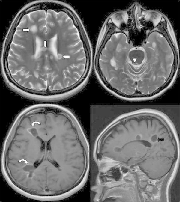
Four-panel brain MRI series demonstrating diagnostic features of Multiple Sclerosis (MS). Top row: Axial T2-weighted images showing multiple hyperintense lesions (arrows) within the bilateral periventricular white matter, the body of the corpus callosum, and the right periaqueductal grey matter (arrowhead). Bottom left: Axial T1-weighted image post-gadolinium contrast administration highlights active inflammatory demyelination, characterized by prominent ring and incomplete open-ring enhancement (curved arrows). Bottom right: Sagittal T1-weighted view reveals characteristic ovoid plaques oriented perpendicularly to the lateral ventricles, consistent with Dawson's fingers (black arrow). The imaging findings illustrate spatial and temporal dissemination according to the McDonald criteria, specifically highlighting active white matter pathology and typical anatomical distributions for central nervous system demyelinating disease.
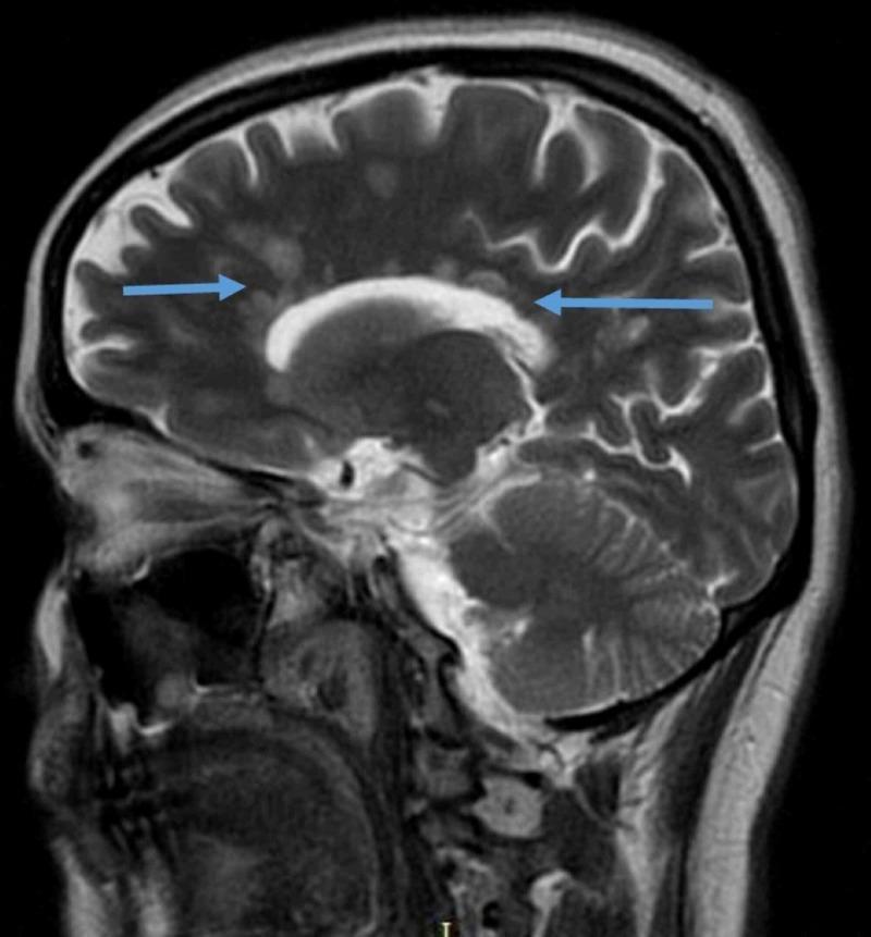
Diagnostic imaging of the human brain featuring a sagittal T2-weighted fluid-attenuated inversion recovery (FLAIR) MRI sequence. The image demonstrates hallmark radiological signs of a demyelinating disease, likely Multiple Sclerosis. Key findings include multiple hyperintense, ovoid-shaped lesions located in the periventricular white matter. Blue arrows specifically highlight 'Dawson's fingers,' which are inflammatory plaques oriented perpendicular to the long axis of the corpus callosum along the path of medullary veins. The FLAIR sequence provides high contrast by suppressing the signal from the cerebrospinal fluid (CSF), which appears dark, making the bright white matter abnormalities more prominent. Visible anatomical landmarks include the cerebral cortex with clearly defined gyri and sulci, the corpus callosum, brainstem, and cerebellum. This image serves as a classic educational example for teaching the neuroimaging criteria used in the diagnosis of central nervous system demyelination.
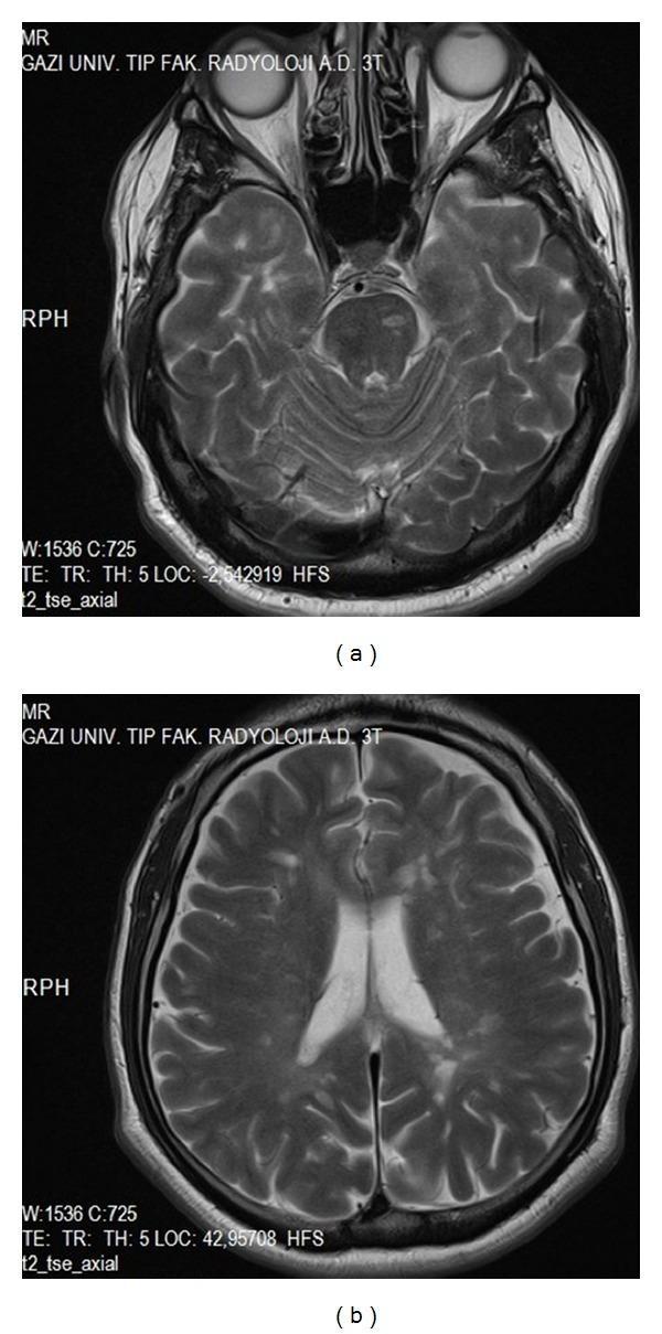
This composite diagnostic image consists of two T2-weighted axial sections from a 3T brain MRI demonstrating clinical features of a demyelinating disease, such as Multiple Sclerosis (MS). Image (a) is an axial section at the level of the posterior fossa, showing a focal area of increased signal intensity (hyperintensity) within the pons, consistent with an infratentorial demyelinating plaque. Image (b) shows a higher axial section at the level of the lateral ventricles, revealing multiple small, discrete hyperintense lesions within the periventricular white matter. These periventricular plaques are characteristic findings in neuroinflammatory disorders. The presence of lesions in both the brainstem and the supratentorial white matter illustrates dissemination in space, a key diagnostic criterion for multiple sclerosis. These images serve as a classic radiological representation of demyelination in the central nervous system for medical educational purposes in neurology and radiology.
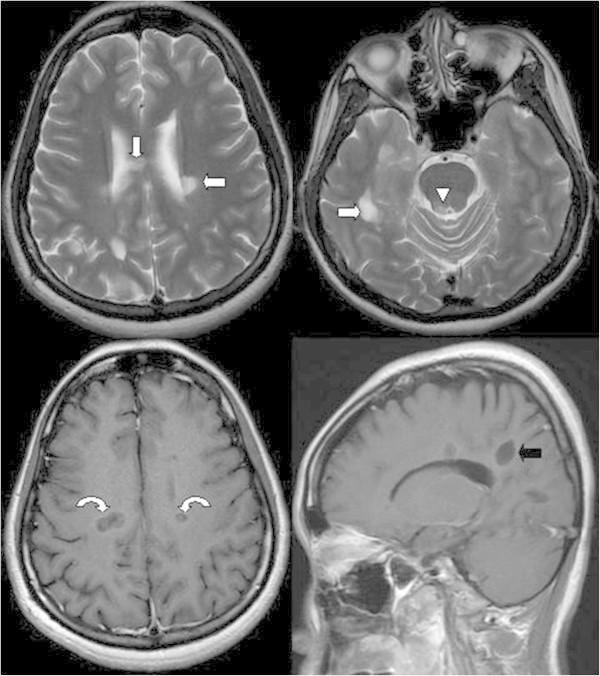
This composite diagnostic image presents Brain Magnetic Resonance Imaging (MRI) findings diagnostic of Multiple Sclerosis (MS). Top Row: Axial T2-weighted images demonstrating multiple hyperintense foci located in the bilateral periventricular white matter, the body of the corpus callosum (arrows), and the periaqueductal grey matter (arrowhead). Bottom Left: Axial T1-weighted image with gadolinium contrast showing distinctive ring-enhancing lesions (curved arrows), indicating active inflammatory demyelination and blood-brain barrier breakdown. Bottom Right: Sagittal T1-weighted image highlighting MS plaques oriented perpendicularly to the callososeptal interface. This characteristic morphology, known as 'Dawson’s fingers,' follows the distribution of the medullary veins. The combination of these findings, particularly the presence of enhancing and non-enhancing lesions, supports the McDonald criteria for dissemination in space and time. This image is an essential educational resource for neuroradiology and neurology, illustrating classic radiographic signatures of demyelinating disease in the central nervous system.
multiple sclerosis disease modifying therapy
Multiple Sclerosis (MS)
Definition
-
Prevalence: ~1 per 1,000 in the United States and Europe (incidence appears to be increasing)
-
Women affected twice as often as men
-
Typical onset: 20-40 years; rare before 10 or after 50 years
-
Robbins & Kumar Basic Pathology, p. 1652; Harrison's 22E
Etiology and Risk Factors
Genetic Factors
| Relationship | Risk of MS |
|---|---|
| Identical (monozygotic) twin affected | 1 in 3 |
| Fraternal twin affected | 1 in 15 |
| Sibling affected | 1 in 25 |
| Parent or half-sibling affected | 1 in 50 |
| First cousin affected | 1 in 100 |
| No family history | 1 in 1,000 |
- Strongest genetic signal: HLA-DRB1*1501 (formerly DR2) - each copy confers ~3-fold increased risk; accounts for ~10% of overall disease risk
-
230 additional susceptibility variants identified (IL-2 receptor, IL-7 receptor, CD58/LFA-3 genes - all immune-related)
- MS severity loci (separate from risk loci) operate within the nervous system rather than the immune system
Environmental Triggers
-
Epstein-Barr virus (EBV) infection - strongly associated; possibly the most important environmental trigger
-
Vitamin D deficiency - latitude effect (MS more prevalent further from equator correlates with lower sunlight exposure)
-
Cigarette smoking
-
Obesity in adolescence
-
Higher risk in individuals who migrated from low-risk to high-risk geographic areas before age ~15
-
Harrison's 22E, p. 2552-2554
Pathogenesis
Immunopathology of an MS Plaque
- Breakdown of the blood-brain barrier (BBB) - perivenular cuffing by T lymphocytes and macrophages; vessel wall is preserved (distinguishes from vasculitis)
- CD4+ Th1 cells secrete IFN-γ, activating macrophages
- CD4+ Th17 cells recruit additional leukocytes
- CD8+ cytotoxic T cells found at the leading edge of lesions - likely responsible for direct axonal damage
- B cells and antibodies: myelin-specific autoantibodies on degenerating myelin, complement activation; success of B cell-depleting therapies (ocrelizumab, ofatumumab) confirms their importance
- Activated macrophages phagocytose myelin debris
- Result: sharply demarcated zones of demyelination (plaques) with relative axonal preservation early, but irreversible axonal loss in chronic lesions
Remyelination
Pathology (Morphology)
Gross
- Plaques (the hallmark lesion): discrete, slightly depressed, gray-tan, glassy-appearing lesions
- Distribution: periventricular white matter (most characteristic), optic nerves and chiasm, brainstem, cerebellar peduncles, spinal cord (ascending and descending tracts)
- Lesions have sharply defined borders
Microscopy
| Phase | Appearance |
|---|---|
| Active plaque | Macrophages stuffed with myelin debris; perivascular lymphocytic cuffing; relative axonal preservation |
| Inactive plaque | Inflammation resolves; dense gliosis (astrocyte proliferation); little/no myelin; axonal loss |
| Shadow plaque | Thin, incomplete remyelination around a plaque |
- Robbins Basic Pathology, p. 1663-1665
Clinical Types and Course
1. Relapsing-Remitting MS (RRMS) - 85%
- Most common form; multiple discrete attacks (relapses) with intervening partial or complete recovery (remissions)
- Over time, accumulating neurologic deficits
- Risk of conversion to SPMS: ~3%/year pre-treatment era; now <1%/year with modern DMTs
2. Secondary Progressive MS (SPMS)
- Initial RRMS phase followed (typically after 10-20 years) by progressive worsening without remissions
- May have occasional superimposed relapses ("active SPMS")
3. Primary Progressive MS (PPMS) - ~10%
- Steady decline from onset, no relapses
- More equal sex distribution; onset ~10 years later (mean ~40 years)
- Faster disability accumulation relative to first symptom
- Approved treatment: ocrelizumab
Special Categories
- Clinically Isolated Syndrome (CIS): First demyelinating episode; not yet MS by definition
- Radiologically Isolated Syndrome (RIS): Incidental MRI findings consistent with MS, no symptoms
- Active progressive MS: Progressive MS (SPMS or PPMS) with new relapses or new MRI lesions
Disability Mechanisms
-
RAW (Relapse-Associated Worsening): Disability from incomplete relapse recovery
-
PIRA (Progression Independent of Relapsing Activity): Insidious neurodegeneration without clinical attacks - now recognized as the dominant driver of disability even in RRMS
-
Harrison's 22E, p. 2350-2356
Clinical Features
Most Common Presenting Symptoms
| System | Symptoms |
|---|---|
| Optic | Optic neuritis: unilateral painful visual loss, reduced color vision, relative afferent pupillary defect (RAPD) |
| Brainstem/cerebellar | Diplopia, nystagmus, internuclear ophthalmoplegia (INO) - adduction lag with contralateral nystagmus (MLF lesion); ataxia, dysarthria, intention tremor |
| Spinal cord | Limb weakness (upper motor neuron pattern), spasticity, hyperreflexia, extensor plantar responses |
| Sensory | Numbness, tingling, loss of proprioception and vibration (dorsal column), dysesthesias |
| Bladder | Urgency, frequency, incontinence (spastic bladder) - most distressing long-term symptom |
| Cognitive | Memory, processing speed, executive function impairment |
| Fatigue | One of the most disabling symptoms; disproportionate to weakness |
Characteristic Signs and Phenomena
- Lhermitte sign: Electric shock sensation down the spine and limbs on neck flexion - indicates cervical cord demyelination
- Uhthoff phenomenon: Worsening of symptoms with heat (exercise, fever, hot bath) - due to slowed conduction in partially demyelinated axons (diagnostic clue; NOT a true relapse)
- Trigeminal neuralgia in a young patient: Think MS (demyelination at CN V root entry zone)
- INO in a young adult: Until proved otherwise, MS
Diagnosis: McDonald Criteria (2017, revised)
MRI Criteria for DIS
- Periventricular
- Cortical/juxtacortical
- Infratentorial (brainstem/cerebellum)
- Spinal cord
MRI Criteria for DIT
- Simultaneous presence of gadolinium-enhancing and non-enhancing lesions, OR
- New T2 or Gd-enhancing lesion on follow-up MRI
MRI Findings
| MRI Finding | Significance |
|---|---|
| T2/FLAIR hyperintense lesions | Demyelination and edema (active) or gliosis (chronic) |
| Dawson's fingers | Perivenous demyelination perpendicular to ventricles on sagittal FLAIR |
| Gadolinium-enhancing lesions | Active BBB disruption = active inflammation (lasts <1 month) |
| T1 black holes | Irreversible axonal loss; worse prognosis |
| Brain/spinal cord atrophy | Strongest correlate of long-term disability |
| Central vein sign | Within plaques on susceptibility-weighted sequences; aids specificity |
| Abnormality in >95% of patients | Most lesions are asymptomatic |
CSF
- Oligoclonal bands (OCBs): ≥2 bands in CSF not present in serum - found in >90% of MS patients; may be absent early
- CSF IgG index elevated (intrathecal IgG synthesis)
- Mononuclear pleocytosis (usually <50 cells/μL; if >50, consider alternative diagnosis)
- Total protein: normal or mildly elevated
Evoked Potentials
- Visual evoked potentials (VEPs): Prolonged P100 latency (even in clinically silent optic neuritis) - useful for detecting a second lesion when one clinical attack is present
- Also: somatosensory EPs, brainstem auditory EPs
Treatment
1. Acute Relapses
- High-dose IV methylprednisolone 1 g/day × 3-5 days - speeds recovery but does not change long-term disability
- Plasma exchange for severe steroid-unresponsive relapses
2. Disease-Modifying Therapies (DMTs)
Moderate-Efficacy DMTs (Injectables/Oral)
| Drug | Mechanism | Route | Key Notes |
|---|---|---|---|
| Interferon beta-1a/1b (Avonex, Rebif, Betaseron) | Immunomodulation, reduces T-cell trafficking | SC/IM | First approved DMTs; ~30% relapse reduction |
| Glatiramer acetate (Copaxone) | Antigen competition, Th2 shift | SC | Well tolerated; no flu-like side effects |
| Teriflunomide (Aubagio) | Inhibits pyrimidine synthesis (anti-proliferative) | Oral | Teratogenic; monitoring required |
| Dimethyl fumarate (Tecfidera) | Nrf2 pathway activation, lymphocyte trafficking | Oral | Flushing, GI upset; monitor lymphocyte counts |
High-Efficacy DMTs
| Drug | Mechanism | Key Notes |
|---|---|---|
| Natalizumab (Tysabri) | Anti-α4 integrin; blocks lymphocyte entry into CNS | ~68% relapse reduction; risk of PML (JC virus reactivation); JC antibody testing mandatory |
| Ocrelizumab (Ocrevus) | Anti-CD20; B-cell depletion | Approved for both RRMS and PPMS (first ever for PPMS); IV 600 mg q6 months |
| Ofatumumab (Kesimpta) | Anti-CD20; B-cell depletion | Subcutaneous monthly injections; home administration advantage |
| Fingolimod (Gilenya) | S1P receptor modulator; traps lymphocytes in lymph nodes | First oral high-efficacy DMT; requires first-dose cardiac monitoring (bradycardia risk) |
| Siponimod, Ozanimod, Ponesimod | Selective S1P1/S1P5 modulators | Do not require first-dose monitoring; approved for active SPMS (siponimod) |
| Cladribine (Mavenclad) | Lymphocyte depletion (purine analog) | Annual pulse courses × 2 years; long-lasting effect |
| Alemtuzumab (Lemtrada) | Anti-CD52; depletes lymphocytes and monocytes | Highly effective but serious autoimmune complications (thyroid disease ~25%, ITP 1-3%) |
Recent evidence: A 2025 network meta-analysis (Köhler et al., PMID 40783682) compared DMTs in highly active RRMS despite prior treatment, confirming anti-CD20 agents and natalizumab as top performers. A 2024 Cochrane review (Ridley et al., PMID 39254048) addressed immunomodulators for progressive MS specifically.
3. Symptom Management
| Symptom | Treatment |
|---|---|
| Spasticity | Baclofen, tizanidine, intrathecal baclofen |
| Fatigue | Amantadine, modafinil; aerobic exercise |
| Bladder urgency | Oxybutynin, tolterodine, mirabegron; intermittent self-catheterization if retention |
| Neuropathic pain/dysesthesias | Gabapentin, pregabalin, amitriptyline, duloxetine |
| Depression | SSRIs; counseling |
| Cognitive impairment | Cognitive rehabilitation |
| Walking disability | Dalfampridine (Ampyra) - potassium channel blocker that improves walking speed in ~35% of patients |
| Uhthoff/heat sensitivity | Cooling vests, avoidance of heat |
Differential Diagnosis
| Condition | Key Distinction |
|---|---|
| Neuromyelitis optica spectrum disorder (NMOSD) | Anti-AQP4 or anti-MOG antibodies; more severe attacks; area postrema lesions (intractable vomiting); longitudinally extensive spinal cord lesions (≥3 vertebral segments); OCBs usually absent |
| ADEM (Acute disseminated encephalomyelitis) | Monophasic; usually post-infectious/post-vaccination; prominent encephalopathy; children > adults |
| CNS vasculitis | Vessel wall involvement on MRI; CSF pleocytosis often >50; systemic features |
| Sarcoidosis | Leptomeningeal enhancement; hilar adenopathy; elevated ACE |
| Vitamin B12/copper deficiency | Subacute combined degeneration; no brain white matter plaques; responds to supplementation |
| Sjogren syndrome | Dry eyes/mouth; anti-Ro/La antibodies |
| CADASIL | Temporal pole and external capsule involvement; NOTCH3 mutation; family history of strokes |
Prognosis Markers
Key Points Summary
- Autoimmune CNS demyelination; Th1/Th17 T cells + B cells attack myelin; HLA-DRB1*1501 strongest genetic risk
- 85% RRMS → can evolve to SPMS; 10% PPMS (no relapses from onset)
- Hallmarks: optic neuritis, INO, Lhermitte sign, Uhthoff phenomenon, bladder dysfunction, fatigue
- MRI: T2 periventricular plaques, Dawson's fingers, Gd-enhancing active lesions; >95% have abnormal MRI
- CSF: oligoclonal bands in >90%; elevated IgG index
- McDonald criteria: DIS + DIT
- Acute relapse: IV methylprednisolone
- DMTs: escalation vs. early high-efficacy; anti-CD20 (ocrelizumab/ofatumumab) and natalizumab are highest efficacy; ocrelizumab only approved DMT for PPMS
- PIRA (silent progression without relapses) is now recognized as the dominant driver of long-term disability even in RRMS
From guyton
Multiple Sclerosis - From Guyton and Hall Textbook of Medical Physiology
1. MS as an Autoimmune Disease (Chapter 35 - Immunity)
"Failure of the Tolerance Mechanism Causes Autoimmune Diseases. Sometimes, people lose immune tolerance of their own tissues... Over 100 diseases that result from autoimmunity have been described..."
"Multiple sclerosis (MS), in which the immune system attacks the myelin that covers nerve fibers, disrupting nervous system communication."
Guyton's Key Physiological Point Here
2. Why Myelin Matters - Saltatory Conduction and the Consequence of Demyelination (Chapter 5 - Membrane Potentials and Action Potentials)
The Myelin Sheath - Normal Function
- Every large nerve fiber is wrapped in a myelin sheath formed by Schwann cells (PNS) or oligodendrocytes (CNS)
- The Schwann cell rotates around the axon many times, laying down multiple layers of sphingomyelin-rich membrane
- Sphingomyelin is an excellent electrical insulator - it decreases ion flow through the membrane approximately 5,000-fold
- The sheath is interrupted every 1 to 3 millimeters at gaps called nodes of Ranvier - the only sites where ions can flow freely
Saltatory Conduction - The Normal Mechanism
"Action potentials occur only at the nodes. Yet, the action potentials are conducted from node to node by saltatory conduction... That is, electrical current flows through the surrounding extracellular fluid outside the myelin sheath, as well as through the axoplasm inside the axon from node to node, exciting successive nodes one after another. Thus, the nerve impulse jumps along the fiber."
| Advantage | Details |
|---|---|
| Speed | Increases conduction velocity 5- to 50-fold compared to unmyelinated fibers |
| Energy efficiency | Only nodes depolarize - ~100 times less ion exchange, therefore far less Na⁺/K⁺-ATPase energy required to restore gradients |
- Large myelinated fibers: up to 100 m/sec
- Small unmyelinated fibers: as little as 0.25 m/sec
What Happens in MS (Demyelination)
- Saltatory conduction fails - impulses can no longer jump from node to node
- The affected segment loses its insulation; electrical current leaks out across the entire axon membrane
- Conduction slows dramatically or blocks entirely
- This explains the fatigable, fluctuating neurological deficits in MS - partial demyelination can still allow slow conduction at rest, but fails under repetitive stimulation (Uhthoff phenomenon)
3. MS and the Pupillary Light Reflex (Chapter 51 - The Eye)
"A few central nervous system diseases damage nerve transmission of visual signals from the retinas to the Edinger-Westphal nucleus, thus sometimes blocking the pupillary reflexes. Such blocks may occur as a result of disorders including central nervous system syphilis, chronic alcoholism, encephalitis, multiple sclerosis, and Lyme disease."
The Physiological Mechanism
- Normally, light signals travel: Retina → Optic nerve → Pretectal nucleus → Edinger-Westphal nucleus → Constrictor pupillae
- The final fibers into the Edinger-Westphal nucleus are predominantly inhibitory
- MS lesions in the pretectal region of the brainstem or in small optic nerve fibers block these pathways
- Loss of normal inhibition → Edinger-Westphal nucleus becomes chronically active → pupils remain persistently constricted + fail to respond to light
- However, pupils can still constrict via the accommodation reflex (near triad: accommodation + convergence + miosis)
Clinical Correlation: Argyll Robertson Pupil
"A pupil that fails to respond to light but does respond to accommodation and is also very small... is an important diagnostic sign of a central nervous system disease such as syphilis."
4. Demyelination from Rapid Sodium Correction (Chapter 25 - Renal Regulation)
"When hypertonic solutions are added too rapidly to correct chronic hyponatremia, this intervention can outpace the brain's ability to recapture the solutes lost from the cells and may lead to osmotic injury of the neurons that is associated with demyelination, a loss of the myelin sheath from nerves."
- Safe correction limit: <10-12 mmol/L in 24 hours, and <18 mmol/L in 48 hours
- This is osmotic demyelination syndrome (ODS) / central pontine myelinolysis - a different cause of demyelination from MS, but demonstrates the same principle: loss of myelin = loss of saltatory conduction = neurological deficit
Summary: What Guyton Teaches About MS
| Topic | Guyton's Teaching |
|---|---|
| Definition | Autoimmune attack on CNS myelin, disrupting nerve communication |
| Mechanism of disease | Failure of immune tolerance → autoreactive immune cells attack myelin sheath |
| Why myelin is critical | Enables saltatory conduction - 5-50× faster, 100× more energy-efficient |
| Consequence of demyelination | Loss of saltatory conduction → slowed or blocked nerve impulse transmission |
| Pupillary involvement | MS brainstem lesions block pretectal pathway → light reflex lost, accommodation reflex preserved |
| Myelin and sodium | Rapid correction of hyponatremia can also cause demyelination (ODS) - illustrates myelin vulnerability |
Note: For detailed clinical features, McDonald criteria, MRI findings, CSF analysis, disease types, and DMT treatment - these are not covered by Guyton. For those, the comprehensive answer from Harrison's 22E, Robbins Pathology, and Bradley-Daroff's Neurology was provided above.