
im a medical student. I have to make a ppt about eosinophilic granulomatosis with polyangitis for my rheumatology class. give me all detailed information from valid and reliable sources which I should put in my ppt
Reading File
Now let me search the medical library and PubMed in parallel for comprehensive EGPA information.
Finding Sources
Finding Sources
Searching PubMed
"eosinophilic granulomatosis with polyangiitis"[MeSH Terms]
Excellent results. Now let me read the most authoritative sources in parallel - Harrison's, Goldman-Cecil, Andrews' Diseases of the Skin, and Adams & Victor's Neurology.
Reading File
Reading File
Reading File
Searching PubMed
"eosinophilic granulomatosis with polyangiitis" treatment
Excellent data. Let me now read the remaining key sections - Fishman's Pulmonary, Adams & Victor's Neurology, and Braunwald's cardiac sections, and also get the Harrison's treatment section fully.
Reading File
Reading File
Reading File
Reading File
Now let me get the Fitzpatrick's dermatology section for images and the ENT (Scott-Brown's) section, plus details from the PubMed guidelines paper and the benralizumab RCT.
Reading File
Reading File
Reading Article
PMID: 37161084
Reading Article
PMID: 38393328
Excellent! I now have comprehensive data from multiple authoritative sources. Let me also fetch the key images and get the Five Factor Score (FFS) details from the Washington Manual.
Reading File
Loading Image
Finding Sources
Let me grab the Five Factor Score details and the Murray & Nadel EGPA section.
Reading File
Reading File
I now have extremely comprehensive data from multiple authoritative sources. Let me compile the full, detailed PPT content for this medical student.
Here is a complete, slide-by-slide breakdown for your EGPA presentation, drawn from Harrison's Principles of Internal Medicine 22E, Goldman-Cecil Medicine, Braunwald's Heart Disease, Andrews' Diseases of the Skin, Fishman's Pulmonary Diseases, Murray & Nadel's Respiratory Medicine, Adams & Victor's Neurology, Scott-Brown's Otorhinolaryngology, Fitzpatrick's Dermatology, and the Washington Manual, plus the 2023 Nature Reviews Rheumatology guidelines (PMID 37161084) and the 2024 NEJM MANDARA trial (PMID 38393328).
SLIDE 1 - Title Slide
Eosinophilic Granulomatosis with Polyangiitis (EGPA)
Formerly known as Churg-Strauss Syndrome
Presenter name | Rheumatology Seminar | [Date]
SLIDE 2 - Historical Background & Nomenclature
- First described in 1951 by Jacob Churg and Lotte Strauss who reported necrotizing vasculitis in multiple organs with eosinophilic tissue inflammation and extravascular granulomas in asthmatic patients with fever and peripheral eosinophilia
- Earlier in 1939, Rackemann and Greene had described a subgroup of polyarteritis nodosa patients with concomitant allergic disease
- Renamed from "Churg-Strauss Syndrome" (CSS) to EGPA by the 2012 Revised International Chapel Hill Consensus Conference Nomenclature for vasculitides
- Belongs to the ANCA-associated vasculitides (AAV) group, alongside Granulomatosis with Polyangiitis (GPA) and Microscopic Polyangiitis (MPA)
Sources: Harrison's 22E; Fishman's Pulmonary; Scott-Brown's ENT
SLIDE 3 - Epidemiology
| Parameter | Data |
|---|---|
| Annual incidence | 0.5-6.8 per million population |
| Prevalence | 10-14 per million (general population); up to 64 per million in asthmatic patients |
| Mean age of onset | 38-50 years |
| Gender | Female-to-male ratio 1.2:1 (no clear predominance in some series) |
| Rare in | Children, adolescents, and age >65 years |
- Uncommon but may be underdiagnosed due to variable clinical presentation
- Can occur at any age except infants
Sources: Harrison's 22E; Fishman's Pulmonary; Murray & Nadel
SLIDE 4 - Classification (Where EGPA Sits)
EGPA is classified as a small-vessel vasculitis under the AAV umbrella:
ANCA-Associated Vasculitides
├── Granulomatosis with Polyangiitis (GPA) — c-ANCA/PR3
├── Microscopic Polyangiitis (MPA) — p-ANCA/MPO
└── Eosinophilic GPA (EGPA) — p-ANCA/MPO (~40-50%)
Key distinguishing features from other AAV:
- Prominent asthma (nearly universal, 96-100%)
- Marked peripheral eosinophilia (>10% or >1000 cells/µL)
- ANCA is only positive in ~40-50% of cases (vs. higher rates in GPA/MPA)
- Lower rate of renal involvement compared to GPA and MPA
- Unique cardiac involvement not typical of other AAV
Sources: Harrison's 22E; Fitzpatrick's Dermatology; Braunwald's Heart Disease
SLIDE 5 - Pathology & Pathogenesis
Vessels involved: Small and medium-sized muscular arteries, capillaries, venules, and arterioles
Three hallmark histopathologic features (the "triad"):
- Necrotizing vasculitis - involving small to medium vessels with fibrinoid necrosis
- Eosinophilic tissue infiltration - diffuse infiltration of tissues by eosinophils
- Extravascular granulomas - palisaded granulomas, often present within vessel walls; differ from GPA granulomas in that they generally lack multinucleated giant cells and have an eosinophil-rich core
Pathogenesis (not fully elucidated):
- Strong association with asthma and atopy points to aberrant immunologic phenomena
- Eosinophils release major basic protein and eosinophil-derived neurotoxin causing direct tissue injury (especially cardiac)
- Th2 immune skewing with IL-4, IL-5, IL-13 elevation drives eosinophilia
- IL-5 is central - drives eosinophil production, survival, and activation
- ANCA (anti-MPO) present in ~40-50% - may contribute to neutrophil activation
- Increased rates of arterial and venous thrombosis linked to dense eosinophilic infiltrates
Two clinical sub-phenotypes (ANCA-based):
- ANCA-positive (p-ANCA/MPO): Higher incidence of renal involvement, peripheral nerve disease, alveolar hemorrhage; features more like "classic" AAV vasculitis
- ANCA-negative: More likely to have cardiopulmonary complications (myocarditis, cardiomyopathy)
Sources: Harrison's 22E; Andrews' Diseases of the Skin; Braunwald's Heart Disease; Fishman's Pulmonary
SLIDE 6 - The Three Clinical Phases
EGPA classically evolves through three sequential (but overlapping) phases:
Phase 1: Prodromal / Allergic Phase
- Duration: Months to many years (typically 8-10 years before EGPA recognition; up to 30 years reported)
- Features: Allergic rhinitis, nasal polyposis, late-onset asthma (mean onset age 35 years, unlike childhood allergic asthma), drug sensitivities
- The asthma is often severe, poorly controlled, and adult-onset
- Asthma typically precedes vasculitis by 3-6 years
Phase 2: Eosinophilic Phase
- Remarkably high peripheral blood eosinophilia (can reach 20-90%)
- Eosinophilic tissue infiltration of: lungs (eosinophilic pneumonia), GI tract, skin, and other organs
Phase 3: Vasculitic Phase
- Systemic necrotizing vasculitis affecting multiple organs
- Heralded by constitutional symptoms: fever, malaise, weight loss, arthralgias, myalgias
- Onset typically ~3 years after initial symptoms
- Asthma may persist, worsen, or temporarily improve during this phase
Important: Phases overlap and may not appear in strict order; asthma/rhinosinusitis rarely arise after vasculitic manifestations.
Sources: Goldman-Cecil Medicine; Andrews' Diseases of the Skin; Fishman's Pulmonary; Scott-Brown's ENT
SLIDE 7 - Clinical Manifestations (System-by-System)
Pulmonary (Most Dominant - Nearly Universal)
- Asthma: Present in 96-100%; the most consistent feature
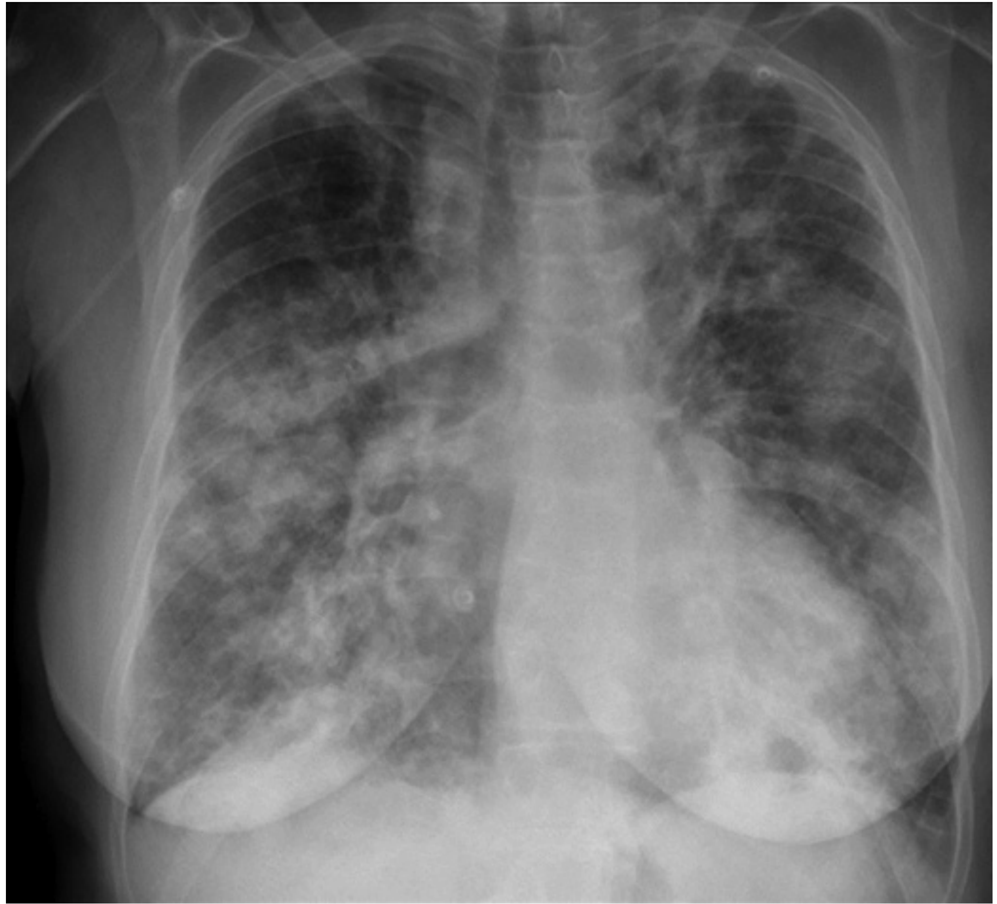
- Pulmonary infiltrates: Present in 30-70% on chest X-ray (transient, migratory, bilateral)
- Eosinophilic pneumonia
- Diffuse alveolar hemorrhage (DAH) is rare in EGPA (<5%)
- Pleural effusions (less common)
Neurological (2nd Most Common)
- Mononeuritis multiplex: Occurs in up to 63-93% of patients; most common form of neuropathy
- Acute, painful, asymmetrical peripheral neuropathy
- Involves both sensory and motor fibers
- Mechanism: necrotizing vasculitis of the vasa nervorum (identical to polyarteritis nodosa histologically, but with more intense eosinophilic infiltration)
- Cranial nerve involvement is less common
ENT / Upper Airway
- Allergic rhinitis and sinusitis: Up to 61% of patients; often the first manifestation
- Nasal polyps: ~50% of patients; characteristically recur after surgery unless immunosuppressive therapy is given
- Paranasal sinusitis present in ACR classification criteria
Cardiac (Major Cause of Mortality)
- Occurs in 14-60% of patients (variable by series)
- Spectrum includes: pericarditis, myocarditis, coronary arteritis, myocardial infarction, restrictive/dilated/congestive cardiomyopathy, valvular dysfunction, arterial thrombosis
- Mechanism: Eosinophilic infiltration of myocardium + granulomatous inflammation + direct toxicity from major basic protein and eosinophil-derived neurotoxin
- Cardiomyopathy from ischemia secondary to arteritis of intramyocardial arteries
- Cardiac disease is the most frequent cause of death (responsible for 39% of mortality)
- ANCA-negative patients are at greater risk for cardiac complications
Skin (~51-78% of patients)
- Palpable purpura: ~50% (reflects vasculitis histologically)
- Subcutaneous nodules: ~30%, on extensor surfaces of extremities and scalp
- Eosinophilic dermatitis, granulomatous dermatitis producing erythematous macules, papules, nodules
- Urticaria, livedo reticularis, solar urticaria
- Plaques resembling Wells syndrome (eosinophilic cellulitis)
- Firm, nontender papules on fingertips (may mimic septic emboli)
- Skin biopsy: small-vessel vasculitis with intense eosinophilic infiltrate; palisaded granulomas with eosinophil-rich core (no multinucleated giant cells)
Renal
- Less common than in GPA or MPA
- Focal segmental necrotizing glomerulonephritis (with or without crescents)
- Renal insufficiency in a subset of patients
- More common in ANCA-positive patients
Gastrointestinal
- Abdominal pain, diarrhea, GI bleeding
- Eosinophilic gastroenteritis
- GI involvement is a poor prognostic factor
Constitutional
- Fever, malaise, anorexia, weight loss (nonspecific but characteristic of multisystem disease)
Sources: Harrison's 22E; Braunwald's Heart Disease; Adams & Victor's Neurology; Fishman's Pulmonary; Andrews' Diseases of the Skin; Fitzpatrick's Dermatology
SLIDE 8 - Laboratory Findings
| Test | Finding |
|---|---|
| Peripheral eosinophilia | >1000 cells/µL in >80% of patients; often 5000-9000/µL; correlates with disease severity |
| ESR / CRP | Elevated in >80% during active disease |
| Fibrinogen, α2-globulins | Elevated (evidence of systemic inflammation) |
| ANCA | Positive in ~40-50%; nearly always p-ANCA (anti-MPO); rarely c-ANCA (anti-PR3) |
| IgE | Elevated (reflects atopic background) |
| Urinalysis | Hematuria, proteinuria if renal involvement |
| Creatinine | Elevated if renal insufficiency |
| BNP/NT-proBNP | Elevated with cardiac involvement |
| Chest X-ray/CT | Bilateral, transient pulmonary infiltrates; no cavitation (unlike GPA) |
| ECG + Echocardiography | Should be performed in ALL newly diagnosed patients |
| Cardiac MRI | Most sensitive for detecting myocardial involvement |
Key point: ANCA status affects phenotype:
- ANCA+: More renal, peripheral nerve, DAH involvement
- ANCA-: More cardiac, pulmonary involvement
Sources: Harrison's 22E; Fishman's Pulmonary; Braunwald's Heart Disease; Washington Manual
SLIDE 9 - Diagnostic Criteria (ACR 1990 Classification)
The 1990 American College of Rheumatology (ACR) classification criteria for EGPA (6 criteria):
| Criterion | Description |
|---|---|
| 1. Asthma | History of wheezing or diffuse high-pitched rales on expiration |
| 2. Eosinophilia | Eosinophils >10% on differential white blood cell count |
| 3. Mononeuritis multiplex or polyneuropathy | Attributable to systemic vasculitis |
| 4. Pulmonary infiltrates (non-fixed) | Migratory or transient pulmonary infiltrates |
| 5. Paranasal sinus abnormality | History or radiographic evidence of sinusitis |
| 6. Extravascular eosinophils | Biopsy showing eosinophils in extravascular areas |
Sensitivity 85%, Specificity 99.7% when ≥4 criteria are present.
For diagnosis to be made, a patient should have evidence of:
- Asthma
- Peripheral blood eosinophilia
- Clinical features consistent with vasculitis
Histologic confirmation: Optimally by biopsy, but pathognomonic features may not occur simultaneously - clinical + lab + biopsy together are most reliable.
Note: The 2022 ACR/EULAR classification criteria (updated) are now used in research settings; the diagnosis remains clinical in practice.
Sources: Harrison's 22E; Scott-Brown's ENT; Andrews' Diseases of the Skin
SLIDE 10 - Differential Diagnosis
| Condition | Key Distinguishing Features |
|---|---|
| GPA (Granulomatosis with Polyangiitis) | Upper airway destruction, cavitary lung lesions, c-ANCA/PR3, NO eosinophilia or asthma |
| MPA (Microscopic Polyangiitis) | p-ANCA+, no granulomas, no eosinophilia or asthma |
| Hypereosinophilic Syndrome (HES) | Less aggressive, diffuse eosinophilic tissue infiltration without true vasculitis; lacks asthma as a defining feature |
| Eosinophilic Pneumonia | Pulmonary eosinophilia without systemic vasculitis |
| Polyarteritis Nodosa (PAN) | Medium vessels, renal/bowel infarctions, no eosinophilia, no lung involvement |
| Parasitic Infestation | (especially helminths) - causes eosinophilia; must exclude |
| Viral infections | CMV, Hepatitis B and C - exclude before diagnosing EGPA |
| Leukoproliferative disorder | Eosinophilia without vasculitis |
| Allergic bronchopulmonary aspergillosis | Asthma + eosinophilia but specific Aspergillus serology |
Sources: Braunwald's Heart Disease; Adams & Victor's; Fishman's
SLIDE 11 - Prognosis - Five Factor Score (FFS)
The French Vasculitis Study Group validated the Five Factor Score (FFS) to stratify prognosis and guide treatment intensity.
FFS: Add 1 point for each of the following:
- Renal insufficiency (serum creatinine >1.58 mg/dL)
- Clinically significant GI disease (bleeding, perforation, infarction, pancreatitis)
- Cardiac symptoms (cardiomyopathy, infarction)
- Age ≥65 years
- Absence of upper airway (ENT) involvement (its presence is protective - indicates a more limited phenotype)
| FFS Score | 5-Year Mortality |
|---|---|
| 0 | Low (~11%) |
| 1 | Intermediate (~26%) |
| ≥2 | High (~46%) |
Prognostic pearls:
- Cardiac or GI involvement = worse prognosis
- Without treatment: 5-year survival 25%
- With treatment: 78-month actuarial survival 72%; remission in >90% of patients
Sources: Murray & Nadel; Fishman's Pulmonary; Harrison's 22E
SLIDE 12 - Treatment Overview
General Principle
Treatment intensity is determined by disease severity (FFS score), not diagnosis alone.
Step 1: Induction of Remission
All patients: High-dose glucocorticoids (first-line)
- Prednisone 1 mg/kg/day (typically 40-60 mg/day) orally, or IV methylprednisolone pulses for severe disease
- Achieves remission in >80-90% of patients as monotherapy for mild-moderate disease
- Tapering is often limited by persistent asthma
Addition of cyclophosphamide (CYC) for severe/life-threatening disease:
- Indicated when: neurological involvement, cardiac involvement, renal involvement, severe GI disease, CNS involvement
- Oral daily or IV pulse cyclophosphamide + prednisone
- After remission (3-6 months), replace CYC with azathioprine or methotrexate
Step 2: Maintenance of Remission
- Azathioprine or Methotrexate (or leflunomide) as steroid-sparing agents
- Rituximab - increasingly used especially in ANCA-positive patients with glomerulonephritis or severe small-vessel vasculitis (data still emerging for EGPA specifically)
- Mycophenolate mofetil (MMF) - alternative option
Step 3: Biologic Therapy - Anti-IL-5 Agents (MAJOR ADVANCE)
Mepolizumab (Anti-IL-5 monoclonal antibody)
- FDA-approved for EGPA (2017)
- Dose: 300 mg subcutaneously every 4 weeks
- Phase 3 RCT: More weeks in remission (28% vs 3%), higher remission rate (32% vs 3%) vs. placebo
- Significantly reduces oral glucocorticoid requirements
- Should NOT be used for severe/life-threatening disease
- Best role: Relapsing or resistant asthma requiring glucocorticoids, sinus disease, mild-moderate eosinophilic disease, mild vasculitis
Benralizumab (Anti-IL-5Rα monoclonal antibody) - 2024 NEJM MANDARA Trial
- Dose: 30 mg subcutaneously every 4 weeks
- Phase 3 RCT (MANDARA, 2024, NEJM, PMID 38393328): Benralizumab was noninferior to mepolizumab for induction of remission in relapsing/refractory EGPA
- Remission at weeks 36 and 48: Benralizumab 59% vs. Mepolizumab 56%
- Complete oral glucocorticoid withdrawal in 41% (benralizumab) vs. 26% (mepolizumab) - advantage for benralizumab
- Eosinophil counts reduced to near-zero with benralizumab (32 vs 72/µL at week 52)
- Serious adverse events: 6% (benralizumab) vs. 13% (mepolizumab)
Omalizumab (Anti-IgE)
- Beneficial for EGPA with predominantly asthma and sinonasal disease
Other agents used in refractory/special cases:
- IFN-α, IVIG - used in some refractory cases
- Leukotriene receptor antagonists - may be beneficial but note association with EGPA unmasking
- Anti-TNF agents (infliximab, etanercept) - limited reports
Treatment Summary Table
| Disease Severity | Induction | Maintenance |
|---|---|---|
| Mild (FFS=0) | Glucocorticoids alone | Taper GC + mepolizumab |
| Moderate-Severe (FFS≥1) | GC + cyclophosphamide | AZA or MTX + mepolizumab |
| Relapsing/Refractory | Rituximab ± GC | Anti-IL-5 therapy |
Sources: Harrison's 22E; Goldman-Cecil; Andrews' Diseases of the Skin; Washington Manual; Braunwald's Heart Disease; PMID 38393328 (NEJM 2024); PMID 37161084 (Nat Rev Rheumatol 2023)
SLIDE 13 - Cardiac Management (Special Focus)
- Echocardiography + 12-lead ECG should be performed in ALL newly diagnosed patients
- Cardiac MRI is the most sensitive tool for myocardial involvement
- Endomyocardial biopsy if diagnosis is in doubt (low yield due to patchy disease)
- Common echo findings: LV dilatation in 30%, reduced shortening fraction, increased wall echogenicity
- High-dose glucocorticoids typically produce good cardiac response (90% remission)
- For severe cardiac involvement: pulsed IV cyclophosphamide is first choice
- Congestive heart failure from granulomatous myocardial inflammation = most frequent cause of death
- Increased risk of arterial and venous thrombosis - consider anticoagulation
Sources: Braunwald's Heart Disease; Harrison's 22E
SLIDE 14 - Neurological Manifestations (Special Focus)
- Neuropathy occurs in ~three-quarters of patients
- Mononeuritis multiplex is the most common pattern: acute, painful, asymmetrical
- Histology: vasculitic changes in nerve biopsies similar to PAN, but more intense eosinophilic infiltration
- Fever and weight loss typically precede neuropathy
- c-ANCA/p-ANCA found in >50% of cases (note: Adams & Victor states c-ANCA but most current sources favor p-ANCA/MPO)
- Distinguishing from Hypereosinophilic Syndrome (HES): HES causes diffuse nerve infiltration by eosinophils (not vasculitis), producing a painful diffuse sensorimotor neuropathy
- Association with leukotriene receptor antagonist (zafirlukast) use has been reported - may be drug-triggered or unmasking of disease
- Treatment: corticosteroids initially; rituximab, AZA, MTX, cyclophosphamide for fulminant/refractory neuropathy
Sources: Adams & Victor's Neurology 12E
SLIDE 15 - Association with Asthma Medications
An important and often-tested clinical pearl:
- The development of EGPA has been associated with several asthma therapies:
- Leukotriene receptor antagonists (LTRAs): e.g., zafirlukast, montelukast
- 5-lipoxygenase inhibitors
- Inhaled glucocorticoids
- Omalizumab
- Macrolide antibiotics (e.g., azithromycin), estrogen, carbamazepine
- Debated mechanism: These steroid-sparing agents may unmask pre-existing EGPA by allowing reduction of systemic corticosteroid dose, rather than directly causing the disease
- A short duration between asthma onset and vasculitis is associated with increased severity of vasculitis
Sources: Fishman's Pulmonary; Adams & Victor's Neurology
SLIDE 16 - Prognosis & Follow-Up
- Untreated: 5-year survival ~25% (poor prognosis)
- With treatment: >90% achieve clinical remission; 78-month actuarial survival ~72%
- Relapses occur in 25-40%, usually heralded by:
- Return of peripheral blood eosinophilia
- Worsening asthma during steroid taper
- Most patients cannot fully discontinue glucocorticoids primarily due to persistent asthmatic symptoms
- 5-year mortality by FFS: 0 = ~11%, 1 = ~26%, ≥2 = ~46%
- Main cause of death: Myocardial involvement (39% of deaths)
- Monitoring: Serial eosinophil counts, ESR/CRP, renal function, echo (cardiac monitoring), clinical assessment of asthma and neuropathy
- 2023 Nature Reviews Rheumatology guidelines (PMID 37161084) recommend a multidisciplinary approach, with 16 evidence-based statements covering diagnosis, staging, treatment, and follow-up
Sources: Harrison's 22E; Goldman-Cecil; Murray & Nadel; PMID 37161084
SLIDE 17 - Summary Slide
| Feature | EGPA |
|---|---|
| Former name | Churg-Strauss Syndrome |
| Type of vasculitis | Small-vessel, AAV |
| Key hallmark | Asthma + Eosinophilia + Vasculitis |
| Three phases | Prodromal (allergic) → Eosinophilic → Vasculitic |
| ANCA | ~40-50% positive (p-ANCA/MPO) |
| Most common organ | Lungs (asthma, infiltrates) |
| Most dangerous complication | Cardiac (leading cause of death) |
| Most common neuropathy | Mononeuritis multiplex (~72-93%) |
| Histology triad | Vasculitis + Eosinophilic infiltration + Extravascular granulomas |
| Diagnosis | ACR criteria (≥4/6) + clinical context |
| 1st-line treatment | Glucocorticoids ± cyclophosphamide |
| Key biologic | Mepolizumab 300 mg SC q4w (FDA-approved) |
| Newest evidence | Benralizumab noninferior to mepolizumab (MANDARA trial, NEJM 2024) |
| Prognostic tool | Five Factor Score (FFS) |
SLIDE 18 - References (Key Sources)
Textbooks:
- Fauci AS et al. Harrison's Principles of Internal Medicine, 22nd Ed. (2025) - Ch. on Vasculitis
- Goldman L. Goldman-Cecil Medicine, International Ed. (2025)
- Zipes DP. Braunwald's Heart Disease, 12th Ed. - Cardiovascular manifestations of EGPA
- Weedon D. Andrews' Diseases of the Skin, 13th Ed. - Cutaneous vasculitis
- Fishman AP. Fishman's Pulmonary Diseases and Disorders, 6th Ed. - EGPA chapter
- Murray JF. Murray & Nadel's Textbook of Respiratory Medicine, 7th Ed.
- Ropper AH. Adams and Victor's Principles of Neurology, 12th Ed.
- Cummings CW. Scott-Brown's Otorhinolaryngology Head & Neck Surgery
Key Recent Evidence:
- Emmi G et al. "Evidence-Based Guideline for the diagnosis and management of EGPA." Nature Reviews Rheumatology, 2023. PMID 37161084
- Wechsler ME et al. "Benralizumab versus Mepolizumab for EGPA (MANDARA Trial)." NEJM, 2024;390:911-921. PMID 38393328
- Solans-Laqué R et al. "Red flags for clinical suspicion of EGPA." Eur J Intern Med, 2024. PMID 38880725
Tips for Your PPT
- Use the three-phase diagram as a visual on Slide 6 - draw an arrow timeline with phases labeled; it's the most frequently tested concept
- Highlight the ANCA phenotype split (ANCA+ vs. ANCA-) on Slides 5 and 8 - this is a high-yield distinction
- Cardiac involvement deserves its own slide - examiners love asking about the most common cause of death
- Include the FFS table with 5-year mortality - makes for a clean, memorable slide
- The MANDARA 2024 trial (benralizumab vs. mepolizumab, NEJM) is very recent and will impress your audience - include the key numbers (59% vs. 56% remission, 41% vs. 26% glucocorticoid withdrawal)
- Use the CXR image from Scott-Brown's (above) showing bilateral infiltrates to illustrate the pulmonary phase
- Distinguish from GPA with a comparison table - commonly tested in rheumatology exams
Create a comparison table: EGPA vs. GPA vs. MPA for my study guide
Let me pull the GPA and MPA data from the medical library to make this comparison accurate and textbook-sourced.
Finding Sources
Finding Sources
Reading File
Reading File
Reading File
Reading File
Reading File
I now have comprehensive data from all three AAV conditions directly from Harrison's, Goldman-Cecil, Andrews', and other authoritative sources. Here is the complete comparison table:
ANCA-Associated Vasculitides: EGPA vs. GPA vs. MPA
Sources: Harrison's 22E; Goldman-Cecil Medicine; Andrews' Diseases of the Skin; Fishman's Pulmonary; Murray & Nadel; Braunwald's Heart Disease; Robbins & Kumar Basic Pathology
PART 1 - Basic Identification
| Feature | EGPA | GPA | MPA |
|---|---|---|---|
| Former name | Churg-Strauss Syndrome | Wegener's Granulomatosis | - (always called MPA) |
| Vessel size | Small + medium | Small + medium | Small only |
| Granulomas | YES - extravascular, eosinophil-rich core, NO multinucleated giant cells | YES - necrotizing, palisaded with multinucleated giant cells, neutrophil-rich core | NO |
| Eosinophilia | YES - hallmark (>1000 cells/µL in >80%) | No | No |
| Asthma | YES - 96-100% (defining feature) | No | No |
| Atopy / Allergic history | YES - allergic rhinitis, nasal polyps, drug sensitivities | No | No |
PART 2 - Epidemiology
| Feature | EGPA | GPA | MPA |
|---|---|---|---|
| Incidence | 0.5-6.8 per million/year | ~11.8 per million person-years (UK) | Less common than GPA; north-south gradient (southern Europe 3-4x higher) |
| Mean age of onset | 38-50 years | 40-55 years; peak 55-69 | 40-60 years |
| Gender | Slight female predominance (F:M = 1.2:1) | Slight male predominance (M:F = 1.3:1) | Male predominance |
| Race | Any | More common in Caucasians | Any |
PART 3 - ANCA Profile (Most High-Yield)
| Feature | EGPA | GPA | MPA |
|---|---|---|---|
| ANCA positivity | ~40-50% | ~90% | ~70% |
| Dominant ANCA type | p-ANCA (anti-MPO) | c-ANCA (anti-PR3) ~75-80%; p-ANCA (anti-MPO) ~10-15% | p-ANCA (anti-MPO) |
| ANCA-negative disease | Yes (ANCA-negative = more cardiac phenotype) | Yes (~10-25% are ANCA-negative) | Less common |
| ANCA guides treatment? | Partially (ANCA+ → more like classic AAV) | ANCA titers do NOT reliably guide treatment changes | Similar to GPA |
Memory trick: GPA = C (c-ANCA/PR3), MPA & EGPA = P (p-ANCA/MPO)
PART 4 - Organ Involvement
| Organ System | EGPA | GPA | MPA |
|---|---|---|---|
| Upper airway (ENT) | Allergic rhinitis, nasal polyps (~50%), recurrent sinusitis. No destruction | Hallmark - destructive sinusitis, nasal mucosal ulcers, nasal septal perforation → saddle-nose deformity, subglottic stenosis, "strawberry gums" (hypertrophic gingivitis). Present in 95% | Mild ENT involvement in some patients; no destructive disease |
| Lungs | Asthma (100%), transient migratory infiltrates (30-70%), eosinophilic pneumonia. DAH rare (<5%). No cavitation | Pulmonary nodules/infiltrates in 85-90%, cavitation is hallmark on CT, cough, hemoptysis, dyspnea. DAH may occur | Pulmonary capillaritis in 12-29%, DAH more prominent than GPA. Interstitial lung disease (worse outcome). NO cavitation |
| Kidneys | Less common; focal segmental GN. More common in ANCA+ patients | Pauci-immune crescentic GN in 85% - was leading cause of death before cyclophosphamide | Crescentic GN in 80-90% - most common/severe renal involvement of the three |
| Heart | Most severe cardiac disease of all AAV - pericarditis, myocarditis, cardiomyopathy, coronary arteritis. Occurs in 14-60%. Leading cause of death | Cardiac involvement in ~12% - pericarditis, coronary vasculitis, cardiomyopathy | Cardiac involvement reported but less prominent |
| Peripheral nervous system | Mononeuritis multiplex in 63-93% - most common after lungs | Mononeuritis multiplex in ~23% | Vasculitic neuropathy in ~58% |
| Eyes | Less common | Eye involvement in 52% - conjunctivitis, episcleritis, scleritis, proptosis (retroorbital mass) | Eye involvement can occur |
| Skin | Palpable purpura (~50%), subcutaneous nodules (30%), eosinophilic dermatitis, urticaria, livedo reticularis | Palpable purpura, papules, nodules (ulcerating), vesicles; biopsy shows palisaded granulomas with multinucleated giant cells + neutrophil-rich core. 45-46% of patients | Purpuric papules/macules, livedo reticularis, retiform purpura, cutaneous ulcers, digital ischemia. 44% of patients |
| GI tract | Yes - eosinophilic gastroenteritis; GI involvement = poor prognostic factor | Yes - can involve GI tract | Yes |
PART 5 - Histopathology
| Feature | EGPA | GPA | MPA |
|---|---|---|---|
| Vasculitis type | Necrotizing small-vessel + medium-vessel vasculitis | Necrotizing granulomatous vasculitis | Necrotizing small-vessel vasculitis; no granulomas |
| Granulomas | Extravascular; palisaded; eosinophil-rich core; lack multinucleated giant cells; flame figures may be present | Necrotizing; palisaded with multinucleated giant cells; neutrophil/debris-rich core | Absent |
| Eosinophilic infiltrate | Prominent, diffuse | Minimal | Absent |
| Renal histology | Focal segmental necrotizing GN | Pauci-immune crescentic GN; granuloma formation rarely seen on biopsy | Pauci-immune crescentic/segmental necrotizing GN |
| Immune complexes | Absent (pauci-immune) | Absent (pauci-immune) | Absent (pauci-immune) |
PART 6 - Key Investigations
| Investigation | EGPA | GPA | MPA |
|---|---|---|---|
| ANCA | p-ANCA/MPO ~40-50% | c-ANCA/PR3 ~75-80% | p-ANCA/MPO ~70% |
| Eosinophil count | >1000/µL (>80% of patients); often 5000-9000/µL | Normal | Normal |
| IgE | Elevated | Normal | Normal |
| ESR/CRP | Elevated | Elevated | Elevated |
| Chest X-ray/CT | Bilateral transient infiltrates; no cavitation | Nodules, bilateral cavitary lesions (hallmark) | Bilateral infiltrates; no cavitation |
| Biopsy (most useful site) | Skin, nerve, lung | Nasal mucosa, lung, kidney | Kidney, skin |
| Echo/Cardiac MRI | Mandatory in all newly diagnosed patients | Not mandatory | Not usually required |
| Urinalysis | Mild hematuria/proteinuria (if renal involved) | Hematuria, casts, proteinuria | Hematuria, casts, proteinuria (prominent) |
PART 7 - Treatment
| Treatment | EGPA | GPA | MPA |
|---|---|---|---|
| Induction (mild/moderate) | Glucocorticoids alone (prednisone 1 mg/kg/day) | GC + Rituximab or GC + Cyclophosphamide | GC + Rituximab or GC + Cyclophosphamide |
| Induction (severe/life-threatening) | GC + Cyclophosphamide (cardiac, CNS, renal, GI) | GC + Rituximab (preferred) or GC + Cyclophosphamide | GC + Rituximab or GC + Cyclophosphamide |
| Rituximab role | Emerging; preferred in ANCA+ with GN or severe vasculitis | Well-established; non-inferior to CYC; superior in relapsing disease | Well-established |
| Avacopan (C5aR inhibitor) | Not established | Yes - approved; reduces GC requirements in severe disease | Yes - approved |
| Maintenance | Azathioprine or Methotrexate; Anti-IL-5 therapy | Rituximab (q6mo - superior to azathioprine); Azathioprine or MTX | Rituximab; Azathioprine or MTX |
| Unique biologic therapy | Mepolizumab 300 mg SC q4w (FDA-approved) and Benralizumab (MANDARA trial, NEJM 2024) | No disease-specific biologic beyond rituximab | No disease-specific biologic |
| Anti-IL-5 therapy | Yes - central role | No | No |
| Plasma exchange | Rarely (ANCA+ with severe pulmonary-renal syndrome) | No benefit in most (PEXIVAS trial); consider if anti-GBM co-positive | Same as GPA |
| Methotrexate (limited disease) | Yes (maintenance) | Yes (limited, non-organ-threatening disease) | Yes (mild/limited disease) |
PART 8 - Prognosis
| Feature | EGPA | GPA | MPA |
|---|---|---|---|
| Untreated prognosis | 5-year survival ~25% | Fatal within months (mean survival ~5 months historically) | Poor |
| With treatment | >90% achieve remission; 72% survival at 78 months | Good if remission maintained | 5-year survival ~75%; 7-year ~62% |
| Relapse rate | 25-40% | ~50% (rituximab reduces relapse vs. AZA) | ~70% (highest relapse rate) |
| Main cause of death | Cardiac (39%) | Renal failure (historically); infection (treatment-related) | Renal failure; infection |
| Prognostic scoring | Five Factor Score (FFS) | BVAS (Birmingham Vasculitis Activity Score) | BVAS |
PART 9 - Quick Memory Summary
| EGPA | GPA | MPA | |
|---|---|---|---|
| One-line definition | Asthma + eosinophilia + vasculitis | Granulomatous upper + lower airway + kidney | Small-vessel vasculitis, no granulomas |
| Single most distinctive feature | Asthma + eosinophilia | Saddle nose + cavitary lung lesions | Pauci-immune GN without granulomas |
| ANCA | p-ANCA/MPO (40-50%) | c-ANCA/PR3 (75-80%) | p-ANCA/MPO (70%) |
| Cavitation on imaging | No | Yes | No |
| Granulomas | Yes (eosinophil-rich, no giant cells) | Yes (giant cells, neutrophil core) | No |
| Most dangerous organ | Heart | Upper airway / Kidney | Kidney |
| Unique treatment | Anti-IL-5 (mepolizumab/benralizumab) | Rituximab (well-established) | Rituximab |
Sources: Harrison's Principles of Internal Medicine 22E (2025) - Ch. 375; Goldman-Cecil Medicine; Andrews' Diseases of the Skin 13E; Fishman's Pulmonary Diseases; Murray & Nadel's Respiratory Medicine; Braunwald's Heart Disease 12E; Robbins & Kumar Basic Pathology.
she has asked me to begin the ppt with a case[simple concise] then begin with the disease
Your credits are exhausted. Your daily credits reset tomorrow, or you can purchase a top-up for more.
CAN YOU GIVE ME BOTH CLINICAL DIAGNOSTIC CRITERIA
Your credits are exhausted. Your daily credits reset tomorrow, or you can purchase a top-up for more.
This is a shared conversation. Sign in to Orris to start your own chat.