
sex
- Human sexuality / sexual health
- Biological sex (genetics, anatomy)
- Sex education
- A specific medical or clinical topic
- Something else entirely
csom
chronic suppurative otitis media CSOM tympanic membrane perforation
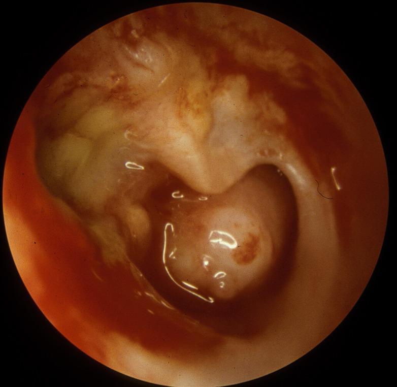
This endoscopic clinical photograph depicts the right tympanic membrane and middle ear cavity, demonstrating hallmark features of Chronic Suppurative Otitis Media (CSOM). A large subtotal perforation is visible, through which the structures of the middle ear are exposed. The middle ear mucosa appears erythematous and thickened, indicative of chronic inflammation. There is evidence of mucoid drainage, presenting as a viscous, translucent fluid within the cavity. Crucially, the long process of the incus shows significant erosion, a common sequela of long-standing inflammatory processes in the middle ear. The remaining tympanic membrane remnants appear thickened and opaque. This image serves as a classic educational example of middle ear pathology and the destructive potential of chronic infectious processes on the ossicular chain.
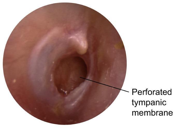
This clinical photograph shows an otoscopic view of the right ear, demonstrating a large, subtotal perforation of the tympanic membrane located primarily in the posterior and inferior quadrants. The perforation is patent, revealing the underlying middle ear space, which appears dark and recessed. The remaining tympanic membrane tissue is erythematous and thickened, suggesting chronic inflammation, with a loss of the normal translucent, pearly-gray appearance and light reflex. The handle of the malleus is visible but obscured by the retracted and inflamed tissue. The external auditory canal appears relatively normal without signs of stenosis or active otorrhea at the time of imaging. This presentation is characteristic of chronic suppurative otitis media (CSOM), a condition defined by persistent tympanic membrane perforation and a history of intermittent or continuous discharge.
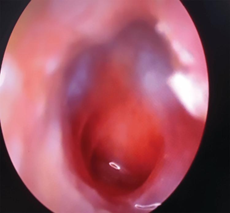
This clinical photograph, obtained via otoscopy/endoscopy, depicts a human tympanic membrane with a large central perforation. The perforation occupies a significant portion of the pars tensa, appearing as a dark, well-demarcated void that reveals the underlying middle ear space. The surrounding tympanic membrane remnant is erythematous and thickened, indicating active or chronic inflammation. The middle ear mucosa visible through the defect appears congested and reddish. Key anatomical landmarks, such as the handle of the malleus and the light reflex, are obscured or distorted due to the extensive tissue loss and inflammatory changes. This visual is characteristic of chronic suppurative otitis media (CSOM) or significant acoustic/mechanical trauma. The image serves as an educational example of middle ear pathology and the visual assessment of tympanic membrane integrity in an otolaryngology context.
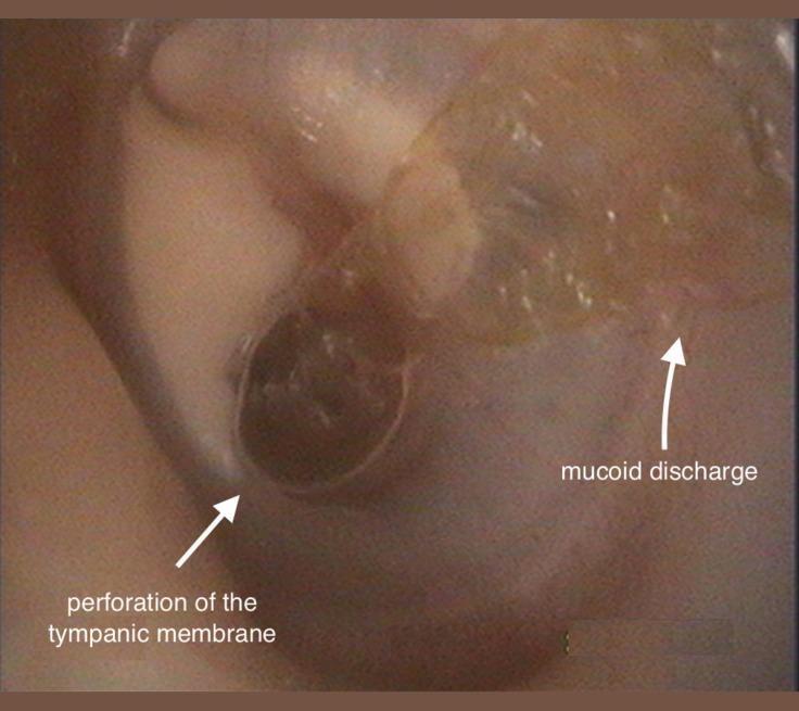
This clinical otoscopic photograph of a left ear demonstrates signs of chronic otitis media (COM). The image clearly shows a large, central perforation of the tympanic membrane, appearing as a dark, circular void where the membrane should be intact. Adjacent to this perforation, there is visible mucoid discharge, which presents as a translucent, glistening, and somewhat viscous fluid resting on the membrane surface. Additionally, white, opaque areas consistent with tympanosclerotic plaques are visible on the remaining portions of the tympanic membrane, indicating previous inflammation or scarring. The external auditory canal appears mildly erythematous. This visual is characteristic of chronic suppurative otitis media, an important educational example for distinguishing types of membrane perforations and associated middle ear pathology. The presence of discharge and structural changes in the membrane are key diagnostic markers used by otorhinolaryngologists to assess disease chronicity and planning for potential tympanoplasty.
Chronic Suppurative Otitis Media (CSOM)
Definition
Classification
| Feature | Tubotympanic (Safe/Benign) Type | Atticoantral (Unsafe/Dangerous) Type |
|---|---|---|
| Location | Central (pars tensa) perforation | Marginal / attic perforation |
| Cholesteatoma | Absent | Present (common) |
| Discharge | Mucoid/mucopurulent, profuse | Scanty, foul-smelling, purulent |
| Hearing loss | Conductive (mild-moderate) | Conductive or mixed |
| Bone erosion | Rare | Common |
| Complications | Uncommon | Frequent (hence "unsafe") |
Pathophysiology
- ET dysfunction → persistent negative middle-ear pressure → middle ear effusion (MEE)
- MEE causes mucosal edema, submucosal gland formation (converts to secretory mucosa), and granulation tissue
- Bacterial toxins and inflammatory mediators rupture the TM's basement membrane → lamina propria extrudes into the middle ear lumen → granulation tissue with angiogenic/epithelial growth factors → fibroblast recruitment, neovascularization, polyp formation
- Enzymes in the granulation tissue break down the TM's collagen skeleton → TM weakening → retraction pockets / perforation
- Deep retraction pockets + perforations set the stage for cholesteatoma formation
Microbiology
- Aerobic: Pseudomonas aeruginosa (most common, ~31%), Staphylococcus aureus (~19%), Proteus mirabilis, E. coli, Klebsiella
- Anaerobic: Peptococcus, Bacteroides sp., Peptostreptococcus, Fusobacterium, Clostridium, Propionibacterium
- Fungal: Candida species (seen in ~10-35% after prolonged topical antibiotic use)
Clinical Features
- Otorrhea - intermittent or continuous, sometimes foul-smelling (especially with cholesteatoma)
- Hearing loss - usually conductive; >30 dB suggests ossicular erosion
- Otalgia and headache are uncommon - their presence should raise suspicion for intracranial complication or malignancy
- Vertigo raises suspicion for labyrinthitis or labyrinthine fistula
- TM perforation (central in tubotympanic; marginal/attic in atticoantral)
- Discharge in the external auditory canal (EAC)
- Middle ear mucosa may be erythematous, thickened, or show granulation tissue
- Polyps may protrude through the EAC
- Scutal erosion, ossicular erosion visible through perforation
Clinical Images
Audiological Assessment
- Full audiometry is imperative preoperatively
- Conductive hearing loss is most common
- Sensorineural hearing loss (SNHL) of 5-33 dB may also coexist (document preoperatively)
- Occasionally hearing is preserved despite ossicular erosion due to sound transmission via the cholesteatoma directly to the oval window
Complications
Intratemporal
- Conductive hearing loss (TM perforation, ossicular erosion - most common: incus long process)
- Sensorineural hearing loss (suppurative labyrinthitis, cochlear hair cell loss)
- Labyrinthine fistula - mainly horizontal semicircular canal; causes vertigo and SNHL
- Facial nerve paralysis - acute (infective) or chronic (pressure from cholesteatoma in fallopian canal)
- Mastoiditis
- Tympanosclerosis
Intracranial (life-threatening - "unsafe" type)
- Meningitis
- Extradural/subdural abscess
- Brain abscess (temporal lobe, cerebellum)
- Lateral sinus thrombophlebitis
- Otitic hydrocephalus
- Brain hernia / CSF leakage (tegmen erosion)
Management
Medical Treatment
- Aural toilet (microsuction/mopping) - the single most important step; removes debris and allows topical agents to reach target
- Topical antibiotics - fluoroquinolones (ciprofloxacin/ofloxacin ear drops) are first-line; acetic acid/alcohol drops also used
- Systemic antibiotics - reserved for complications or when topical therapy fails; based on culture/sensitivity
Surgical Indications
- Cholesteatoma combined with medically refractory CSOM - nearly absolute indication
- Failed multiple attempts at medical treatment
- Symptoms suggesting complications (vertigo, facial weakness, headache)
- Deep retraction pockets
Surgical Options
- Tympanoplasty (myringoplasty) - TM repair; often sufficient for tubotympanic disease
- Mastoidectomy:
- Canal wall up (CWU) - preserves posterior EAC; reduced postoperative convalescence, no water restrictions; higher recurrence rate reported
- Canal wall down (CWD) - removes posterior EAC; wider access, lower recurrence; open cavity requires lifelong follow-up and debridement
- Tympanomastoidectomy - combined approach for extensive disease
- Eustachian tube obliteration + mastoid obliteration - considered before cochlear implantation
Special Consideration: CSOM and Cochlear Implantation
- First stage: radical mastoidectomy + ET obliteration + mastoid obliteration
- Second stage: cochlear implantation 2-6 months later (once the ear is dry)