
Okazakı fragments 2. EAA, Isoelectric pH, BUN & Zwitterion 3. Folate trap & MSUD 4. Multiple myeloma (B-J proteins) 5. Functions of plasma proteins (albumin) 6. Orotic aciduria & Lesch-Nyhan syndrome & SCID 7. Wobbles hypothesis
I'll research all 7 topics from your list using the medical library. Let me read the skill file first, then run parallel searches.
Searching sources...
Reading File
Now let me run parallel searches for all 7 topics at once:
Finding Sources
Finding Sources
Finding Sources
Finding Sources
Finding Sources
Finding Sources
Finding Sources
Finding Sources
Good results. Now let me read the key sections to get full content:
Reading File
Reading File
Reading File
Reading File
Reading File
Reading File
Reading File
Reading File
Excellent - I now have rich content for all 7 topics. Let me compile the full comprehensive answer:
Biochemistry Notes: All 7 Topics
1. Okazaki Fragments
Okazaki fragments arise because DNA polymerase can only synthesize new DNA in the 5' → 3' direction. At the replication fork, one strand (leading strand) is synthesized continuously, while the other (lagging strand) must be synthesized discontinuously in short segments - these are Okazaki fragments.
Mechanism:
- Helicase unwinds the double helix; topoisomerase relieves torsional strain ahead of the fork; single-strand binding proteins (SSBPs) stabilize the exposed strands.
- Primase synthesizes a short RNA primer (~10 nt) on the lagging strand template.
- Pol α extends the primer by ~20 deoxyribonucleotides.
- Pol δ (processive enzyme) takes over and extends the fragment further.
- The RNA primer is removed by RNase H (or Flap endonuclease 1, FEN1), and the gap is filled in by DNA polymerase.
- DNA ligase joins adjacent Okazaki fragments.
Key facts:
- Size: ~200 nucleotides in eukaryotes vs. 1,000-2,000 nt in prokaryotes
- Eukaryotic Okazaki fragment size is roughly equivalent to the length of DNA wrapped around one nucleosome
| Polymerase | Role | Exonuclease |
|---|---|---|
| Pol α | Starts primer extension (low processivity) | None |
| Pol δ | Lagging strand synthesis | 3'→5' (proofreading) |
| Pol ε | Leading strand synthesis | 3'→5' (proofreading) |
| Pol β | DNA repair only | None |
| Pol γ | Mitochondrial replication | 3'→5' |
- Basic Medical Biochemistry - A Clinical Approach, 6e
2. Essential Amino Acids (EAA), Isoelectric pH, BUN & Zwitterion
Essential Amino Acids (EAA)
Amino acids that cannot be synthesized by the body and must be obtained from the diet. Mnemonic: PVT TIM HaLL (or "10 EAA"):
| EAA | Note |
|---|---|
| Phenylalanine | Precursor to Tyr (Tyr becomes conditionally essential if Phe deficient) |
| Valine | BCAA |
| Threonine | |
| Tryptophan | Precursor to serotonin, niacin |
| Isoleucine | BCAA |
| Methionine | Precursor to Cys (Cys conditionally essential) |
| Histidine | Essential in infants; required in adults |
| Arginine | Conditionally essential (e.g., in growth, recovery) |
| Leucine | BCAA |
| Lysine |
BCAAs (Val, Leu, Ile) are catabolized primarily in muscle (not liver). They are unique in that they are the only amino acids whose initial catabolism occurs outside the liver.
Zwitterion & Isoelectric pH (pI)
A zwitterion is the form of an amino acid that carries both a positive and a negative charge simultaneously, with a net charge of zero. It is the predominant form at the amino acid's isoelectric point (pI).
- At pH < pI: the amino acid is positively charged (protonated)
- At pH = pI: net charge = 0 (zwitterion, no migration in electric field)
- At pH > pI: the amino acid is negatively charged (deprotonated)
pI calculation:
pI = (pKa1 + pKa2) / 2 For an amino acid with a charged R group, pI uses the two pKa values flanking the zwitterionic form.
Special pI values:
- Most amino acids: pI ~5-6 (acidic range due to the carboxyl group)
- Basic AAs (Arg, Lys, His): pI > 7 (positively charged at physiological pH)
- Acidic AAs (Asp, Glu): pI < 3
BUN (Blood Urea Nitrogen)
- Amino acid catabolism produces ammonia (NH₃), which is toxic
- In the urea cycle (liver), ammonia is converted to urea for excretion via kidney
- BUN reflects protein catabolism and renal filtration
- Normal BUN: ~8-20 mg/dL
- Elevated BUN (azotemia): seen in high protein intake, dehydration, GI bleeding, or renal failure
- BUN:Creatinine ratio >20:1 suggests pre-renal cause; 10-20:1 normal
3. Folate Trap & MSUD
Folate Trap
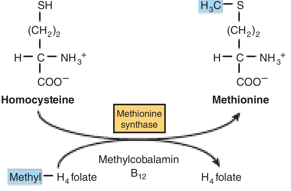
The "folate trap" (also called the methyl-folate trap) occurs in Vitamin B12 deficiency.
Normal pathway:
- Methyltetrahydrofolate (methyl-THF) donates its methyl group to homocysteine to form methionine, catalyzed by methionine synthase (a B12-dependent enzyme)
- The THF released is then available for other one-carbon transfer reactions (especially dTMP synthesis)
In B12 deficiency:
- Methionine synthase is impaired
- Methyl-THF cannot be converted back to THF
- Folate accumulates as methyl-THF and is "trapped" in this unusable form
- Result: functional folate deficiency even when dietary folate is adequate
- dUMP → dTMP conversion (via thymidylate synthase) is impaired
- Leads to megaloblastic anemia (same as dietary folate deficiency)
Clinical distinction: B12 deficiency also causes subacute combined degeneration of the spinal cord (myelin damage) - folate deficiency does NOT. This is key for differentiating both causes of megaloblastic anemia.
If folate (not B12) is given to a B12-deficient patient, it partially corrects the anemia (by bypassing the trap) but does not correct the neurological damage. - Basic Medical Biochemistry, 6e
MSUD (Maple Syrup Urine Disease)
- Autosomal recessive disorder (1:185,000)
- Defect in branched-chain α-keto acid dehydrogenase (BCKD) complex - the mitochondrial enzyme that oxidatively decarboxylates BCAAs (Leu, Ile, Val) and their α-keto acids
- Leu, Ile, Val and their corresponding α-keto acids accumulate in blood and urine
Clinical features:
- Feeding problems, vomiting, ketoacidosis
- Altered muscle tone, neurological deterioration, coma (primarily due to elevated leucine)
- Maple syrup odor in urine (due to isoleucine accumulation)
- Fatal if untreated; causes intellectual disability if treatment delayed
Treatment: Synthetic formula free of BCAAs + carefully monitored supplementation of Leu, Ile, Val. Thiamine-responsive variant responds to high-dose thiamine (B1).
- Biochemistry, Lippincott 8e; Harper's Illustrated Biochemistry 32e
4. Multiple Myeloma & Bence-Jones Proteins
Multiple myeloma is a clonal plasma cell malignancy in which a single abnormal plasma cell clone proliferates in the bone marrow and produces a monoclonal immunoglobulin or immunoglobulin fragment.
Bence-Jones (B-J) Proteins
- Free immunoglobulin light chains (either κ or λ) produced in excess by the malignant plasma cells
- Small enough to be filtered by the glomerulus and appear in urine
- Classic property: precipitate at 40-60°C and redissolve at 100°C (heat test - historical)
- Incidence in MM: 50-80% of patients
- Both free light chains AND serum M protein present in ~60-70% of patients
- ~20% of patients have only free light chains (no full M protein)
- ~1% of myelomas are nonsecretory (absence of M protein does not exclude MM)
Diagnosis
In 99% of patients, labs show increased immunoglobulin in blood and/or light chains in urine.
Most common M proteins:
- IgG: ~55% of cases
- IgA: ~25% of cases
- IgM, IgD, IgE: rare
CRAB criteria (diagnostic hallmarks):
| C | Hypercalcemia (bone destruction) |
| R | Renal dysfunction (cast nephropathy from B-J proteins) |
| A | Anemia (marrow replacement) |
| B | Bone lesions ("punched-out" lytic lesions) |
Myeloma kidney (cast nephropathy): B-J proteins form casts in tubules → tubular damage → renal failure. B-J proteins are also directly toxic to tubular epithelium.
Serum protein electrophoresis: Shows a sharp "M spike" (monoclonal band) in the gamma region vs. the broad band in normal sera. - Robbins Pathology, 10e
5. Functions of Plasma Proteins - Focus on Albumin
Plasma proteins are classified into albumin, globulins, and fibrinogen.
Site of synthesis: Liver produces essentially all albumin, all fibrinogen, and 50-80% of globulins. Remaining globulins (gamma globulins = immunoglobulins) are made by lymphoid tissue (plasma cells).
Liver can produce up to 30 g/day of plasma proteins when needed.
Albumin - Functions
| Function | Detail |
|---|---|
| Colloid osmotic (oncotic) pressure | Primary determinant of plasma oncotic pressure (~25 mmHg); prevents fluid leak from capillaries. Hypoalbuminemia → edema (nephrotic syndrome, cirrhosis, malnutrition) |
| Transport protein | Carries fatty acids, bilirubin, bile salts, thyroid hormones (T3/T4), steroid hormones, Ca²⁺, Mg²⁺, drugs (warfarin, aspirin, penicillin, diazepam) |
| Drug binding | Binds many drugs - affects their free (active) concentration; drug interactions occur through competitive binding |
| Acid-base buffer | Acts as a plasma buffer |
| Nutritional reserve | Acts as a labile protein store; macrophages can pinocytose plasma proteins → catabolize → release amino acids for tissue use |
| Antioxidant | Binds free copper; reduces oxidative damage |
Normal serum albumin: 3.5-5.0 g/dL
Clinical note: In liver cirrhosis, albumin synthesis drops → decreased oncotic pressure → generalized edema and ascites. - Guyton & Hall Textbook of Medical Physiology; Tietz Textbook of Laboratory Medicine, 7e
Other Plasma Proteins Summary
| Protein | Function |
|---|---|
| Globulins (α1, α2, β) | Transport (transferrin, haptoglobin, ceruloplasmin), enzymatic activity |
| Gamma globulins | Immunoglobulins (antibody-mediated immunity) |
| Fibrinogen | Polymerizes to fibrin - blood coagulation |
| α1-antitrypsin | Protease inhibitor |
| Haptoglobin | Binds free hemoglobin |
| Transferrin | Iron transport |
| Ceruloplasmin | Copper transport; ferroxidase activity |
| Complement proteins | Innate immunity |
6. Orotic Aciduria, Lesch-Nyhan Syndrome & SCID
All three are disorders of purine or pyrimidine metabolism.
Hereditary Orotic Aciduria
- Defect: Mutation in UMP synthase (a bifunctional enzyme with OPRTase and ODCase activities) - required in pyrimidine de novo synthesis
- Pathway block: Orotic acid cannot be converted to UMP → orotic acid accumulates in blood and urine
- Features: Megaloblastic anemia (not corrected by B12 or folate), failure to grow, crystalluria (orange crystals in urine)
- No hyperammonemia (unlike CPS I/OTC deficiency which also causes orotic aciduria - distinguish by ammonia levels)
- Treatment: Uridine supplementation (bypasses the enzyme defect, provides pyrimidines, and also suppresses de novo synthesis via feedback)
Orotic aciduria without hyperammonemia = UMP synthase defect (hereditary orotic aciduria). Orotic aciduria WITH hyperammonemia = OTC deficiency (urea cycle defect).
Lesch-Nyhan Syndrome
- Defect: Absent or severely deficient HGPRT (hypoxanthine-guanine phosphoribosyltransferase) - a purine salvage enzyme
- X-linked recessive (affects males)
- HGPRT normally salvages hypoxanthine and guanine → IMP and GMP. Without it, hypoxanthine/guanine are degraded → excess uric acid
- PRPP (normally consumed by salvage) accumulates → stimulates de novo purine synthesis → more uric acid
Clinical triad:
- Hyperuricemia / gout (joint deposits, tophi, nephrolithiasis)
- Intellectual disability (neurological damage)
- Self-mutilation - compulsive biting of lips and fingers (pathognomonic)
Treatment: Allopurinol (xanthine oxidase inhibitor) controls uric acid but does not correct neurological manifestations.
SCID (Severe Combined Immunodeficiency)
Most common biochemical cause relevant to purine metabolism:
ADA Deficiency (Adenosine Deaminase):
- ADA normally converts adenosine → inosine and deoxyadenosine → deoxyinosine
- Without ADA: deoxyadenosine (dAdo) and dATP accumulate
- dATP is toxic to lymphocytes (especially T cells): inhibits ribonucleotide reductase → blocks DNA synthesis in lymphocytes
- Result: Loss of both T and B cell function = SCID
PNP Deficiency (Purine Nucleoside Phosphorylase):
- Causes partial immunodeficiency - predominantly T-cell loss; B cells relatively spared
- dGTP accumulates → toxic to T cells
Summary table:
| Disease | Enzyme Defect | Cell Type Affected | Key Feature |
|---|---|---|---|
| SCID (ADA deficiency) | ADA | T + B cells (complete) | dATP accumulation |
| PNP deficiency | PNP | T cells (partial) | dGTP accumulation |
| Lesch-Nyhan | HGPRT | Not immune cells | Self-mutilation, gout |
| Hereditary orotic aciduria | UMP synthase | Not immune cells | Megaloblastic anemia |
- Basic Medical Biochemistry - A Clinical Approach, 6e
7. Wobble Hypothesis
Proposed by Francis Crick (1966) to explain how fewer than 61 tRNA species are needed to read 61 sense codons.
The Problem It Solves
The genetic code has 64 codons: 61 code for amino acids, 3 are stop codons. Strictly, 61 tRNA species would be required - but cells actually use far fewer (~45 in humans). The wobble hypothesis explains this economy.
The Hypothesis
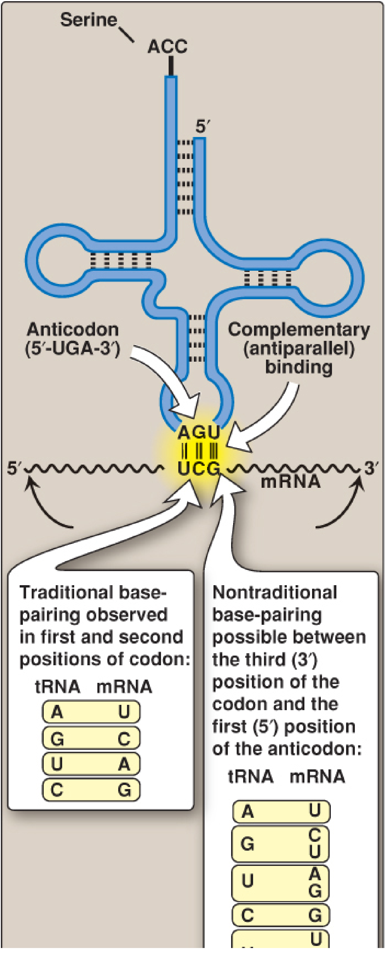
Strict Watson-Crick pairing applies to the 1st and 2nd positions of the codon (5' → 3') / 3rd and 2nd positions of the anticodon (3' → 5').
Wobble occurs at the 3rd base of the codon (= 5' base of the anticodon): the spatial constraint is relaxed, allowing non-Watson-Crick base pairing.
| 5' Anticodon base (wobble position) | Can pair with 3' codon base |
|---|---|
| C | G only |
| A | U only |
| U | A or G |
| G | C or U |
| Inosine (I) | U, C, or A (most flexible - can read 3 codons!) |
Inosine is the deamination product of adenosine (hypoxanthine base) and is commonly found at the wobble position of anticodons. Its flexibility is the most important example of wobble pairing.
Consequences
- A single tRNA can recognize 2-3 synonymous codons for the same amino acid
- Explains degeneracy of the genetic code (especially variation in the 3rd codon base)
- The code is degenerate but unambiguous: multiple codons specify one amino acid, but each codon specifies only one amino acid
- In mitochondria, the wobble rules are even more relaxed - only 22 tRNAs decode all mitochondrial codons (vs. 31 predicted by Crick's original rules)
Example: The anticodon 3'-IGC-5' (inosine) in tRNA^Ala reads codons GCU, GCC, and GCA (all code for Ala).
- Biochemistry, Lippincott Illustrated Reviews 8e; Basic Medical Biochemistry, 6e
Quick Reference Summary
| Topic | Key Point to Remember |
|---|---|
| Okazaki fragments | Lagging strand; ~200 nt in eukaryotes; Pol δ + RNA primer; ligase joins |
| EAA | PVT TIM HaLL; BCAAs catabolized in muscle; pI = pH of zero net charge |
| Folate trap | B12 deficiency → methionine synthase impaired → methyl-THF trapped → functional folate deficiency |
| MSUD | BCKD defect; BCAAs accumulate; maple syrup odor; leucine causes neurotoxicity |
| Multiple myeloma (B-J) | Free light chains in urine; CRAB criteria; IgG most common; myeloma kidney |
| Albumin | Oncotic pressure + transport + buffer + nutritional reserve |
| Orotic aciduria | UMP synthase defect; megaloblastic anemia; no hyperammonemia |
| Lesch-Nyhan | HGPRT defect; X-linked; self-mutilation + gout + intellectual disability |
| SCID | ADA deficiency → dATP toxic to T+B cells; PNP deficiency → T cells only |
| Wobble hypothesis | 3rd codon base / 5' anticodon base; inosine reads U, C, A; fewer tRNAs needed |
Phase 1 and phase 2 reactions
Phase 1 reactions introduce or unmask a functional group (oxidation, reduction, hydrolysis) - mainly via CYP450 enzymes. Phase 2 reactions conjugate that group to a polar molecule (glucuronide, sulfate, glutathione, etc.) making the drug water-soluble for excretion. Phase 1 can activate or inactivate drugs; Phase 2 almost always inactivates.
Searching sources...
Reading File
Finding Sources
Finding Sources
Finding Sources
Good - now let me read the key dedicated sections from Lippincott Pharmacology and Goodman & Gilman in parallel:
Reading File
Reading File
Reading File
Reading File
Now let me get more detail on Phase II conjugation reactions and CYP enzymes specifically:
Reading File
Finding Sources
Reading File
Reading File
I now have comprehensive material from multiple pharmacology textbooks. Here is the full organized answer:
Phase I and Phase II Drug Metabolism (Biotransformation)
Purpose: The kidney cannot efficiently excrete lipophilic drugs - they cross cell membranes easily and are reabsorbed in the distal tubule. Biotransformation converts these lipid-soluble drugs into more polar (hydrophilic) metabolites so they can be excreted in urine or bile.
Primary site: Liver (hepatic microsomes - smooth ER). Also: GI tract, kidney, lung, plasma.
Phase I Reactions - "Functionalization"
Goal: Introduce or unmask a polar functional group (-OH, -NH₂, -SH, -COOH) to make the drug more reactive/polar.
Types of reactions:
- Oxidation (most common)
- Reduction
- Hydrolysis
Effect on Pharmacologic Activity
Phase I metabolism may:
- Inactivate an active drug (most common outcome)
- Activate a prodrug → active metabolite (e.g., enalapril → enalaprilat; codeine → morphine via CYP2D6; clopidogrel → active thienopyridine via CYP2C19)
- Produce a toxic metabolite (e.g., acetaminophen → NAPQI via CYP2E1)
- Have no effect on activity
The CYP450 System - Main Phase I Engine
The cytochrome P450 (CYP) system is a superfamily of heme-containing monooxygenases located primarily in hepatocyte smooth ER and intestinal mucosa. These catalyze the vast majority of Phase I oxidative reactions.
Mechanism of CYP oxidation:
Drug + O₂ + NADPH → Drug-OH + H₂O + NADP⁺
CYP Nomenclature: Family (number) → Subfamily (letter) → Specific isoform (number)
- e.g., CYP3A4 = Family 3, Subfamily A, Isoform 4
Key CYP Isoforms
| Isoform | % of Drug Metabolism | Important Substrates | Inhibitors | Inducers |
|---|---|---|---|---|
| CYP3A4/5 | ~50% (most important) | Statins, CCBs, cyclosporine, tacrolimus, HIV PIs, midazolam, erythromycin | Ketoconazole, itraconazole, erythromycin, clarithromycin, ritonavir, grapefruit juice | Rifampin, phenobarbital, carbamazepine, St. John's Wort |
| CYP2D6 | ~25% | Codeine, tricyclic antidepressants, fluoxetine, paroxetine, propranolol, haloperidol, timolol | Fluoxetine, paroxetine, quinidine, bupropion | None known |
| CYP2C9 | ~15% | Warfarin, phenytoin, NSAIDs (ibuprofen, celecoxib), losartan, glipizide | Amiodarone, fluconazole | Rifampin, phenobarbital, carbamazepine |
| CYP2C19 | Omeprazole, clopidogrel (prodrug), mephenytoin | Omeprazole, ritonavir, fluoxetine | Rifampin | |
| CYP1A2 | Theophylline, caffeine, clozapine, warfarin (R) | Fluvoxamine, ciprofloxacin | Tobacco smoke, omeprazole | |
| CYP2E1 | Ethanol, acetaminophen, isoniazid | Ethanol, isoniazid |
CYP3A4 alone metabolizes >50% of all prescription drugs - Katzung's Basic and Clinical Pharmacology, 16e
Other Phase I Enzymes (Non-CYP)
- Monoamine oxidase (MAO) - oxidizes catecholamines, tyramine
- Flavin-containing monooxygenases (FMO) - oxidize N- and S-containing drugs
- Alcohol dehydrogenase / aldehyde dehydrogenase - ethanol metabolism
- Xanthine oxidase - purine catabolism; inactivates 6-MP
- Esterases/Amidases - hydrolysis (e.g., aspirin → salicylate; succinylcholine hydrolysis by plasma cholinesterase)
Phase II Reactions - "Conjugation"
Goal: Attach a large, polar, endogenous molecule to the Phase I product (or sometimes directly to the parent drug if it already has a suitable functional group), rendering it highly water-soluble and almost always pharmacologically inactive.
Key feature: Requires high-energy activated intermediates (e.g., UDPGA for glucuronidation, PAPS for sulfation, acetyl-CoA for acetylation).
Phase II Reaction Types
| Reaction | Enzyme | Co-substrate | Product | Notes |
|---|---|---|---|---|
| Glucuronidation | UDP-glucuronosyltransferase (UGT) | UDPGA (UDP-glucuronic acid) | Glucuronide conjugate | Most common Phase II reaction; adds -glucuronic acid; products excreted in bile or urine; can undergo enterohepatic recirculation |
| Sulfation | Sulfotransferase (SULT) | PAPS (3'-phosphoadenosine-5'-phosphosulfate) | Sulfate conjugate | Second most common; high affinity, low capacity (saturates at low doses); e.g., paracetamol at therapeutic doses |
| Acetylation | N-acetyltransferase (NAT) | Acetyl-CoA | Acetyl conjugate | Genetic polymorphism - "fast" vs. "slow" acetylators; relevant for isoniazid, hydralazine, procainamide; acetylated products may be less water-soluble |
| Methylation | Methyltransferases (COMT, TPMT) | SAM (S-adenosylmethionine) | Methyl conjugate | COMT inactivates catecholamines; TPMT inactivates 6-MP/azathioprine (TPMT deficiency → toxicity) |
| Glutathione conjugation | Glutathione-S-transferase (GST) | Glutathione (GSH) | Mercapturic acid (excreted) | Detoxifies reactive electrophiles (e.g., NAPQI from acetaminophen); protective mechanism |
| Amino acid conjugation | Acyltransferases | Glycine, glutamine, taurine | Amino acid conjugate | e.g., bile acids, salicylate + glycine → salicyluric acid |
UGT and CYP3A4 together account for the metabolism of >75% of all drugs in clinical use - Katzung, 16e
Important Note on "Direct" Phase II
Some drugs bypass Phase I entirely and go directly to Phase II if they already contain a suitable functional group. Examples:
- Oxazepam, lorazepam (benzodiazepines) - directly glucuronidated (safe in liver disease and elderly)
- Morphine - direct glucuronidation → morphine-6-glucuronide (active) and morphine-3-glucuronide
Summary Comparison Table
| Feature | Phase I | Phase II |
|---|---|---|
| Reaction type | Oxidation, reduction, hydrolysis | Conjugation (glucuronidation, sulfation, acetylation, methylation, GSH, amino acid) |
| Key enzymes | CYP450s, MAO, FMO, esterases | UGT, SULT, NAT, COMT, TPMT, GST |
| Effect on polarity | Moderately increased | Greatly increased (highly water-soluble) |
| Effect on activity | Variable (may activate, inactivate, or no change) | Almost always inactivates |
| Energy requirement | NADPH + O₂ (for CYP) | High-energy co-substrates (UDPGA, PAPS, Acetyl-CoA, SAM, GSH) |
| Product | Polar metabolite (intermediate) | Polar conjugate (final excretable product) |
| Location | Hepatic smooth ER (microsomes) | Cytosol + microsomes |
Enzyme Induction vs. Inhibition
Inducers (↑ CYP synthesis → ↑ drug metabolism → ↓ plasma levels):
- Classic broad inducers: Rifampin, Phenobarbital, Carbamazepine, Phenytoin, St. John's Wort
- Mechanism: Enhanced transcription via nuclear receptors (PXR, AhR)
- Effect: Therapeutic failure of co-administered drugs; accelerated self-metabolism (autoinduction)
Inhibitors (↓ CYP activity → ↓ drug metabolism → ↑ plasma levels → toxicity risk):
- Classic: Ketoconazole/itraconazole, Erythromycin/clarithromycin, Ritonavir, Grapefruit juice, Amiodarone, Cimetidine, Chloramphenicol
- Mechanisms: Substrate competition, competitive inhibition, noncompetitive inhibition
Genetic Polymorphisms - "Poor" vs. "Ultrarapid" Metabolizers
Especially important for CYP2D6 and NAT:
| Phenotype | CYP2D6 | Clinical Impact |
|---|---|---|
| Poor metabolizer | No/minimal activity | Codeine → no morphine formed (no analgesia); higher TCA levels → toxicity |
| Ultrarapid metabolizer | Multiple gene copies | Codeine → excess morphine → respiratory depression (dangerous in breastfeeding mothers) |
NAT slow acetylators: Isoniazid accumulates → peripheral neuropathy risk; hydralazine → drug-induced lupus.
TPMT deficiency: Azathioprine / 6-MP cannot be methylated → accumulates → severe bone marrow toxicity.
Practical Clinical Points
- Prodrugs depend on Phase I activation - poor metabolizers get no effect (codeine, clopidogrel)
- Narrow therapeutic index drugs (warfarin, phenytoin, cyclosporine) are highly susceptible to CYP drug interactions
- Lorazepam/oxazepam are preferred benzodiazepines in liver disease - they undergo direct Phase II glucuronidation, bypassing Phase I
- Acetaminophen toxicity: At therapeutic doses, glucuronidation and sulfation are the main pathways. CYP2E1 produces small amounts of toxic NAPQI, neutralized by GSH. In overdose, GSH depletes → NAPQI accumulates → hepatic necrosis
- First-pass metabolism: CYP3A4 in the intestinal mucosa + liver can extensively metabolize orally absorbed drugs before systemic circulation (e.g., nitroglycerin, cyclosporine, midazolam)

- Lippincott Illustrated Reviews: Pharmacology; Goodman & Gilman's Pharmacological Basis of Therapeutics; Katzung's Basic and Clinical Pharmacology, 16e; Harrison's Principles of Internal Medicine, 22e
This is a shared conversation. Sign in to Orris to start your own chat.