
intermuscular spaces of arm
Create a study guide on arm anatomy and compartments
arm
arm compartments intermuscular septa cross section anatomy diagram
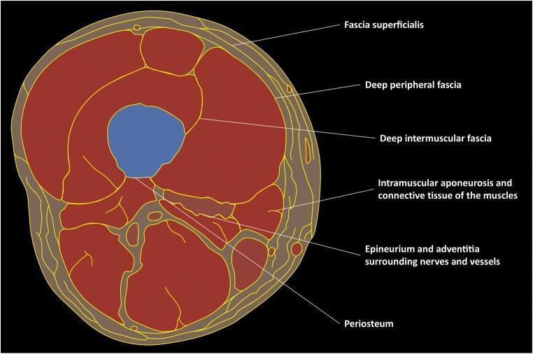
This medical schematic illustrates a transverse cross-section of the human thigh, focusing on the concentric and compartmental organization of the fascial system. Centrally, the femur is depicted in blue, outlined by the periosteum. Moving peripherally, the muscular tissue (red) is organized into functional compartments separated by the deep intermuscular fascia. Within the muscle bodies, the diagram identifies the intramuscular aponeurosis and connective tissue. Enveloping the entire muscular mass is the deep peripheral fascia, which serves as the interface between the deep musculature and the hypodermis. The outermost fascial layer shown is the fascia superficialis, located within the subcutaneous tissue. Between these fascial layers, the diagram highlights neurovascular bundles encapsulated by epineurium and adventitia. This illustration serves as an educational tool for understanding musculoskeletal anatomy, specifically the relationship between connective tissue layers, muscle compartments, and neurovascular pathways essential for diagnostic imaging and surgical planning.
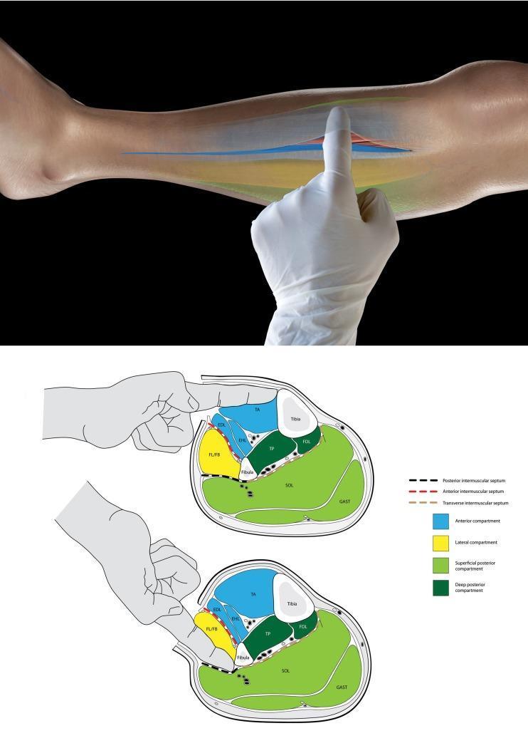
This educational graphic consists of a clinical photograph and two anatomical cross-sections illustrating the surgical technique for identifying leg compartments during a fasciotomy. The top photograph shows a gloved surgeon's finger inserted into an incision in the lower leg's fascia to palpate internal structures. Below, two schematic transverse cross-sections of the mid-leg categorize the four compartments: anterior (blue), lateral (yellow), superficial posterior (light green), and deep posterior (dark green). The top diagram demonstrates the 'tibia test': the finger enters the anterior compartment and palpates the medial tibia to confirm correct localization. The bottom diagram demonstrates the identification of the lateral (peroneal) compartment by palpating the fibula. The diagrams highlight the role of the intermuscular septa (anterior, posterior, and transverse) as physical barriers that prevent cross-compartment palpation, serving as a vital intraoperative check to ensure all compartments are adequately decompressed in cases of acute compartment syndrome.
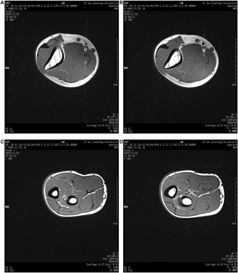
The image consists of four axial T1-weighted magnetic resonance imaging (MRI) cross-sections of the human upper limb, used for anatomical modeling. Panels A and B display cross-sections of the mid-upper arm, characterized by a single large central humerus (dark cortical bone with bright marrow signal) and expansive, encircling muscle groups. Panels C and D show the mid-forearm, clearly distinguished by the presence of two distinct bones: the radius and the ulna. Across all images, there is a clear demarcation of tissue layers including the outermost skin, a bright subcutaneous adipose layer, dark skeletal muscle compartments, and vascular structures. The contrast between the high-signal fat and intermediate-signal muscle allows for the identification of intermuscular septa and neurovascular bundles. These diagnostic images provide the anatomical foundation for understanding musculoskeletal structure, fluid distribution (relevant to lymphedema studies), and the spatial relationship between cortical bone and surrounding soft tissue compartments in the extremities.
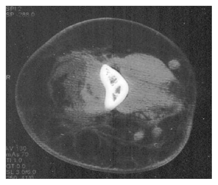
**Imaging Modality:** Computed Tomography (CT) scan, axial cross-section. **Anatomical Region:** Upper extremity, specifically the mid-shaft region of the arm (humerus). **Observed Pathology:** Extensive soft tissue inflammatory process characterized by a large, poorly circumscribed fluidic collection within the deep compartments. There is significant heterogeneous rearrangement and increased attenuation of the subcutaneous and intermuscular fatty tissue, consistent with fat stranding and cellulitis. **Characteristic Visual Features:** * **Muscular Involvement:** Evidence of muscular necrosis and edema, visualized as loss of normal myofascial architecture and low-attenuation areas within the muscle bellies. * **Fascial Changes:** Marked thickening of the intermuscular septa and deep fascia. * **Fluid Collection:** A localized, hypodense fluid accumulation suggestive of abscess formation or necrotizing infection. * **Bony Landmark:** The humerus is centrally located with a preserved cortical outline, though surrounding soft tissue changes are severe. **Diagnostic Features:** The combination of diffuse fat stranding, thickened septa, and muscular breakdown is highly suggestive of a severe necrotizing soft tissue infection (NSTI) or phlegmonous transformation. Neutral bone appearance helps differentiate primarily soft-tissue pathology from primary osteomyelitis with secondary extension.
Arm Anatomy and Compartments - Study Guide
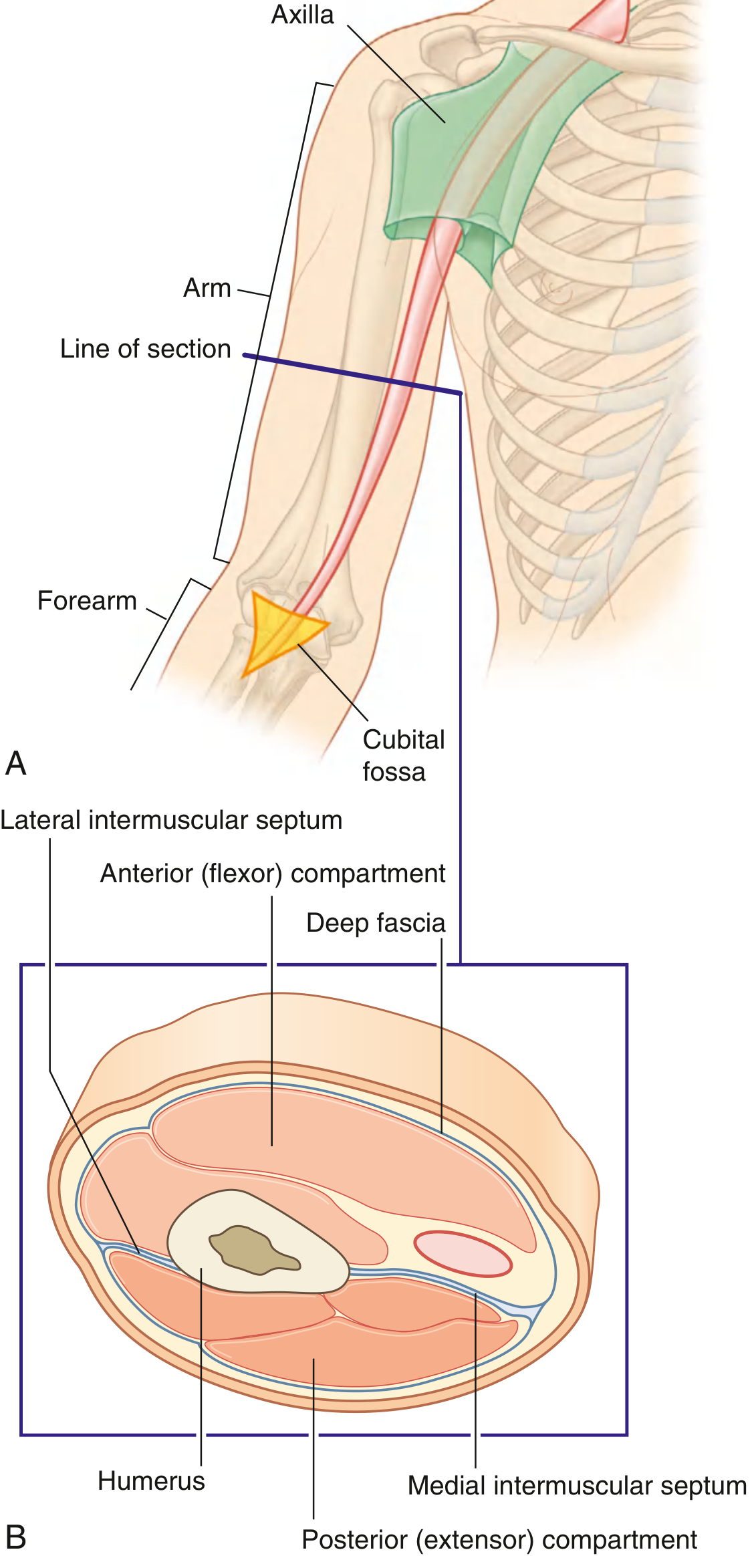
Overview
Fascial Framework
| Structure | Description |
|---|---|
| Brachial fascia | Dense fibrous sheath surrounding all arm muscles; continuous proximally with pectoral/axillary fascia, distally with antebrachial fascia |
| Lateral intermuscular septum | Projects from brachial fascia to lateral border/supracondylar ridge of humerus; separates anterior from posterior compartment laterally |
| Medial intermuscular septum | Projects from brachial fascia to medial border/supracondylar ridge of humerus; separates anterior from posterior compartment medially |
- Gray's Anatomy for Students, pp. 869-870
Three Anatomic Compartments of the Shoulder-Arm Region
- Deltoid compartment - subdivided into anterior, middle, and posterior subcompartments; deltoid fascia is continuous with the brachial fascia and lateral intermuscular septum distally
- Anterior brachial compartment
- Posterior brachial compartment
Anterior Compartment (Flexor)
Muscles
| Muscle | Origin | Insertion | Innervation | Action |
|---|---|---|---|---|
| Biceps brachii (long head) | Supraglenoid tubercle of scapula | Radial tuberosity + bicipital aponeurosis | Musculocutaneous (C5, C6) | Flexion of elbow; supination of forearm (most powerful when elbow flexed); weak shoulder flexion |
| Biceps brachii (short head) | Coracoid process (with coracobrachialis) | Radial tuberosity + bicipital aponeurosis | Musculocutaneous (C5, C6) | Same as above |
| Brachialis | Distal half of anterior humerus | Coronoid process/tuberosity of ulna | Musculocutaneous (C5, C6); small radial nerve contribution to lateral part | Flexion of forearm - the main elbow flexor |
| Coracobrachialis | Coracoid process tip | Medial midshaft humerus | Musculocutaneous (C6, C7) - nerve pierces through this muscle | Flexion and adduction of arm at shoulder |
Key point: Brachialis is the largest contributor to elbow flexion. The musculocutaneous nerve penetrates coracobrachialis before passing between biceps and brachialis in the midarm. - Imaging Anatomy Atlas
Neurovascular Supply - Anterior Compartment
- Runs medially in the arm
- Gives off the profunda brachii (deep artery of the arm), which accompanies the radial nerve into the posterior compartment
| Nerve | Course in Arm | Branches in Arm |
|---|---|---|
| Musculocutaneous | Pierces coracobrachialis; runs between biceps and brachialis | All three anterior compartment muscles; continues as lateral cutaneous nerve of forearm |
| Median | Enters from axilla lateral to brachial artery; crosses to medial side distally | None (passes through to forearm) |
| Ulnar | Enters medially with brachial vessels; at mid-arm pierces medial intermuscular septum to enter posterior compartment; then passes behind medial epicondyle | None in arm |
Posterior Compartment (Extensor)
Muscle
| Muscle | Origin | Insertion | Innervation | Action |
|---|---|---|---|---|
| Triceps brachii (long head) | Infraglenoid tubercle of scapula | Olecranon of ulna | Radial nerve (C6-C8) | Extension of elbow; adduction/extension of arm |
| Triceps brachii (lateral head) | Posterior humerus superior to radial groove | Olecranon | Radial nerve | Extension of elbow |
| Triceps brachii (medial head) | Posterior humerus inferior to radial groove | Olecranon | Radial nerve (branch arises before posterior compartment entry) | Extension of elbow |
Neurovascular Supply - Posterior Compartment
- Gives off posterior radial collateral artery, which courses posterior to brachioradialis along the lateral intermuscular septum
- Originates from posterior cord of brachial plexus
- Enters arm posterior to brachial artery
- Enters posterior compartment via the triangular interval (space between long head of triceps, teres major, and humeral shaft)
- Runs diagonally in the radial (spiral) groove - directly on bone
- Passes anteriorly through the lateral intermuscular septum to enter anterior compartment
- Runs between brachialis and brachioradialis anterior to the lateral epicondyle
- Enters forearm deep to brachioradialis
Muscular branches of radial nerve in arm: triceps brachii (all heads), brachioradialis, extensor carpi radialis longus, and lateral part of brachialis
Intermuscular Spaces (Gaps/Intervals) of the Arm
Triangular Interval (Lower Triangular Space)
- Boundaries: Long head of triceps (medial), teres major (superior), shaft of humerus (lateral)
- Contents: Radial nerve + profunda brachii artery
- These structures enter the posterior compartment here
Triangular Space (Upper Triangular Space / Medial Triangular Space)
- Boundaries: Teres minor (superior), teres major (inferior), long head of triceps (lateral)
- Contents: Circumflex scapular artery (branch of subscapular artery)
Quadrangular Space (Quadrilateral Space)
- Boundaries: Teres minor (superior), teres major (inferior), long head of triceps (medial), surgical neck of humerus (lateral)
- Contents: Axillary nerve + posterior circumflex humeral artery
Arcade of Struthers
- A thickening of the brachial fascia extending from the medial head of triceps to the medial intermuscular septum
- The ulnar nerve passes under this arcade approximately 8 cm above the medial epicondyle as it pierces the medial intermuscular septum
- Clinically relevant: can cause ulnar nerve entrapment; the nerve may be kinked following triceps muscle transposition procedures
Key Nerve Transitions Through Septa
| Nerve | Septum Crossed | Direction | Clinical Relevance |
|---|---|---|---|
| Radial nerve | Lateral intermuscular septum | Posterior → Anterior | Injured in midshaft humerus fracture (radial groove) → wrist drop |
| Ulnar nerve | Medial intermuscular septum | Anterior → Posterior | Passes posterior to medial epicondyle; vulnerable to compression at arcade of Struthers |
Superficial Veins
| Vein | Course |
|---|---|
| Basilic vein | Passes vertically in distal half of arm, penetrates deep fascia, becomes axillary vein at lower border of teres major |
| Cephalic vein | Ascends anterolaterally on the arm, passes through anterior wall of axilla to join axillary vein |
- Gray's Anatomy for Students, p. 879
Clinical Pearls
- Radial nerve palsy / wrist drop - midshaft humeral fracture injures the nerve in the radial groove; always test radial nerve in suspected humeral shaft fractures
- Musculocutaneous nerve damage - causes loss of elbow flexion (biceps, brachialis) and loss of sensation on lateral forearm (lateral cutaneous nerve of forearm)
- Ulnar nerve entrapment at arcade of Struthers - causes medial forearm and hand sensory loss (ring/little fingers) and intrinsic hand weakness
- Compartment syndrome of the arm - elevated pressure in anterior or posterior compartment compromises neurovascular structures; the lateral intermuscular septum must be released during fasciotomy
- Bicipital aponeurosis - the flat sheet of connective tissue fanning off the biceps tendon medially to blend with the forearm deep fascia; protects the brachial artery and median nerve during venepuncture at the cubital fossa