
carbohydrate chemistry
I'll read the medical library skill first since carbohydrate chemistry is a biochemistry/medical topic.
Reading File
Now let me search the medical library for carbohydrate chemistry content, running parallel searches:
Finding Sources
Finding Sources
Let me search for a dedicated biochemistry section on carbohydrate chemistry with more specific headings, and also pull the best sources:
Finding Sources
Finding Sources
Excellent - I found the best source: Lippincott's Biochemistry and Harper's Illustrated Biochemistry have dedicated carbohydrate chemistry sections. Let me read the core content now in parallel:
Reading File
Reading File
Good content. Let me now fetch images and read more on disaccharides, polysaccharides, and key reactions in parallel:
Loading Image
Reading File
Reading File
Excellent content. Let me now fetch more images and read the section on important polysaccharides and monosaccharide metabolism:
Loading Image
Loading Image
Reading File
Now let me also read the Harper's section on important polysaccharides and the section on glycosidic bonds + disaccharides:
Reading File
Reading File
Now let me fetch key images and read more from Harper's on polysaccharides:
Loading Image
Reading File
I now have comprehensive content from two authoritative biochemistry textbooks. Let me compile the full answer:
Carbohydrate Chemistry
Carbohydrates are the most abundant organic molecules in nature, with the empirical formula (CH₂O)ₙ (n ≥ 3), hence "hydrates of carbon." They serve as energy sources, energy storage polymers, structural materials (cellulose, chitin), and cell-surface recognition molecules.
1. Classification
By Carbon Number
| Class | Carbons | Aldose Example | Ketose Example |
|---|---|---|---|
| Triose | 3 | Glyceraldehyde | Dihydroxyacetone |
| Tetrose | 4 | Erythrose | Erythrulose |
| Pentose | 5 | Ribose | Ribulose |
| Hexose | 6 | Glucose, Galactose, Mannose | Fructose |
| Heptose | 7 | - | Sedoheptulose |
By Carbonyl Group
- Aldoses - have an aldehyde (-CHO) as the carbonyl group (e.g., glucose, galactose)
- Ketoses - have a keto (C=O) group (e.g., fructose, ribulose)
By Polymer Length
- Monosaccharides - single sugar units
- Disaccharides - 2 units (e.g., lactose, sucrose, maltose)
- Oligosaccharides - 3-10 units (mostly not digested by human enzymes)
- Polysaccharides - >10 units, can be hundreds of residues (starch, glycogen, cellulose)
2. Structure of Glucose - Three Representations
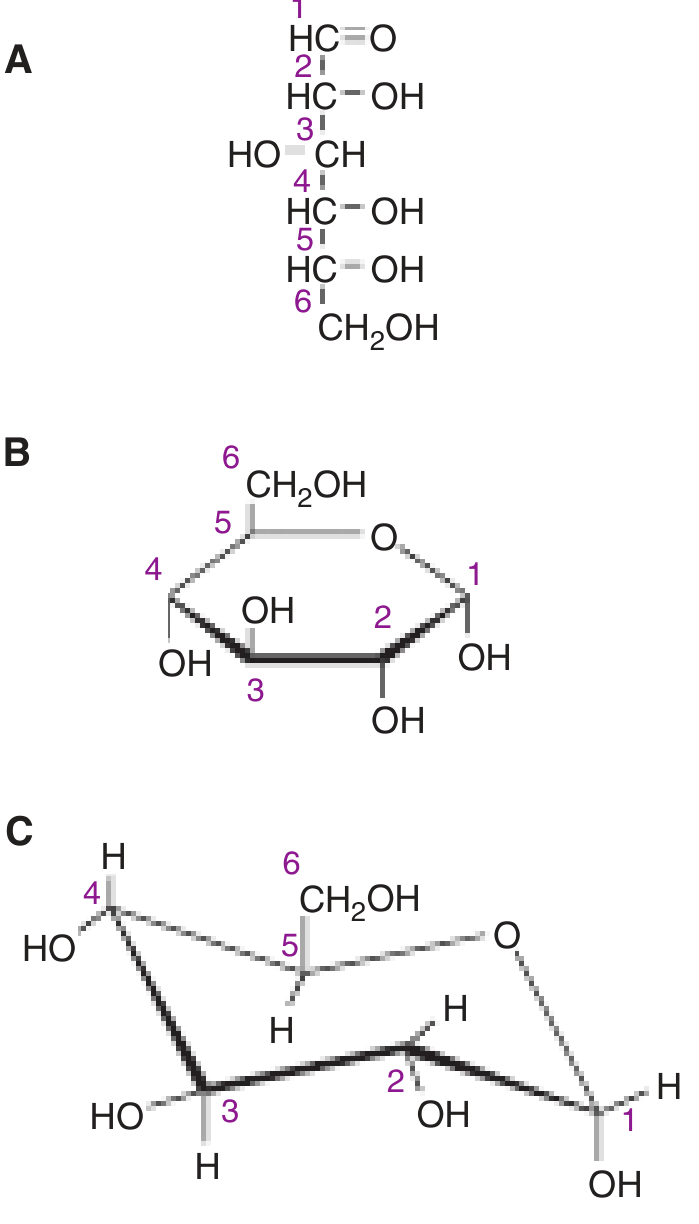
D-Glucose shown as (A) straight-chain aldehyde form, (B) α-D-glucopyranose (Haworth projection), and (C) chair conformation. - Harper's Illustrated Biochemistry, 32nd Ed.
3. Stereochemistry and Isomerism
D- and L-Isomers (Enantiomers)
- The designation D or L is based on the orientation of the -OH on the asymmetric carbon farthest from the carbonyl carbon (C-5 in a hexose)
- D-isomer: -OH on the right in a Fischer projection
- L-isomer: -OH on the left
- Virtually all biologically active monosaccharides in humans are D-isomers
- Most enzymes are stereospecific for either D or L forms; isomerases can interconvert them
Epimers
- Compounds that differ in configuration around only one carbon (not the carbonyl carbon) are epimers
- Glucose and galactose are C-4 epimers (differ at C-4 only)
- Glucose and mannose are C-2 epimers (differ at C-2 only)
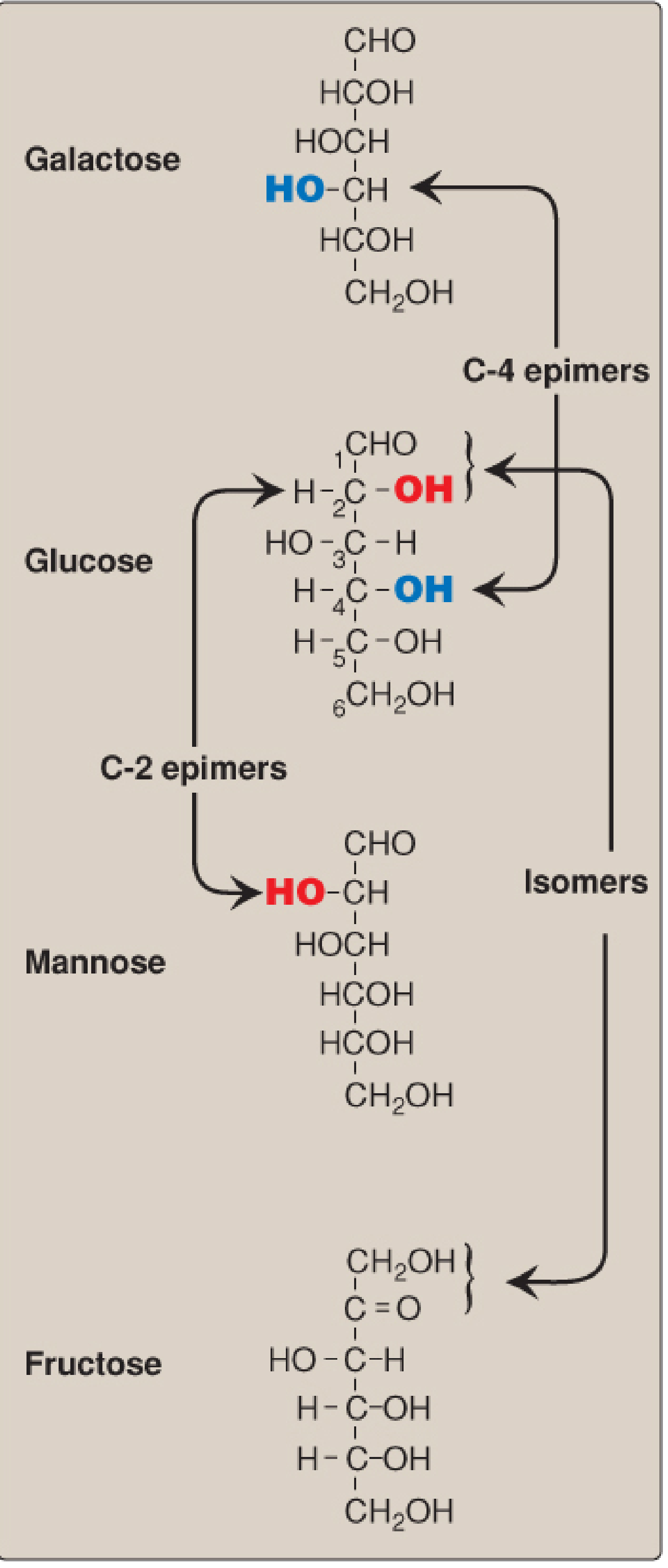
C-2 and C-4 epimers and isomers of glucose. - Lippincott's Biochemistry, 8th Ed.
Optical Activity
- D/L designation refers to spatial configuration, not direction of light rotation
- Glucose is dextrorotatory (+), sometimes called "dextrose"
- Fructose is levorotatory (-), so it is D(-)-fructose
- Hydrolysis of sucrose yields an "invert sugar" because the strongly levorotatory fructose inverts the dextrorotatory action of sucrose
4. Ring Structures (Cyclization)
Less than 1% of monosaccharides with 5+ carbons exist in the open-chain form in solution. They cyclize spontaneously when the carbonyl group reacts with a hydroxyl group on the same molecule:
- Pyranose - 6-membered ring (5C + 1O), analogous to pyran; over 99% of glucose in solution
- Furanose - 5-membered ring (4C + 1O), analogous to furan; common form of fructose and ribose
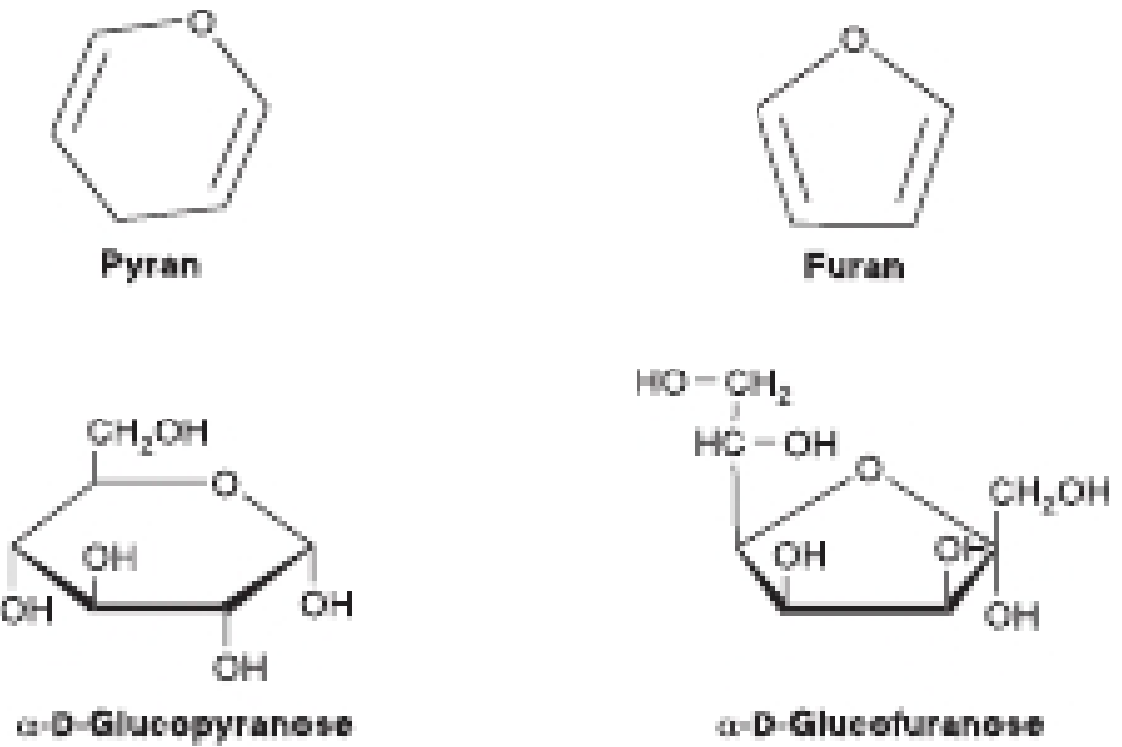
Pyranose and furanose forms of glucose. - Harper's Illustrated Biochemistry, 32nd Ed.
Anomeric Carbon and Anomers
- Cyclization makes the former carbonyl carbon asymmetric - this is the anomeric carbon
- Creates two new diastereomers called α and β anomers:
- α form: -OH on the anomeric carbon is axial (same side as the ring in a modified Fischer projection; trans to CH₂OH in Haworth)
- β form: -OH on the anomeric carbon is equatorial (opposite side)
- For glucose: α form = 36%, β form = 64% at equilibrium
Mutarotation
- The α and β anomers interconvert spontaneously in solution through the open-chain form
- This process is called mutarotation
- Biologically, the anomeric configuration matters: glycogen is built from α-D-glucopyranose; cellulose is built from β-D-glucopyranose
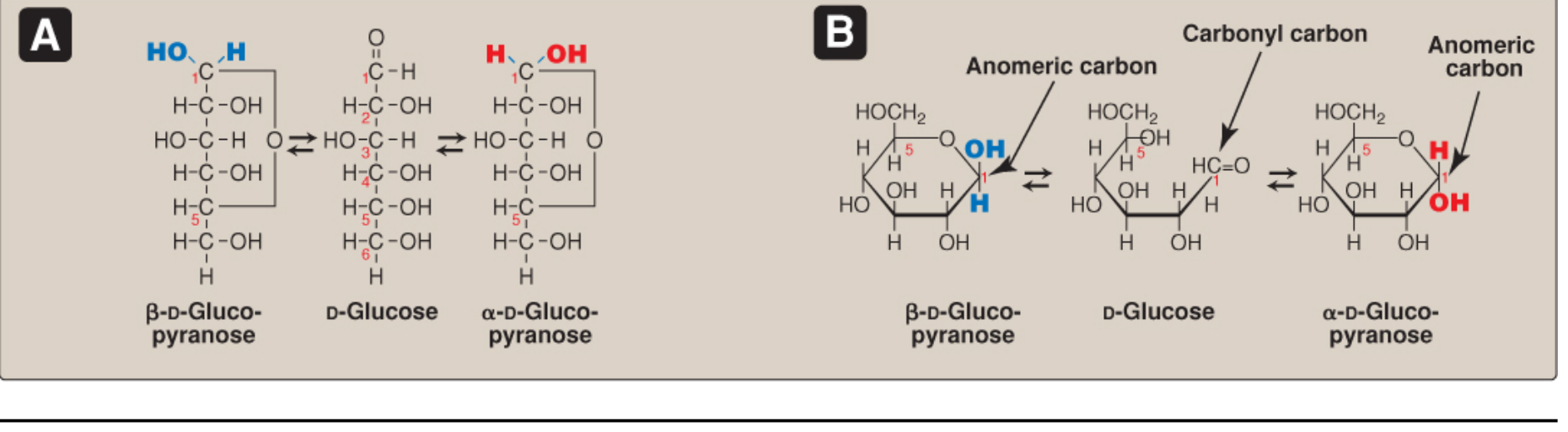
Mutarotation of glucose. - Lippincott's Biochemistry, 8th Ed.
5. Reducing Sugars
- If the anomeric carbon is free (not involved in a glycosidic bond), the ring can open, exposing the aldehyde group
- This aldehyde can reduce chromogenic agents like Benedict's reagent (Cu²⁺ is reduced, changing color)
- All monosaccharides are reducing sugars
- Among disaccharides: lactose and maltose are reducing (one free anomeric -OH); sucrose is non-reducing (both anomeric carbons are engaged in the glycosidic bond)
- Fructose (a ketose) is also a reducing sugar because it isomerizes to an aldose
Clinical note: A positive Benedict's test in urine indicates glycosuria, which is not normal and warrants follow-up testing to identify the specific sugar.
6. Glycosidic Bonds
Monosaccharides are joined by glycosidic bonds, formed enzymatically by glycosyltransferases using nucleotide sugars (activated donors, e.g., UDP-glucose) as substrates.
- Named by the carbons involved and the anomeric configuration: e.g., β(1→4) linkage in lactose (C-1 of β-galactose joined to C-4 of glucose)
- α(1→4) bonds: found in starch (amylose, amylopectin), glycogen, and maltose
- α(1→6) bonds: branch points in glycogen and amylopectin
- β(1→4) bonds: found in cellulose and lactose
- α(1→2)β bond: sucrose (both anomeric carbons engaged)
N- and O-Glycosidic Bonds to Non-Carbohydrates
- N-glycosidic bond: sugar links to -NH₂ group (e.g., nucleosides, N-linked glycoproteins)
- O-glycosidic bond: sugar links to -OH group (e.g., O-linked glycoproteins, steroids)
7. Important Monosaccharide Derivatives
| Derivative | Structure/Feature | Significance |
|---|---|---|
| Glucosamine | Glucose with -NH₂ at C-2 | Component of heparin, hyaluronic acid |
| Galactosamine | Galactose with -NH₂ at C-2 | Component of cartilage (chondroitin sulfate) |
| N-Acetylglucosamine | Glucosamine with N-acetyl group | Component of glycoproteins, chitin |
| N-Acetylneuraminic acid (sialic acid) | 9-carbon amino sugar | Cell-surface glycoprotein/glycolipid terminal residue |
| Glucuronic acid | Glucose with oxidized C-6 → -COOH | Detoxification reactions (UDP-glucuronate), proteoglycans |
8. Important Disaccharides
| Disaccharide | Components | Linkage | Reducing? | Source |
|---|---|---|---|---|
| Maltose | Glucose + Glucose | α(1→4) | Yes | Starch hydrolysis, malt |
| Isomaltose | Glucose + Glucose | α(1→6) | Yes | Glycogen/amylopectin branch hydrolysis |
| Lactose | Galactose + Glucose | β(1→4) | Yes | Milk |
| Sucrose | Glucose + Fructose | α(1→2)β | No | Sugar cane, sugar beet |
| Trehalose | Glucose + Glucose | α(1→1)α | No | Insect hemolymph, fungi |
9. Polysaccharides
Starch (Plant Storage)
- Amylose (13-20%): linear, unbranched helical chain of glucose with α(1→4) linkages
- Amylopectin (80-87%): branched; chains of 24-30 glucose residues with α(1→4) in chains and α(1→6) at branch points
Glycogen (Animal Storage - "Animal Starch")
- More highly branched than amylopectin; chains of 12-15 glucose residues with α(1→4) links and α(1→6) branch points
- Liver glycogen: maintains blood glucose between meals
- Muscle glycogen: local fuel for contraction
- Rapidly mobilized by phosphorylase
Cellulose (Plant Structural)
- Linear polymer of glucose with β(1→4) linkages
- Humans cannot digest cellulose (no β(1→4)-glucosidase)
- Major component of dietary fiber
Inulin
- Polymer of fructose; found in artichokes, dahlias, chicory
- Not hydrolyzed by intestinal enzymes; no caloric value
- Used clinically to measure GFR (glomerular filtration rate)
10. Digestion of Dietary Carbohydrates
| Stage | Enzyme | Action |
|---|---|---|
| Mouth | Salivary α-amylase | Hydrolyzes random α(1→4) bonds in starch/glycogen → dextrins + oligosaccharides |
| Stomach | - | Amylase is inactivated by low pH; digestion pauses |
| Small intestine (lumen) | Pancreatic α-amylase | Continues hydrolysis of starch; produces maltose, maltotriose, limit dextrins |
| Small intestine (brush border) | Maltase: maltose → 2 glucose | Final monosaccharide release |
| Lactase (β-galactosidase): lactose → glucose + galactose | Deficiency → lactose intolerance | |
| Sucrase: sucrose → glucose + fructose | Deficiency → osmotic diarrhea | |
| Isomaltase: α(1→6) bonds of dextrins | - |
- Final products (glucose, galactose, fructose) are absorbed by enterocytes via specific transporters (SGLT-1 for glucose/galactose, GLUT-5 for fructose)
11. Clinically Relevant Points
- Lactase deficiency (lactose intolerance): undigested lactose fermented by colonic bacteria → gas, osmotic diarrhea, bloating. Very common (up to 70% of adults globally)
- Sucrase-isomaltase (SI) deficiency: intolerance to sucrose; affects up to 9% of Americans of European descent; treated with dietary restriction and enzyme replacement
- Glycosuria (reducing sugar in urine): positive Benedict's test is pathological; requires follow-up for diabetes, galactosemia, or other metabolic disorders
- Glycemic index: reflects how rapidly a starch raises blood glucose; determined by digestibility and cooking
- Glycogen storage diseases (e.g., von Gierke, Pompe, McArdle diseases): enzyme defects in glycogen synthesis or breakdown
Sources: Lippincott Illustrated Reviews: Biochemistry 8th Ed., Chapter 7; Harper's Illustrated Biochemistry 32nd Ed., Chapter 15
This is a shared conversation. Sign in to Orris to start your own chat.