
Can you make mbbs lavel notes on ulcerative dysplasia and non ulcerative dysplasia in pathology
Reading File
Now let me search the medical library for relevant content on dysplasia topics.
Finding Sources
Finding Sources
Good. Let me now read the key sections from Robbins Pathology and the GI textbooks on dysplasia in ulcerative colitis.
Reading File
Reading File
Reading File
Now let me search for "non-ulcerative dysplasia" and the specific classification in ulcerative colitis (flat vs. raised dysplasia), and also look for cervical/epithelial dysplasia.
Finding Sources
Finding Sources
Let me now read the key Robbins section on colitis-associated neoplasia and the dysplasia section from Clinical GI Endoscopy, and also look at epithelial dysplasia from Cummings.
Reading File
Reading File
Reading File
Let me get more content on the epithelial dysplasia section and also fetch the histology image.
Reading File
Reading File
Now let me fetch the histology image from the Cummings text and also get the Robbins figure on IBD dysplasia.
Loading Image
Finding Sources
I now have sufficient information from the textbooks. Let me compile comprehensive MBBS-level notes on ulcerative dysplasia and non-ulcerative dysplasia in pathology.
MBBS Pathology Notes: Ulcerative Dysplasia & Non-Ulcerative Dysplasia
PART 1 - DYSPLASIA: GENERAL CONCEPTS
Definition: Dysplasia (= "intraepithelial neoplasia") refers to a pre-invasive, reversible (early stages) or potentially irreversible (severe) alteration of epithelial cells characterized by abnormal growth, maturation, and cytologic atypia - without invasion through the basement membrane.
Hallmarks of Dysplasia (General)
| Feature | Description |
|---|---|
| Nuclear enlargement | Nuclei are larger than normal |
| Hyperchromasia | Dark-staining due to increased DNA |
| Pleomorphism | Variation in cell and nuclear size/shape |
| Coarse chromatin | Clumped, irregular chromatin texture |
| Loss of polarity | Normal orientation of cells disrupted |
| Increased mitoses | Including atypical/abnormal mitotic figures |
| Loss of maturation | Cells do not mature as they reach the surface |
Key distinction from reactive/regenerative changes: in inflammation, epithelial cells mature as they reach the mucosal surface; in dysplasia, cells remain cytologically immature at the surface.
- Robbins & Kumar Basic Pathology, p. 557
- Robbins, Cotran & Kumar Pathologic Basis of Disease, p. 749
PART 2 - ULCERATIVE DYSPLASIA
= Dysplasia occurring in the setting of chronic ulcerative colitis (UC) and other inflammatory bowel disease (IBD)
Also called: Colitis-Associated Neoplasia / IBD-associated dysplasia
Pathogenesis
- Long-standing UC causes repeated cycles of mucosal injury and regeneration
- Inflammation-related free radical damage accumulates genetic alterations
- Key molecular events: p53 mutation (early), p16 inactivation, oncogene activation, genomic instability
- Two pathways to cancer: (1) step-wise accumulation of mutations, (2) whole-genome doubling (faster pathway)
Risk Factors for Cancer in UC
- Duration - risk rises sharply after 8-10 years of disease
- Extent - pancolitis > left-sided disease; proctitis alone has no increased risk
- Severity of inflammation - greater frequency and severity of neutrophilic infiltration = higher risk
- Coexisting Primary Sclerosing Cholangitis (PSC) - 4x increased risk; begin surveillance at diagnosis
- Family history of CRC in first-degree relative <50 years
- Male gender, prior history of dysplasia, stricturing disease in UC
Classification of Ulcerative Dysplasia (IBD-Associated)
Grading:
| Grade | Features |
|---|---|
| Indefinite for dysplasia | Cannot distinguish reactive from true dysplasia |
| Low-Grade Dysplasia (LGD) | Mild-moderate cytologic atypia; glandular architecture mildly distorted |
| High-Grade Dysplasia (HGD) | Marked cytologic and architectural atypia; may associate with carcinoma |
Endoscopic Classification (Paris Classification):
- Visible dysplasia - detected in clearly identifiable lesions:
- Polypoid (pedunculated/sessile)
- Non-polypoid (superficially elevated, flat, or depressed)
- Invisible dysplasia - detected only in random biopsy samples, not visible endoscopically
The old terms "DALM" (dysplasia-associated lesion or mass) and "sporadic adenoma" are now abandoned and replaced by "visible" and "invisible" dysplasia.
- Clinical Gastrointestinal Endoscopy Expert Consult 3e, p. 588
Morphology (Histology)
Low-Grade Dysplasia:
- Nuclei are pencil-shaped (elongated), hyperchromatic
- Nuclear stratification confined to lower half of cell
- Cytoplasm shows some maturation towards surface
- Glandular architecture mildly distorted
High-Grade Dysplasia:
-
Nuclei rounded, markedly enlarged, prominent nucleoli
-
Nuclear stratification throughout full thickness of epithelium
-
Loss of cytoplasmic maturation
-
Architectural disarray: cribriform glands, villiform surfaces, back-to-back glands
-
Atypical mitoses prominent
-
Robbins, Cotran & Kumar Pathologic Basis of Disease
Clinical Importance of Ulcerative Dysplasia
- Dysplasia is a precursor of colitis-associated carcinoma
- Dysplasia can occur in flat (endoscopically normal) mucosa - hence need for systematic biopsies
- Interobserver variability is HIGH for LGD (<50% agreement); BETTER for HGD (~85%)
- Second opinion from specialist GI pathologist is mandatory when dysplasia is detected
Surveillance Protocol
- Screening colonoscopy 8-10 years after IBD onset (even in isolated proctitis)
- Annual surveillance for highest risk: PSC + UC, extensive active inflammation, family history CRC <50 yrs, prior dysplasia, stricturing disease
- Random biopsies: 4-quadrant biopsies every 10 cm throughout colon (33 biopsies needed for 90% sensitivity)
- High-definition colonoscopy + chromoendoscopy preferred over random biopsies
- Clinical Gastrointestinal Endoscopy Expert Consult 3e
Management Based on Dysplasia Grade
| Finding | Management |
|---|---|
| Indefinite for dysplasia | Optimize medical therapy, repeat biopsy |
| LGD (focal, polypoid, resectable) | Endoscopic resection + close follow-up |
| LGD (flat/multifocal/nonpolypoid) | Consider total proctocolectomy |
| HGD (any form) | Total proctocolectomy - risk of synchronous invasive carcinoma |
| Invasive carcinoma found under LGD | Colectomy |
PART 3 - NON-ULCERATIVE DYSPLASIA
= Dysplasia occurring in tissues NOT associated with chronic ulceration/IBD
This is a broad category that includes dysplasia in:
- Gastric epithelium (in chronic gastritis, without active ulceration)
- Esophageal/Barrett epithelium
- Squamous epithelium (oral cavity, larynx, cervix, vulva)
3A. GASTRIC DYSPLASIA (Non-Ulcerative)
Background: Arises in the setting of chronic gastritis (H. pylori-associated or autoimmune), intestinal metaplasia, and atrophy - not from active ulceration
Pathogenesis sequence:
Normal gastric mucosa → Chronic gastritis → Atrophy → Intestinal metaplasia → Dysplasia → Adenocarcinoma (Correa cascade)
Morphology:
- Variations in epithelial size, shape, and orientation
- Coarse chromatin, hyperchromasia, nuclear enlargement
- Reactive/regenerative cells mature at the surface; dysplastic cells do not
Low-Grade vs. High-Grade:
- LGD: tubular architecture preserved, mild cytologic atypia
- HGD: loss of architecture, marked nuclear atypia, atypical mitoses
Clinical relevance:
-
Gastric adenoma (10% of gastric polyps) exhibits dysplasia - risk of malignant transformation far greater than colonic polyps
-
Carcinoma found in up to 30% of gastric adenomas at time of excision
-
Risk significantly elevated with lesions >2 cm
-
Robbins & Kumar Basic Pathology, p. 557
3B. ESOPHAGEAL (BARRETT) DYSPLASIA (Non-Ulcerative)
Background: Complicates Barrett esophagus (columnar metaplasia from chronic GERD); not related to ulcerative disease
Categories:
- Negative for dysplasia
- Indefinite for dysplasia
- Low-grade dysplasia (LGD)
- High-grade dysplasia (HGD)
Molecular basis:
- p53 mutation occurs early (in metaplastic Barrett cells)
- Then: p16 inactivation → oncogene activation → genomic instability
- OR: p53-mutant cells undergo whole-genome doubling → rapid cancer formation
Key histologic features:
- Nuclear changes: enlargement, pleomorphism, hyperchromatism, stratification, atypical mitoses
- Loss of cytoplasmic maturation (surface cells remain immature)
- Crowding of tubules and villiform surfaces (architectural distortion)
Diagnostic challenge: LGD is easily confused with reactive reflux esophagitis changes; interobserver agreement for LGD <50%, for HGD ~85%
Management:
-
HGD: radiofrequency ablation (RFA) or esophagectomy
-
LGD: surveillance every 6-12 months, RFA increasingly recommended
-
Sleisenger & Fordtran's GI and Liver Disease
3C. SQUAMOUS EPITHELIAL DYSPLASIA (Oral/Laryngeal - Non-Ulcerative)
Context: Occurs in mucosa of oral cavity, larynx, oropharynx - associated with tobacco, alcohol, HPV, areca nut use - not related to ulceration
Classification by extent of involvement:
| Grade | Histology | Old Terminology |
|---|---|---|
| Mild Dysplasia | Cytologic atypia confined to lower 1/3 (basal layer) | |
| Moderate Dysplasia | Atypical cells involve lower 2/3; upper half retains maturation | |
| Severe Dysplasia | Essentially full-thickness atypia; surface still present | CIS (Carcinoma in Situ) |
| Carcinoma in Situ | Full-thickness epithelial cell atypia ("top-to-bottom effect"); no invasion |
Cytologic features of dysplasia:
- Loss of basal cell polarity, "streaming" of spinous layer toward surface
- Nuclear hyperchromatism, pleomorphism
- Increased mitoses including atypical forms
- Drop-shaped rete ridges
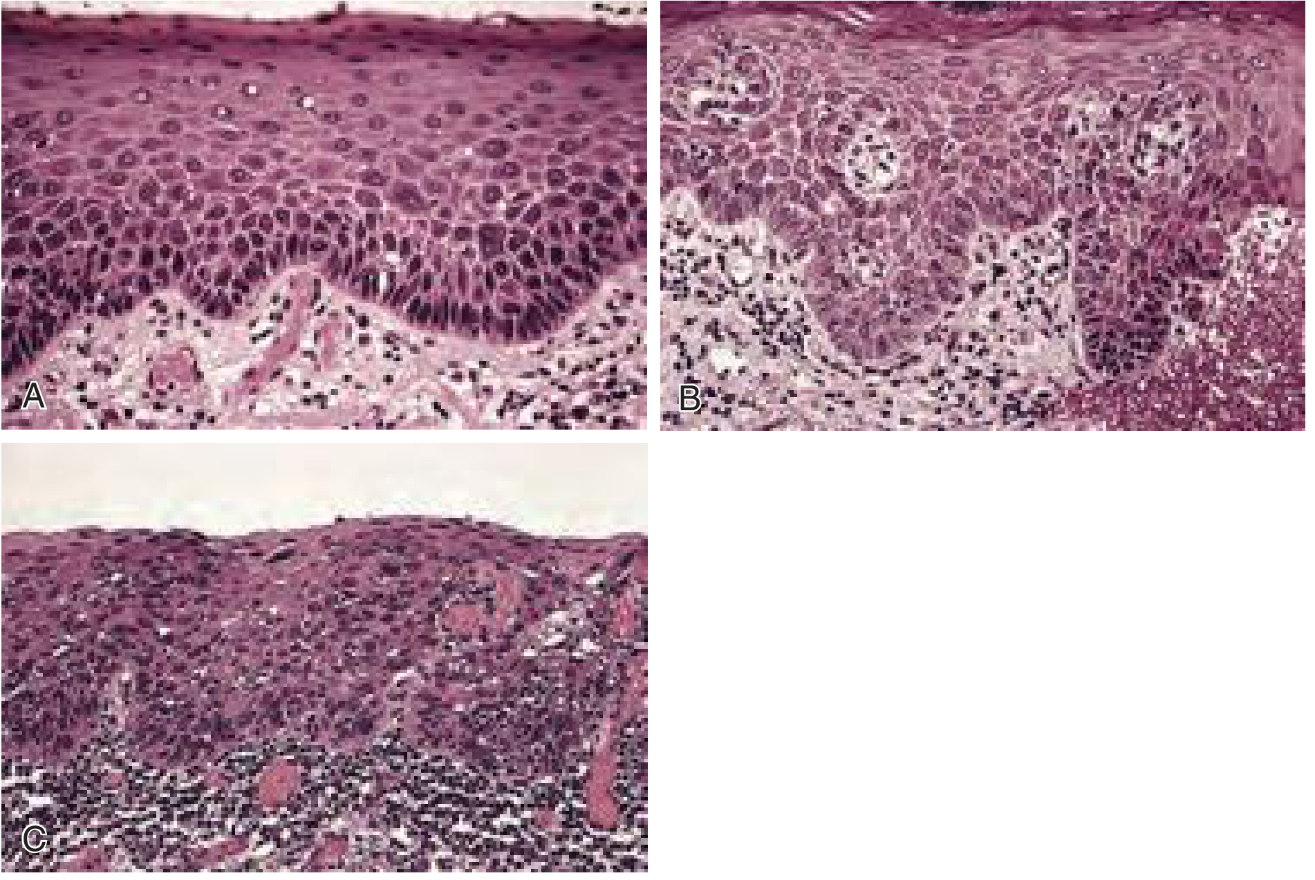
Cummings Otolaryngology, Fig. 87.7
Clinical lesion - Leukoplakia:
- White patch that cannot be wiped off (keratosis)
- Dysplasia/carcinoma in situ/invasive carcinoma found in 17-25% of leukoplakia biopsies
- Risk areas: floor of mouth, lateral tongue, soft palate (high risk)
- Erythematous/granular/ulcerated areas should be biopsied first
Management:
-
Benign/mild dysplasia: observe or elective excision
-
Moderate or worse: mandatory removal (scalpel, laser, cryoablation, electrocautery)
-
No proven chemoprevention strategy (retinoids under study)
-
Cummings Otolaryngology Head & Neck Surgery, p. 1581
3D. CERVICAL/VULVAR DYSPLASIA (CIN/VIN - Non-Ulcerative)
- Cervical Intraepithelial Neoplasia (CIN) / Vulvar Intraepithelial Neoplasia (VIN) - NOT caused by ulceration; caused by HPV
- Graded as CIN I (mild), CIN II (moderate), CIN III (severe/CIS)
- Managed according to grade: observation (CIN I), colposcopy/LEEP/cone biopsy (CIN II-III)
PART 4 - KEY COMPARISON TABLE: ULCERATIVE vs. NON-ULCERATIVE DYSPLASIA
| Feature | Ulcerative Dysplasia (IBD-associated) | Non-Ulcerative Dysplasia |
|---|---|---|
| Setting | Chronic UC / Crohn colitis | Gastritis, Barrett esophagus, oral/laryngeal leukoplakia, chronic inflammation without ulceration |
| Epithelial type | Colonic glandular | Gastric glandular, esophageal columnar, squamous (oral, cervical) |
| Cause | Chronic mucosal ulceration and inflammation | Chronic gastritis (H. pylori), GERD, tobacco, alcohol, HPV, sun exposure |
| Distribution | Flat (invisible) or polypoid; often multifocal | Usually localized; macroscopically visible as polyp or white/red patch |
| Molecular events | p53, p16, genome doubling | p53 (Barrett), HPV E6/E7 (CIN), RAS mutations (gastric) |
| Grading | LGD / HGD (+ "indefinite") | Mild / Moderate / Severe / CIS (squamous); LGD / HGD (glandular) |
| Cancer risk | High if HGD (synchronous cancer common) | Gastric adenoma: up to 30% have carcinoma; Oral: 17-25% |
| Surveillance | Colonoscopy every 1-5 years (IBD programs) | EGD with biopsies (Barrett); colposcopy (CIN); biopsy leukoplakia |
| Treatment | Endoscopic resection (LGD polypoid) or colectomy | Endoscopic resection/ablation (Barrett HGD); excision (oral); LEEP (CIN) |
| Key challenge | Invisible dysplasia; reactive vs. dysplastic histology | Interobserver variability (Barrett LGD) |
PART 5 - HIGH-YIELD EXAM POINTS
- Dysplasia = pre-invasive, cytologically abnormal epithelium without basement membrane breach
- In UC, dysplasia surveillance starts 8-10 years after onset; PSC patients start at time of diagnosis
- "Indefinite for dysplasia" is a valid category in IBD biopsies - confirm with second GI pathologist
- Old DALM terminology is abandoned - use "visible" vs. "invisible" dysplasia instead
- Reactive epithelium matures at the surface; dysplastic epithelium does not
- Squamous dysplasia graded by how many thirds of epithelium are involved (lower 1/3, 2/3, full thickness)
- Barrett LGD: <50% interobserver agreement; HGD: ~85% agreement
- Colitis-associated dysplasia is often multifocal and can be flat/invisible - need systematic biopsies
- Non-ulcerative dysplasia in gastric adenoma carries risk of carcinoma in up to 30% of cases
- HGD in any setting = colectomy (IBD) or ablation/resection (Barrett/oral)
Sources:
- Robbins & Kumar Basic Pathology, p. 557 (gastric dysplasia, general concepts)
- Robbins, Cotran & Kumar Pathologic Basis of Disease (colitis-associated neoplasia; Barrett dysplasia)
- Sleisenger & Fordtran's GI and Liver Disease (Barrett dysplasia pathway, IBD surveillance)
- Clinical Gastrointestinal Endoscopy Expert Consult 3e (IBD dysplasia classification, AGA guidelines)
- Cummings Otolaryngology Head & Neck Surgery, p. 1581 (squamous epithelial dysplasia, oral leukoplakia)
This is a shared conversation. Sign in to Orris to start your own chat.