
Every thing about cardiac tamponad
cardiac tamponade pericardial effusion echocardiogram ECG
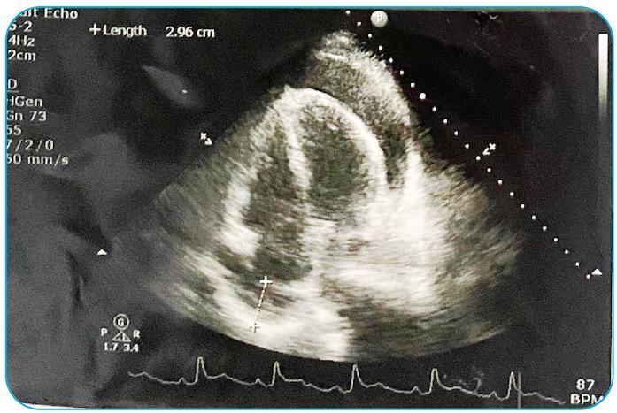
This diagnostic image is a transthoracic echocardiogram (TTE) in an apical four-chamber view. It reveals a significant, circumferentially distributed, echo-free (anechoic) space between the parietal and visceral pericardium, diagnostic of a large pericardial effusion. A measurement call-out on the screen indicates a fluid pocket depth of 2.96 cm. Pathognomonic signs of cardiac tamponade are suggested by the apparent compression of the right-sided heart chambers. At the bottom of the display, a synchronized electrocardiogram (ECG) lead shows the patient's heart rhythm, with a recorded heart rate of 87 BPM (beats per minute). This visual is critical for teaching the ultrasonographic identification of life-threatening pericardial fluid accumulation and its hemodynamic impact on ventricular filling. The clinical relevance involves the immediate need for pericardiocentesis in the setting of cardiovascular compromise.
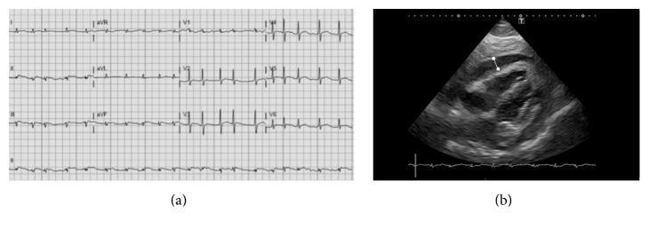
The composite image displays two diagnostic modalities evaluating acute pericarditis progressing to cardiac tamponade. Panel (a) shows a twelve-lead electrocardiogram (ECG) demonstrating atrial fibrillation characterized by an irregularly irregular rhythm and absent P-waves. Notable findings include low-voltage QRS complexes across both limb and precordial leads, which is a clinical sign of electrical insulation due to fluid accumulation. Panel (b) is a subcostal view cardiac ultrasound (echocardiogram) revealing a large circumferential pericardial effusion. A white measurement marker indicates an effusion depth exceeding 20 mm. Within the hypoechoic pericardial space, dense echogenic structures and fibrinous strands are visible, suggesting purulent or exudative pericarditis (e.g., meningococcal origin). The combination of low-voltage ECG and large effusion on ultrasound is highly suggestive of impending cardiac tamponade.
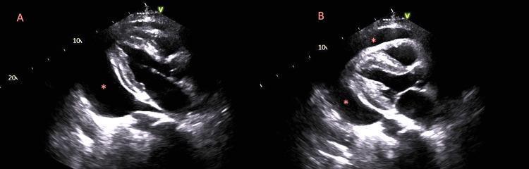
This diagnostic image consists of two side-by-side echocardiogram frames (labeled A and B) in a parasternal long-axis view, demonstrating the pathognomonic signs of cardiac tamponade. The images show a large, circumferential pericardial effusion, visualized as a prominent hypoechoic (black) space surrounding the myocardium, indicated by red asterisks. Panel A illustrates diastolic right ventricular collapse, where the anterior wall of the right ventricle is compressed by the high-pressure fluid. Panel B, compared to Panel A, shows a shift in the heart's position and the relative distribution of the fluid within the pericardial sac. This dynamic shifting of the heart within the large volume of fluid is known as 'swinging heart' physiology. This phenomenon is a critical echocardiographic finding often associated with electrical alternans on an ECG and clinical tamponade, indicating a medical emergency. The visualization is essential for medical students and clinicians to recognize hemodynamic compromise requiring urgent pericardiocentesis.
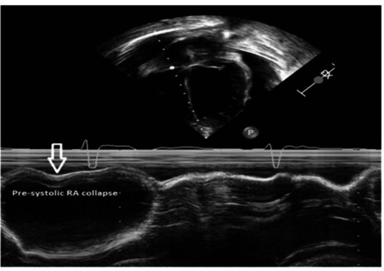
This diagnostic image is a transthoracic echocardiogram combining a B-mode sector view (top) and an M-mode tracing (bottom) to demonstrate findings consistent with cardiac tamponade. The top sector view shows a cross-section of the cardiac chambers surrounded by a large anechoic space, representing a massive pericardial effusion. The bottom M-mode tracing focuses on the right atrial (RA) wall dynamics. A white arrow and text annotation highlight a pre-systolic RA collapse, characterized by an inward invagination or concavity of the RA free wall during late diastole. This finding occurs when intrapericardial pressure exceeds intracavitary right atrial pressure, serving as a highly sensitive sign for impending hemodynamic compromise in the setting of pericardial effusion. An overlaid electrocardiogram (ECG) tracing allows for temporal correlation of wall motion with the cardiac cycle, confirming the timing of the collapse. This educational visual is critical for training in cardiology and emergency medicine to recognize early sonographic markers of cardiac tamponade.
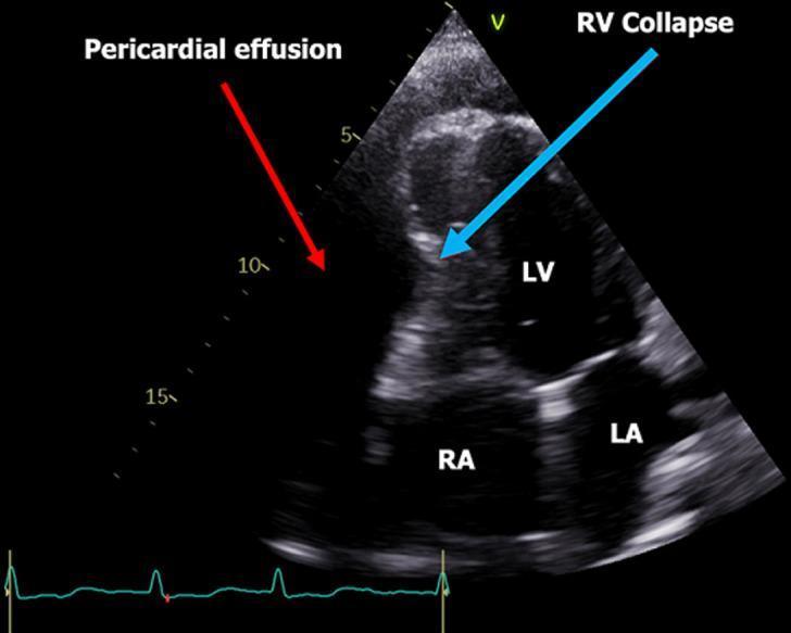
A two-dimensional transthoracic echocardiogram (TTE) in an apical four-chamber view demonstrating a large, circumferential pericardial effusion. The effusion appears as a prominent anechoic (dark) space surrounding the cardiac chambers, highlighted by a red arrow. All four chambers are labeled: Right Atrium (RA), Left Atrium (LA), Left Ventricle (LV), and Right Ventricle (RV). A blue arrow points to the RV, where the free wall shows inward compression or diastolic collapse, a hallmark sign of cardiac tamponade. The image includes a simultaneous electrocardiogram (ECG) tracing at the bottom, which shows sinus rhythm with relatively low voltage complexes. This diagnostic image is used to illustrate the hemodynamic impact of pericardial fluid on right-sided heart chambers, emphasizing the clinical significance of recognizing signs of tamponade physiology through bedside ultrasonography.
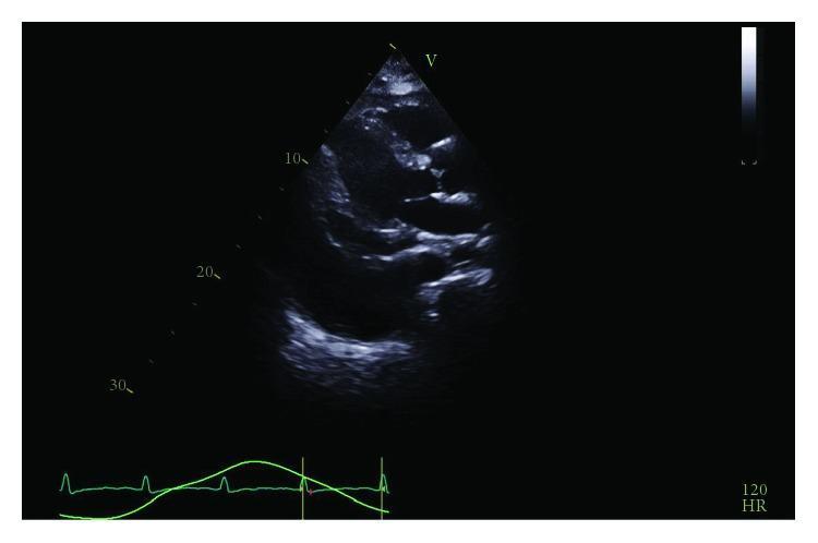
This diagnostic image is a transthoracic echocardiogram (TTE) in a parasternal long-axis view demonstrating a large, circumferential pericardial effusion. The pathology is characterized by a significant echo-lucent (dark) space surrounding the cardiac silhouette, most prominent anteriorly and posteriorly. Visible hemodynamic consequences include compression of the right ventricle, suggestive of cardiac tamponade physiology. Additionally, a large adjacent pleural effusion is noted, appearing as a separate fluid collection posterior to the descending aorta. The heart chambers show restricted diastolic filling due to the external pressure exerted by the pericardial fluid. An ECG trace at the bottom shows sinus tachycardia with a heart rate of 120 bpm, which is a common compensatory finding in patients with tamponade. This image is a classic educational example of restrictive cardiac pathology requiring urgent intervention such as a pericardial window or pericardiocentesis.
"cardiac tamponade"[MeSH Terms] AND management
Cardiac Tamponade - A Complete Overview
Definition
Anatomy & Physiology of the Pericardium
Pathophysiology
- Fluid fills the recesses of the parietal pericardium
- Fluid accumulates faster than the pericardium can stretch
- Fluid exceeds the body's ability to increase blood volume to support right ventricular filling pressure
-
During inspiration, negative pleural pressure transmits to the pericardium, increasing RV venous return and RV filling
-
In tamponade, this RV expansion within the fixed pericardial space shifts the interventricular septum leftward, compressing the LV
-
LV filling further drops as pulmonary venous blood pools in expanding lungs
-
Result: systolic BP drops >10 mmHg with inspiration - pulsus paradoxus
-
Exception: pulsus paradoxus may be absent when LV diastolic pressure is already elevated (e.g., end-stage renal disease, severe aortic regurgitation)
-
Symptom to Diagnosis, p. 452; Miller's Anesthesia 10e, p. 5026
Etiology
| Category | Causes | Notes |
|---|---|---|
| Malignancy | Lung, breast, lymphoma, GI cancer | Most common overall (32%) |
| Infection | Staph, Strep, TB, HIV | 24%; TB important in endemic areas |
| Idiopathic | Often viral-related | 16% |
| Iatrogenic | Post-catheterization, post-cardiac surgery, anticoagulants | 15% |
| Post-MI | Cardiac wall rupture (44% of post-MI cases) | 7%; wall rupture major cause |
| Uremic | Renal failure untreated (50%), dialysis (20%) | Avoid NSAIDs |
| Autoimmune | SLE, rheumatoid arthritis | |
| Hypothyroidism | Slow accumulation, often asymptomatic | |
| Trauma | Penetrating chest wounds | Especially stab wounds |
| Radiation | Post-radiotherapy (breast, Hodgkin) |
- Rosen's Emergency Medicine, p. 1124
Clinical Features
Symptoms
- Dyspnea - most common (87-88%)
- Chest pain, cough, orthopnea
- Anxiety, lethargy, palpitations (present in ≤25%)
Beck's Triad (Classic but Insensitive)
- All three may not be present, especially in rapidly developing tamponade
- Hypotension is only present in ~28% and diminished heart sounds in ~26% of cases
- More common findings: tachycardia, elevated JVP, and pulsus paradoxus (sensitivity 76-82%)
JVP in Tamponade
- Jugular venous distention is typical
- The x-descent is dominant (during ventricular systole, atrial emptying preserved)
- The y-descent is absent or blunted (impaired early diastolic ventricular filling)
- This x-dominant pattern distinguishes tamponade from constrictive pericarditis (where both x and y descents are prominent)
Pulsus Paradoxus
- Defined as a drop in systolic BP of >10 mmHg during normal inspiration
- A drop of >25 mmHg correlates with significant hemodynamic impairment
- Measured with a manual sphygmomanometer in a quiet room
"Low-Pressure" Tamponade
-
In hypovolemic patients or rapidly hemorrhagic tamponade, JVD may be absent
-
Occurs when venous pressure has not had time to rise compensatorily
-
Goldman-Cecil Medicine, p. 716-717; Symptom to Diagnosis, p. 452
Diagnosis
ECG Findings
- Low voltage - electrical insulation by fluid around the heart
- Electrical alternans - alternating QRS axis beat-to-beat due to the heart "swinging" within the pericardial fluid. Highly specific for large effusion/tamponade.
Chest X-Ray
- Normal until effusion exceeds 250 mL
- "Water-bottle" or globular cardiac silhouette - enlarged, rounded heart
- Fat pad sign on lateral view: separation between epicardial and pericardial fat stripes (≥2 mm)
- Oligemic lung fields
Echocardiography (Diagnostic Standard)
- Trivial: only in systole
- Small: <10 mm echo-free space in diastole
- Moderate: 10-20 mm
- Large: >20 mm
- Very large: >25 mm
| Finding | Sensitivity | Specificity | Notes |
|---|---|---|---|
| RA systolic collapse (inversion) | 50-100% | 33-100% | First to appear; nonspecific alone |
| RV early diastolic collapse | 48-60% | 75-90% | More specific; occurs later |
| IVC plethora (non-collapsing with inspiration) | High | Confirmatory | |
| Exaggerated respiratory variation in valve flow | High | High | Doppler finding |
| "Swinging heart" | Pathognomonic | Very high | Associated with electrical alternans |
-
After cardiac surgery, localized clot can compress individual chambers atypically - classic findings may be absent
-
Elevated right-sided pressures (e.g., pulmonary hypertension) may prevent RV collapse despite tamponade
-
Positive pressure ventilation complicates Doppler respiratory variation assessment
-
Braunwald's Heart Disease, p. 2103-2110; Miller's Anesthesia, p. 5026-5027
Catheterization (Invasive Hemodynamics)
- Equalization of diastolic pressures across all chambers (RVEDP = LVEDP = pericardial pressure)
- Exaggerated respiratory variation in LV and RV pressures
- "X-descent-only" pattern in RA pressure tracing
Differential Diagnosis
- Constrictive pericarditis (prominent y-descent, calcified pericardium, no effusion)
- Tension pneumothorax (absent breath sounds, tracheal deviation)
- Massive pulmonary embolism (right heart strain, no effusion)
- Cardiogenic shock (elevated PCWP, no effusion)
- Superior vena cava syndrome (facial plethora, arm swelling)
Management
Immediate Stabilization (Bridge Measures)
- IV fluid bolus - increases right-sided filling pressure to overcome pericardial constriction; useful transiently, especially in hypovolemic tamponade
- Inotropes are generally ineffective - maximal endogenous sympathetic activation is already present
- Avoid mechanical ventilation if possible - positive intrathoracic pressure further impairs ventricular filling and can cause sudden cardiovascular collapse
- Vasopressors - may be used briefly while preparing for drainage
Definitive Treatment: Pericardial Drainage
1. Pericardiocentesis (First-line)
- Echocardiography-guided percutaneous approach is standard
- Indication: ≥1 cm of pericardial fluid anterior to RV free wall throughout diastole, with hemodynamic compromise
- Access: typically subxiphoid approach; ideal entry site defined by minimum distance from skin to fluid without intervening structures
- Needle enters the pericardial space; fluid drained via an indwelling catheter
- Catheter left in place minimizes risk of recurrent effusion
- In dire circumstances (cardiac arrest, PEA): bedside pericardiocentesis without imaging guidance is acceptable
- LDH, protein, cell count (Light's criteria for exudate/transudate)
- Cytology (malignancy)
- PCR for TB
- Culture (bacteria, TB)
2. Surgical Drainage / Pericardial Window
- Effusion <1 cm (cannot safely aspirate)
- Organized, loculated, or focal effusion
- Recurrent effusions (malignant)
- Pericardial tissue needed for diagnosis
- Bacterial (purulent) pericarditis
- Post-cardiac surgery (loculated hematoma)
3. Pericardiostomy / Pericardiectomy
-
Reserved for recurrent cases
-
Often necessary for chronic large malignant effusions
-
Goldman-Cecil Medicine, p. 718; Braunwald's Heart Disease, p. 2158-2170
Special Scenarios
Traumatic Tamponade
- Most often from penetrating chest wounds (stab > gunshot)
- Hemorrhagic fluid; may not show JVD (low-pressure tamponade due to hypovolemia)
- Requires emergency pericardiocentesis or thoracotomy
Post-Cardiac Surgery
- Localized hematoma compressing individual chambers
- Classic echo findings absent; high index of suspicion required
- Often requires surgical re-exploration
Malignant Effusion
- Most common cancers: lung, breast, lymphoma, GI
- High recurrence rate after simple pericardiocentesis
- Pericardial window or percutaneous balloon pericardiotomy preferred
- In-hospital mortality <10% but 75% mortality at 1 year with malignant etiology
Tuberculous Pericarditis
- Drainage followed by early pericardiostomy (4-6 weeks) if signs of tamponade or constriction
- Triple-drug antituberculous regimen required
- Risk of constrictive pericarditis if untreated
Uremic Pericarditis
- Intensive dialysis is first-line
- NSAIDs contraindicated
- Only ~50% respond to dialysis alone; catheter or surgical drainage needed for the rest
Prognosis
-
In-hospital mortality: <10% overall with prompt drainage
-
Subsequent annual mortality:
- Malignant effusion: ~75% at 1 year
- Non-malignant causes: 3-5% annual mortality
-
Long-term prognosis depends on the underlying cause
-
Goldman-Cecil Medicine, p. 718
Key Points Summary
| Feature | Details |
|---|---|
| Mechanism | Elevated intrapericardial pressure compresses chambers, reduces cardiac output |
| Rate > Volume | Rate of accumulation more important than total volume |
| Classic triad | Hypotension + JVD + muffled heart sounds (Beck's triad) - low sensitivity |
| Key signs | Tachycardia, JVD (x-dominant), pulsus paradoxus >10 mmHg |
| ECG | Low voltage + electrical alternans |
| Echo | RA collapse (early) → RV collapse → swinging heart + IVC plethora |
| Treatment | IV fluids (bridge) + urgent pericardiocentesis or surgical drainage |
| Avoid | Positive pressure ventilation, inotropes alone |
| Prognosis | <10% in-hospital mortality; long-term depends on cause |
Recent Evidence Note: A 2024 systematic review (PMID 39210274) specifically addresses cardiac tamponade in HIV-positive patients, finding infectious and neoplastic etiologies predominate in this population. A 2025 systematic review (PMID 39567105) on cardiac point-of-care ultrasound confirms echocardiography remains the cornerstone of tamponade diagnosis, supporting current guidelines.