
Explain about three lead ECG, where all these three leads are placed on the body, how is it different from 12 lead ECG placement, why they are used, what is the importance of the three lead ECG placement?
Reading File
Finding Sources
Finding Sources
Searching Images
three lead ECG monitoring electrode placement torso diagram RA LA LL leads I II III
Searching PubMed
electrocardiography monitoring AND electrodes
Searching the Web
3 lead ECG monitoring electrode placement RA LA LL vs 12 lead ECG American Heart Association
Reading File
Reading File
Reading Web Page
https://www.ahajournals.org/doi/10.1161/01.cir.0000145144.56…
Three-lead ECG: what it is
A three-lead ECG is mainly a continuous cardiac monitoring system, not a full diagnostic ECG. It uses three electrodes to produce the three standard bipolar limb-lead views:
- Lead I: left arm (LA) minus right arm (RA)
- Lead II: left leg (LL) minus right arm (RA)
- Lead III: left leg (LL) minus left arm (LA)
A key distinction: electrodes are the sticky pads placed on skin; a lead is the electrical viewpoint calculated from electrodes.
Where are the three electrodes placed?
For bedside monitoring, the labels refer to the direction of the limb, but electrodes are usually placed on the torso, not necessarily on the wrists and ankle:
| Electrode | Usual monitoring location |
|---|---|
| RA - right arm | Right upper chest, just below the right clavicle |
| LA - left arm | Left upper chest, just below the left clavicle |
| LL - left leg | Left lower chest or left lower abdomen, below the cardiac border, often near the left iliac crest |
A simple layout:
        RA                         LA
   right upper chest          left upper chest
      below clavicle          below clavicle


                     ♥


                         LL
               left lower abdomen / torso
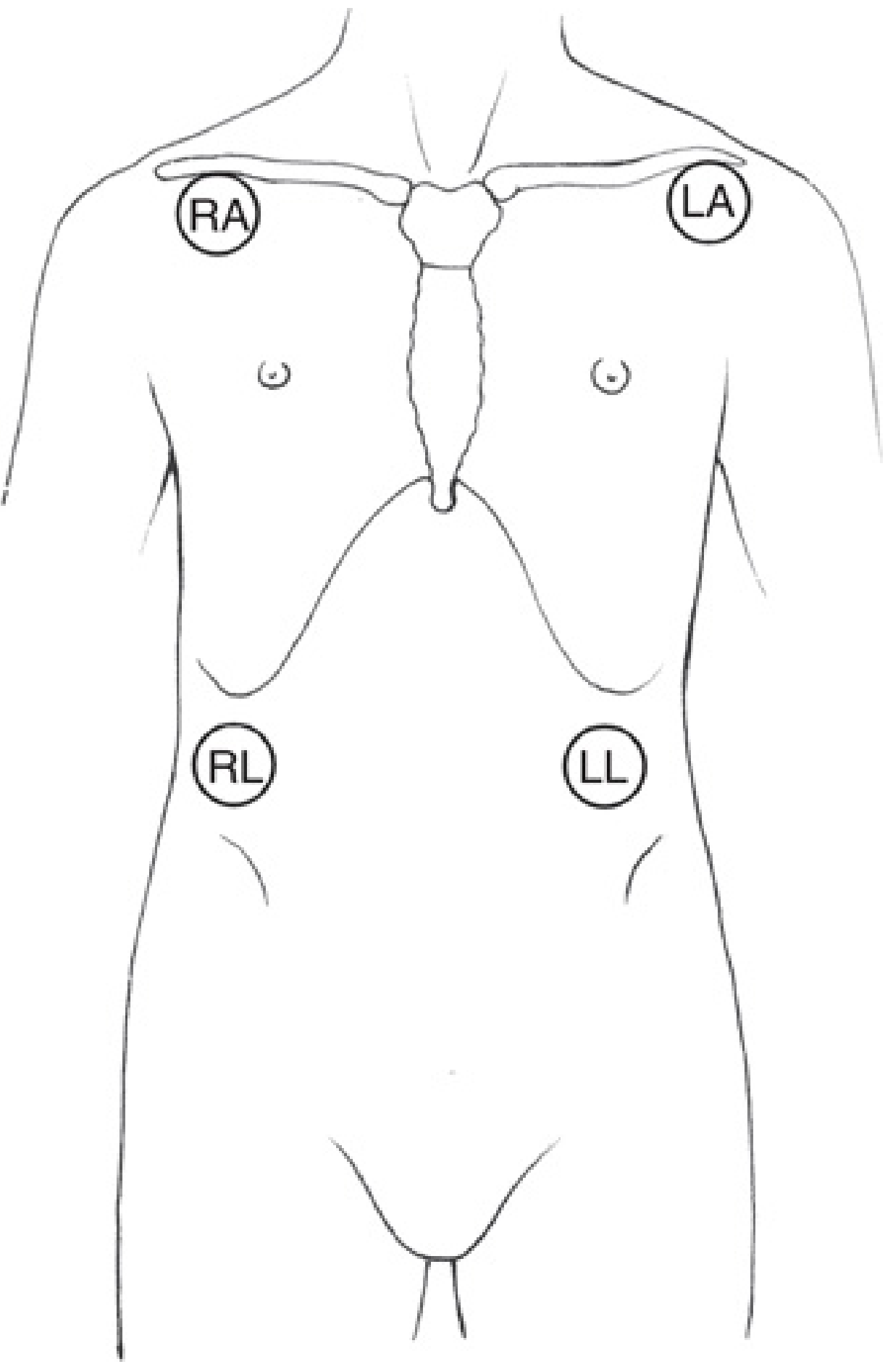
Keep the lower-left electrode below the heart and outside the cardiac silhouette. Moving it up over the precordium can alter Leads II and III, creating a tracing that is not comparable with the usual monitoring position. Miller's Anesthesia, 2-Volume Set, 10e, p. 4715.
Electrode color conventions can vary by country and manufacturer. Confirm the letter label on the cable, not just the color.
What is the difference from a 12-lead ECG?
| Feature | Three-lead ECG | Standard 12-lead ECG |
|---|---|---|
| Number of electrodes | 3 | 10 |
| Number of ECG views | 3 limb-lead views: I, II, III | 12 views: I, II, III, aVR, aVL, aVF, V1-V6 |
| Placement | Usually upper-right torso, upper-left torso, lower-left torso | 4 limb electrodes plus 6 precisely located chest electrodes |
| Main purpose | Continuous rhythm and rate monitoring | Diagnostic assessment of rhythm, conduction, ischemia/infarction, chamber abnormalities, etc. |
| Typical settings | ICU, operating room, recovery room, emergency care, telemetry, transport, defibrillator monitors | Clinic, emergency department, chest-pain assessment, preoperative evaluation, diagnostic workup |
| Ability to detect ischemia/MI | Limited | Much better, though serial ECGs and clinical assessment may still be required |
A 12-lead ECG does not require 12 adhesive pads. It uses:
- Four limb electrodes: RA, LA, RL, LL
- RL is generally a reference/ground electrode.
- Six chest electrodes: V1 to V6.
The chest electrodes provide horizontal-plane views of the heart:
- V1: fourth intercostal space, right sternal border
- V2: fourth intercostal space, left sternal border
- V3: midway between V2 and V4
- V4: fifth intercostal space, left midclavicular line
- V5: left anterior axillary line, level with V4
- V6: left midaxillary line, level with V4
Correct V1-V6 placement matters because even small errors can change the ECG appearance and may mimic or hide abnormalities. Miller's Anesthesia, 2-Volume Set, 10e, p. 4714.
Why is a three-lead ECG used?
It is used because it is quick to apply, practical for prolonged monitoring, and good at showing changes in rhythm over time. Common uses include:
- Continuous heart-rate monitoring
- Detection of bradycardia and tachycardia
- Detection of arrhythmias such as atrial fibrillation, supraventricular tachycardia, ventricular tachycardia, pauses, and frequent ectopic beats
- Observation of the rhythm during anesthesia, procedures, resuscitation, transport, and in ICU/telemetry
- Use on many defibrillator-monitor devices, including when pacing capability may be needed
Lead II is commonly selected for rhythm monitoring because P waves are often relatively easy to see, which helps identify atrial activity and the relationship of P waves to QRS complexes.
Importance of correct three-lead placement
Correct placement is important because it:
-
Produces a more reliable rhythm strip
Incorrect or loose pads can cause artifact that resembles arrhythmia. -
Makes serial monitoring meaningful
If electrodes are placed differently each time, waveform size and shape can change even when the patient's cardiac status has not. -
Improves P-wave visibility and arrhythmia recognition
A well-chosen monitoring lead, often Lead II, helps differentiate sinus rhythm, atrial fibrillation, flutter, AV block, and ectopic rhythms. -
Avoids false alarms and unnecessary treatment
Motion, poor skin contact, dried electrodes, and lead misplacement are common sources of misleading monitor tracings. -
Provides rapid surveillance when a 12-lead ECG is not continuously practical
Continuous monitoring can capture intermittent arrhythmias that a short, resting 12-lead ECG may miss. Ambulatory ECG monitoring is particularly useful for correlating symptoms with transient rhythm events.
Important limitation
A three-lead ECG does not replace a 12-lead ECG when there is:
- Chest pain or suspected acute coronary syndrome
- New ST-segment changes on the monitor
- Suspected myocardial infarction
- New bundle-branch block or conduction abnormality
- A concerning arrhythmia needing full characterization
- Syncope, severe palpitations, or unexplained hemodynamic instability
If ischemia is a concern, a 12-lead ECG is needed because three-lead monitoring gives only limited views of the heart. In higher-risk monitored patients, a precordial monitoring lead such as V5 can increase ischemia detection, but it still does not substitute for a diagnostic 12-lead ECG. Miller's Anesthesia, 2-Volume Set, 10e, p. 4714.
Bottom line: three-lead ECG placement is for fast, continuous rhythm surveillance. Twelve-lead ECG placement is for a more complete, standardized diagnostic picture of cardiac electrical activity.
This is a shared conversation. Sign in to Orris to start your own chat.