
Lurch
What are you asking about?
Lurch in Ortho
Trendelenburg lurch gait orthopaedics
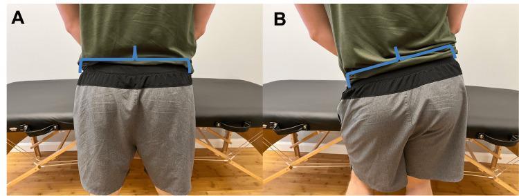
Clinical photograph showing a posterior view of a male subject in two different stances (A and B) to illustrate Trendelenburg gait. Blue graphical overlays are used to indicate pelvic and spinal alignment. Figure A demonstrates normal pelvic stability; the horizontal line across the iliac crests is level and the vertical line through the lumbar spine is perpendicular, indicating effective hip abductor stabilization. Figure B demonstrates a positive Trendelenburg sign, characterized by an inferior pelvic drop on the contralateral (unsupported) side. The horizontal overlay shows a distinct downward tilt toward the left, while the vertical spinal line leans away from the midline, reflecting compensatory truncal shift. This comparison highlights hip abductor weakness, typically involving the gluteus medius and minimus muscles. The image is an educational tool for physical therapy and orthopedic functional assessment, illustrating how pelvic instability alters the kinetic chain and contributes to musculoskeletal issues like knee pain.
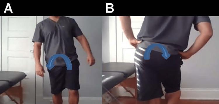
Clinical photograph sequence illustrating a positive Trendelenburg sign and compensated Trendelenburg gait. Image A presents an anterior view of an individual during the stance phase on the left leg. There is a visible lateral trunk lean (compensated gait) toward the stance side, indicated by a blue curved arrow, to maintain pelvic stability despite hip abductor weakness. Image B provides a posterior view demonstrating a significant pelvic drop on the contralateral (right) side while standing on the left leg, indicated by a blue downward-curving arrow. This pelvic tilt reflects insufficiency of the gluteus medius and minimus muscles on the weight-bearing side. The sequence highlights key physical examination findings for hip abductor weakness, typically assessed in sports medicine, orthopedics, and physical therapy to diagnose conditions like gluteal tendinopathy or superior gluteal nerve palsy.
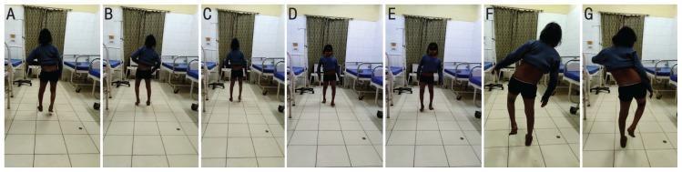
A series of clinical photographs of a 13-year-old female patient demonstrating gait abnormalities and physical examination findings indicative of hip abductor weakness. Panels A through E capture the patient's gait from posterior and anterior views, showing a waddling pattern characterized by lateral trunk oscillations and pelvic instability, consistent with bilateral Trendelenburg gait. Panels F and G display a bilateral positive Trendelenburg test. In these clinical frames, the patient is standing on a single leg; the contralateral side of the pelvis drops (sags) rather than remaining level or rising. To maintain balance and compensate for the pelvic tilt, the patient exhibits a characteristic lateral lean of the torso toward the weight-bearing side. This visual documentation serves as a classic educational representation of gluteus medius and minimus insufficiency, often associated with neuromuscular disorders, proximal myopathies, or hip pathologies. The images clearly illustrate the biomechanical failure of the hip abductor mechanism and the resulting compensatory movements of the upper body and pelvis.
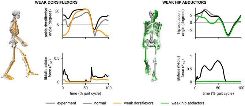
This musculoskeletal biomechanics infographic displays the results of simulated gait adaptations due to muscle weakness, comparing 'normal' tracking (white skeletons) with pathological states. The left panel illustrates 'Weak Dorsiflexors' (orange), demonstrating a drop-foot gait. The skeletal model shows increased ankle plantarflexion during the swing phase, corroborated by the top line graph showing a significant decrease in ankle dorsiflexion angle (degrees) and a bottom graph showing reduced tibialis anterior force throughout the gait cycle. The right panel depicts 'Weak Hip Abductors' (green), characteristic of Trendelenburg gait. The skeletal model exhibits a marked lateral trunk sway toward the stance leg. The accompanying graphs show a reduced hip adduction angle and nearly zero gluteus medius force across the gait cycle compared to normal tracking. Data is plotted against time (% gait cycle), with 'experiment' (gray), 'normal' (black), and pathological conditions (orange/green) highlighted to emphasize the relationship between specific muscle insufficiency and compensatory movement patterns.
Lurch in Orthopaedics
1. Trendelenburg Lurch (Abductor Lurch / Gluteus Medius Lurch)
- Normally, the gluteus medius and minimus (hip abductors) contract during single-leg stance to keep the contralateral pelvis level.
- When abductors are weak or painful, the pelvis drops on the contralateral (swing) side - this is a positive Trendelenburg sign.
- To compensate, the patient leans the trunk over the affected hip - this is the Trendelenburg lurch (also called the compensated Trendelenburg gait).
- Trendelenburg sign = pelvis drops on the unsupported side (seen from behind)
- Trendelenburg lurch = trunk leans toward the supported (affected) side (seen from front/side)
- A patient can have a lurch without a sign (B in Fig. 48.24) if compensation is effective.

- Gluteus medius/minimus tears or atrophy
- Coxa vara
- Hip fracture or dysplasia
- Superior gluteal nerve injury
- Radiculopathy
- Poliomyelitis, myelomeningocele, spinal cord lesions
- Slipped capital femoral epiphysis (SCFE)
- After hip arthroplasty with reduced femoral offset (weakened abductor lever arm)
2. Antalgic Lurch / Coxalgic Gait
- Leaning the trunk over the hip shifts the center of gravity, reducing the moment arm and the compressive load on the joint.
- Distinguished from Trendelenburg lurch in that pain (not pure weakness) is the primary driver.
- Also associated with a shortened stance phase on the affected side (antalgic gait component).
Summary Table
| Feature | Trendelenburg Lurch | Antalgic Lurch |
|---|---|---|
| Trunk leans toward | Affected (stance) side | Affected (painful) side |
| Primary cause | Abductor weakness | Pain |
| Stance phase | Normal or near-normal duration | Shortened on affected side |
| Trendelenburg sign | May be positive | Usually negative |
| Common causes | Nerve injury, muscle tear, fracture | OA, AVN, infection, fracture |
Clinical Relevance
- Gait inspection for abductor lurch is part of the standard hip examination, along with range of motion and special tests - [Campbell's Operative Orthopaedics 15th Ed 2026].
- After total hip arthroplasty, restoring normal hip offset is important; a shortened neck length reduces the abductor lever arm, increasing the risk of a Trendelenburg lurch post-op - [Miller's Review of Orthopaedics 9th Ed].