
Side effects of TB treatment
Reading File
Finding Sources
Finding Sources
Searching PubMed
"antitubercular agents"[MeSH Terms] AND "adverse effects"
Reading File
Reading File
Reading File
Reading File
Reading File
Reading File
Side effects of TB treatment (first-line: HRZE)
By drug
| Drug | Common / important side effects | Key points |
|---|---|---|
| Isoniazid (H) | Hepatitis, peripheral neuropathy, rash, fever. Rarely lupus-like syndrome, optic neuritis or atrophy, seizures, psychiatric symptoms, acne, anemia. | Asymptomatic transient transaminase rise in up to 20%. Symptomatic hepatitis is idiosyncratic and more likely with age, daily alcohol use, and the first 3 months postpartum. Neuropathy occurs in up to 2% at 5 mg/kg because isoniazid interferes with pyridoxine (B6) metabolism. |
| Rifampicin (R) | Rash, pruritus, GI upset, hyperbilirubinemia or hepatitis, pancytopenia. Rarely a hypersensitivity reaction (fever, chills, malaise, rash, occasionally renal or hepatic failure) with intermittent dosing. | Usually mild. It is a strong CYP450 inducer, so it lowers levels of warfarin, oral contraceptives, phenytoin, digoxin, steroids, azoles, methadone, and many antiretrovirals. Harmless orange discoloration of urine, sweat, and tears is expected (this is my addition, not from the retrieved text). |
| Pyrazinamide (Z) | Hepatotoxicity, hyperuricemia, photosensitivity, arthralgia. | Hyperuricemia is common and usually managed conservatively, and clinical gout is rare. Hepatotoxicity is now uncommon at current doses (up to 15% at old, higher doses). Risk rises with older age, liver disease, HIV, and low albumin. |
| Ethambutol (E) | Optic neuritis (reduced acuity, central scotoma, loss of green or red colour vision). Rarely peripheral neuropathy. | Dose dependent, in 1-5% of patients, and more likely with renal impairment. It typically appears months after starting. Stopping the drug early usually allows recovery over months, but deficits may be permanent in the elderly or if recognized late. |
Sources: Harrison's Principles of Internal Medicine 22E, "First-line antituberculosis drugs" (pp. 1465-1466), and Comprehensive Clinical Nephrology 7th Ed, Table 54.3 (pyrazinamide: hyperuricemia, hepatotoxicity, photosensitivity).
Monitoring and management
- Baseline tests: LFTs, and visual acuity, visual fields, and colour vision before ethambutol.
- Repeat tests:
- Check LFTs monthly if the baseline is abnormal or the patient has risk factors (alcohol, hepatitis B or C, HIV, pregnancy or postpartum, age).
- Check visual acuity and colour vision monthly while on ethambutol.
- Stop-drug thresholds (Harrison's): Stop the hepatotoxic drugs if ALT or AST is at least 3 times the upper limit of normal with symptoms or jaundice. Also stop them if it is at least 5 times the upper limit of normal without symptoms.
- Neuropathy prevention: Give pyridoxine 25-50 mg/day to patients at risk, such as those with HIV, diabetes, alcohol use, or malnutrition, and to those on other neurotoxic drugs.
- Ethambutol in young children: It is not routinely used because visual monitoring is difficult.
- Pyrazinamide in pregnancy: WHO and other international bodies recommend it routinely. The US does not, because teratogenicity data are inadequate.
Typical clinical approach (general practice, not from the retrieved text)
- Minor GI upset or itch: Treat symptomatically, give the drugs with food or at bedtime, and continue treatment.
- Drug-induced liver injury: Stop the hepatotoxic drugs, then reintroduce them one at a time once LFTs recover. Follow your local or national protocol for the order and any interim non-hepatotoxic regimen.
- Severe rash or hypersensitivity: Stop all drugs, then reintroduce them sequentially to identify the culprit.
- Visual symptoms: Stop ethambutol immediately and refer to ophthalmology.
Second-line and MDR-TB drugs
These cause a wider range of toxicity. Examples are aminoglycoside ototoxicity and nephrotoxicity, fluoroquinolone tendinopathy and QT prolongation, linezolid myelosuppression and neuropathy, and bedaquiline QT prolongation. I did not verify these against the library in this search.
Recent evidence (PubMed, last 2 years)
Nothing found contradicts the textbook account. Recent reviews include:
- A systematic review and meta-analysis of adverse events with first-line therapy in children (PMID 40127187).
- A meta-analysis of antituberculosis drug-induced liver injury incidence and risk factors in India (PMID 39225936).
- A systematic review of adverse events with bedaquiline- and pretomanid-based MDR regimens (PMID 42121320).
I only read the titles, not the abstracts.
I can go deeper on any of these if you want: the drug-induced liver injury algorithm, MDR-TB regimen toxicities, or HIV co-infection and drug interactions.
How to manage anti-TB drug-induced liver injury step by step?
Reading File
Finding Sources
Finding Sources
Searching PubMed
"antitubercular agents"[MeSH Terms] AND "Chemical and Drug Induced Liver Injury"[MeSH Terms]
Searching the Web
management of anti-tuberculosis drug-induced liver injury guideline ATS 2006 WHO reintroduction sequence ALT 3x 5x
Reading File
Reading File
Step-by-step management of anti-TB drug-induced liver injury (DILI)
This follows Murray & Nadel's Textbook of Respiratory Medicine (Hepatotoxicity section, pp. 1215-1216) and Harrison's 22E. I added the ATS, WHO, BTS, and AASLD points from a web search of guideline summaries. I did not read the primary guidelines themselves.
Step 1: Monitor, and don't over-react to a mild rise
- About 20% of patients on the standard four-drug regimen get an asymptomatic ALT rise. It is frequently due to isoniazid ("hepatic adaptation") and often resolves despite continued treatment.
- If the ALT rise is modest and the patient has no symptoms, do not change therapy. Increase the frequency of clinical and lab monitoring.
- ALT is preferred over AST because it is more specific.
Step 2: Decide whether to stop
Stop all hepatotoxic drugs (H, R, Z) if either of these applies:
- ALT is more than 5 times the upper limit of normal (ULN), with or without symptoms.
- ALT is more than 3 times the ULN with symptoms (nausea, vomiting, abdominal pain, anorexia, jaundice, unexplained fatigue).
Also stop and evaluate promptly if bilirubin rises significantly. Harrison's gives the same thresholds, using "hepatitis symptoms or jaundice" for the 3 times ULN trigger.
Step 3: Exclude other causes
Look for:
- Viral hepatitis (A, B, C, E)
- Biliary disease
- Alcohol
- Other hepatotoxic drugs (including herbal products and paracetamol)
- Ischemia
Do this before labelling it anti-TB DILI.
Step 4: Work out the pattern, which hints at the culprit
- Transaminase-predominant injury is more likely isoniazid or pyrazinamide.
- A disproportionate rise in bilirubin and ALP is more suggestive of rifampicin.
Step 5: Follow the patient until recovery
- Check ALT and INR (or PT) serially until they return to baseline.
- Consult a hepatologist if the patient worsens clinically or biochemically. Watch for jaundice with coagulopathy or encephalopathy, which signals acute liver failure.
- Liver failure may need transplant evaluation (general practice, not stated in the text I retrieved).
Step 6: Bridge the treatment gap if needed
- Whether to give a liver-sparing regimen depends on how infectious the patient is, how severe the TB is, and which treatment phase they are in.
- A typical liver-sparing regimen is ethambutol, a fluoroquinolone (such as levofloxacin), and an aminoglycoside. In a UK series, severe TB was covered with ethambutol plus amikacin.
- If the patient is late in treatment or has a low bacillary burden, it is reasonable to hold all TB treatment until liver function normalizes.
Step 7: Reintroduce drugs once ALT is below 2 times ULN
- For patients with pre-existing liver disease, wait until ALT is back near their baseline.
- Most programs reintroduce drugs one at a time, so a recurrence identifies the culprit.
The usual sequence, from Murray & Nadel:
- Rifampicin first, with or without ethambutol depending on the treatment phase. It is least likely to cause hepatotoxicity.
- Isoniazid after 3-7 days if ALT has not risen.
- Pyrazinamide after another 3-7 days. Whether to restart it depends on the treatment phase and the severity of the injury.
If symptoms recur or ALT rises, stop the last drug added.
Step 8: Adjust the final regimen
- If the hepatitis was severe and rifampicin and isoniazid are tolerated, assume pyrazinamide was responsible and leave it out.
- A prolonged course (several weeks) to normalize also points to pyrazinamide.
- Without pyrazinamide, the total duration may be extended to 9 months, depending on doses taken, disease severity, and bacteriology.
- If isoniazid or rifampicin is the culprit, the regimen is rebuilt around the remaining drugs plus a fluoroquinolone and others. Duration is then usually longer, typically 9 to 18 months depending on which drugs are lost (general practice).
Where guidelines differ
Evidence on the best reintroduction strategy is weak, and the textbook states the optimal approach is unknown.
- ATS and BTS favour sequential reintroduction.
- WHO suggests restarting all drugs together, then going sequential only if a second episode occurs.
- Individualize based on how severe the TB and the liver injury are.
Special situations
- Chronic hepatitis B or C, or HIV: ALT elevations during treatment are several times more likely. In HCV, treating the hepatitis has allowed safe reintroduction of anti-TB drugs (Sleisenger and Fordtran's).
- Pre-existing liver disease: Use baseline ALT as the reference, and monitor more often (monthly or more frequently).
Recent literature
PubMed turned up a few relevant reviews, with no sign they contradict the textbook approach. I only read titles and search snippets:
- Lim et al., BMJ 2023, on anti-TB DILI.
- Lewis et al., 2024, on diagnosis, prevention, and risk management of TB drug DILI (PMID 39212296).
Local guidance (for example, India's national TB programme) may differ in thresholds and reintroduction sequence, so check it for the final protocol.
Detection of leprosy
Reading File
Finding Sources
Finding Sources
Searching Images
leprosy skin lesion hypopigmented anesthetic patch or nerve thickening
Searching PubMed
"leprosy"[MeSH Terms] AND "diagnosis"
Searching the Web
WHO leprosy diagnosis cardinal signs slit skin smear PCR anti-PGL-I Ridley-Jopling classification
Reading File
Reading File
query_images_db
Detection of leprosy (Hansen disease)
Leprosy is diagnosed mainly on clinical grounds. Lab tests are used to confirm and classify the disease, and to help in difficult cases. Sources: Harrison's 22E (Diagnosis section), Park's Textbook of Preventive and Social Medicine (Diagnosis of leprosy), Goldman-Cecil (WHO 2018 criteria), and the WHO leprosy fact sheet.
1. Case suspicion
Suspect leprosy in anyone with:
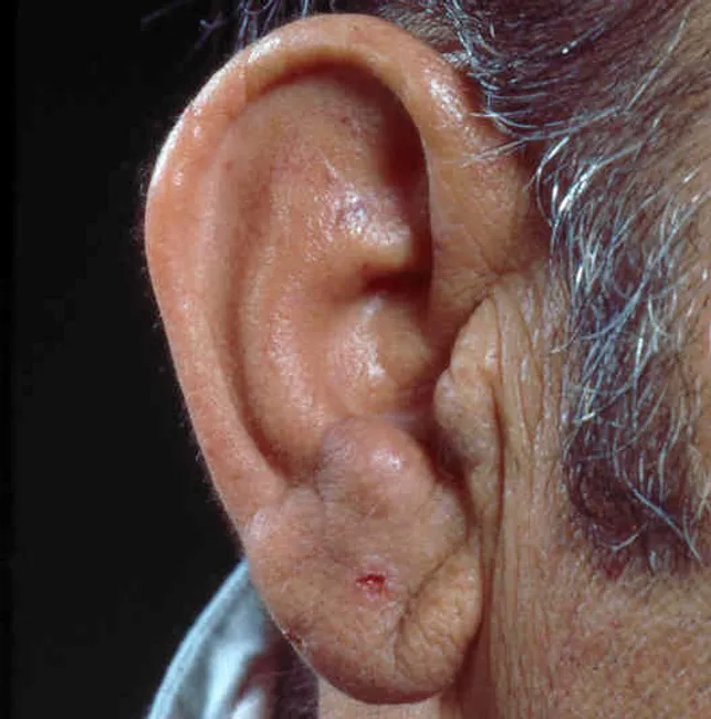
- A skin patch that is pale or reddish, or a plaque or nodule, especially with numbness.
- Nerve pain or tenderness, weakness, or deformity of the hands, feet or eyes.
- Painless ulcers or burns on the hands or feet.
- Contact with a known case.
2. The three cardinal signs
Leprosy is diagnosed when at least one of these is present:
- A hypopigmented or reddish skin lesion with definite loss of sensation.
- Test light touch with cotton, wool or a feather.
- Test pain with the sharp and blunt ends of a toothpick.
- Test temperature if needed.
- Early indeterminate lesions and facial lesions can be hard to judge. Facial innervation is dense, so sensory loss may be subtle.
- A thickened peripheral nerve with sensory loss and/or weakness of the muscles it supplies.
- Palpate the nerve and compare it with the opposite side.
- Note tenderness. In multibacillary disease, thickening is often bilateral.
- Commonly examined nerves: greater auricular, ulnar, radial cutaneous, median, lateral popliteal, posterior tibial, sural.
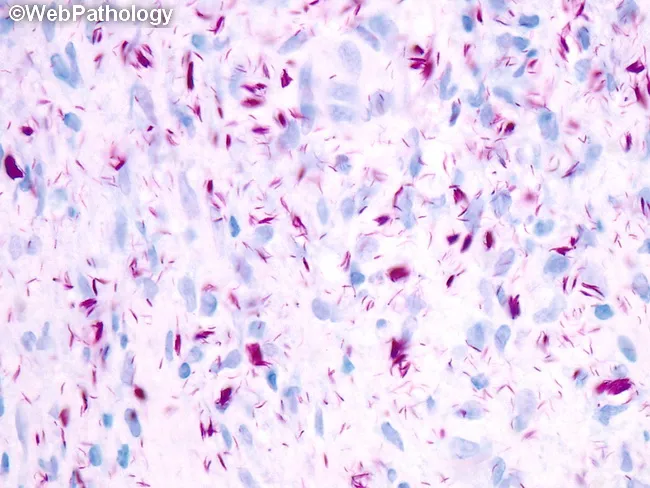
- Acid-fast bacilli on a slit-skin smear, or in a skin biopsy or biopsy PCR.
The first two signs are found by clinical examination alone. The third needs the lab. In practice, a patient with typical numb patches or a thickened nerve can be diagnosed and treated without any lab test.
3. Supporting investigations
| Test | What it shows |
|---|---|
| Slit-skin smear | Taken from the earlobes, forehead, chin, and buttock or thigh (Harrison's). Stained by Ziehl-Neelsen. Gives the bacteriological index (0 to 6) and the morphological index (percentage of solid-staining bacilli). Positive smears mean multibacillary disease. Negative smears are common in tuberculoid disease, so a negative smear does not exclude leprosy. |
| Skin biopsy (histopathology) | Confirms and classifies the disease, supports the diagnosis of reactions, and assesses cure. For macules, take the sample from the middle of the lesion. For plaques, take it from the active indurated edge. Look for granulomas, nerve involvement, and bacilli on Fite-Faraco or Wade-Fite stain. |
| PCR | Detects M. leprae DNA. Useful in paucibacillary or doubtful cases. |
| Anti-PGL-I serology | Detects antibody to phenolic glycolipid-I. It is positive mainly in multibacillary disease, so it is not a diagnostic test and has limited use in paucibacillary disease (my addition, not from the retrieved text). |
| Nerve conduction studies, high-resolution ultrasound | Used to find nerve damage, including "silent" neuropathy. |
| Lepromin test | Not diagnostic. It reflects the host's cell-mediated immunity, and it was historically used to help classify the disease. |
4. Classification after detection
WHO operational classification (for treatment):
- Paucibacillary (PB): 1 to 5 skin lesions, with no bacilli demonstrated in a smear or biopsy.
- Multibacillary (MB): more than 5 lesions, or bacilli on smear or biopsy, or nerve involvement in some schemes.
Ridley-Jopling (immunological spectrum): TT, BT, BB, BL, LL, plus indeterminate leprosy.
5. Look for complications at the time of detection
- Grade disability in the hands, feet and eyes.
- Screen for reactions (type 1 reversal reaction, type 2 erythema nodosum leprosum) and for "silent neuritis". Silent neuritis is progressive nerve impairment without pain or tenderness.
- Screen household contacts, since contact tracing is part of case detection.
6. Differential diagnoses to consider
These include:
- Tinea versicolor
- Vitiligo
- Pityriasis alba
- Post-inflammatory hypopigmentation
- Cutaneous sarcoidosis
- Granuloma annulare
- Cutaneous lymphoma
- Other causes of neuropathy
A 2026 systematic review (PMID 42321940) reported leprosy presenting as lupus, so unexplained neuropathy with skin disease in an endemic area should prompt consideration of leprosy. I read only the titles from PubMed, not the full papers.
Images
I found two relevant images from the library: a lepromatous nodule of the earlobe, and a Wade-Fite stain showing acid-fast bacilli and globi in foamy macrophages.
I can also cover treatment (WHO multidrug therapy) or the management of leprosy reactions if you want either.
This is a shared conversation. Sign in to Orris to start your own chat.