
Mechanism of action of mineralocorticoods
Reading File
Searching sources...
Finding Sources
Finding Sources
Finding Sources
Reading File
Reading File
Finding Sources
Reading File
Finding Sources
Finding Sources
Finding Sources
Reading File
Reading File
Reading File
Reading File
Now I have enough authoritative content to deliver a thorough answer. Let me also fetch the key diagram showing the mechanism.
Loading Image
Loading Image
Here is a complete, textbook-grounded explanation of the mechanism of action of mineralocorticoids.
Mechanism of Action of Mineralocorticoids
Mineralocorticoids (primarily aldosterone) act through both a well-characterized genomic (classical) pathway and a less-understood nongenomic pathway.
1. The Receptor: Mineralocorticoid Receptor (MR)
The MR (gene name NR3C2) is a member of the nuclear receptor superfamily of transcription factors. It has three major functional domains:
- N-terminal transcriptional regulatory domain - regulates transactivation
- Central DNA-binding domain (DBD) - two zinc finger motifs that bind to specific DNA sequences
- C-terminal ligand/hormone-binding domain (LBD) - binds aldosterone
In the absence of hormone, the MR resides in both the cytoplasm and nucleus, complexed with chaperone proteins: hsp90, hsp70, hsp56, and immunophilins (FKBP52, FKBP51, CyP40). This chaperone complex is essential for maintaining high-affinity hormone binding and nuclear trafficking.
"In the absence of hormone, MR is distributed relatively evenly between nuclear and cytoplasmic compartments but, in the presence of hormone, it is highly concentrated in the nucleus." - Brenner and Rector's The Kidney
2. Genomic (Classical) Pathway - Step by Step
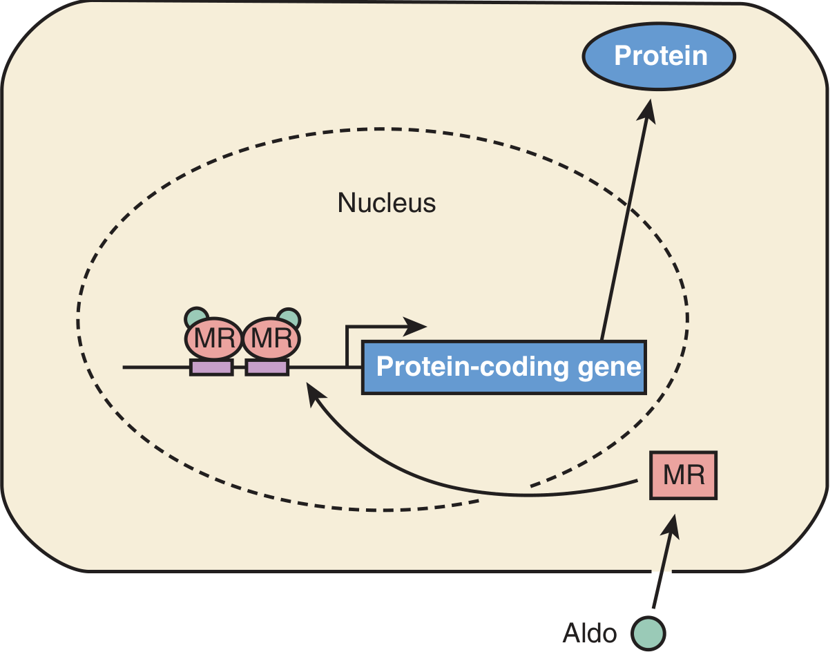
Step 1 - Diffusion into the cell
Aldosterone is a lipid-soluble steroid that freely diffuses across the plasma membrane.
Step 2 - Receptor binding
Aldosterone binds to the cytoplasmic MR with high affinity. This triggers a conformational change that releases the chaperone complex. Nuclear accumulation of the aldosterone-MR complex starts within 30 seconds and is complete by ~10 minutes.
Step 3 - Nuclear translocation
The hormone-MR complex translocates into the nucleus.
Step 4 - Dimerization and DNA binding
The MR-aldosterone complex forms a homodimer (MR/MR) and binds to specific 15-nucleotide DNA sequences called hormone response elements (HREs) located in the promoter regions of target genes.
Step 5 - Transcription of target genes
The MR dimer recruits coactivators and components of the general transcription machinery (e.g., TATA-binding protein), stimulating transcription of key aldosterone-regulated genes.
3. Key Target Genes and Downstream Effects
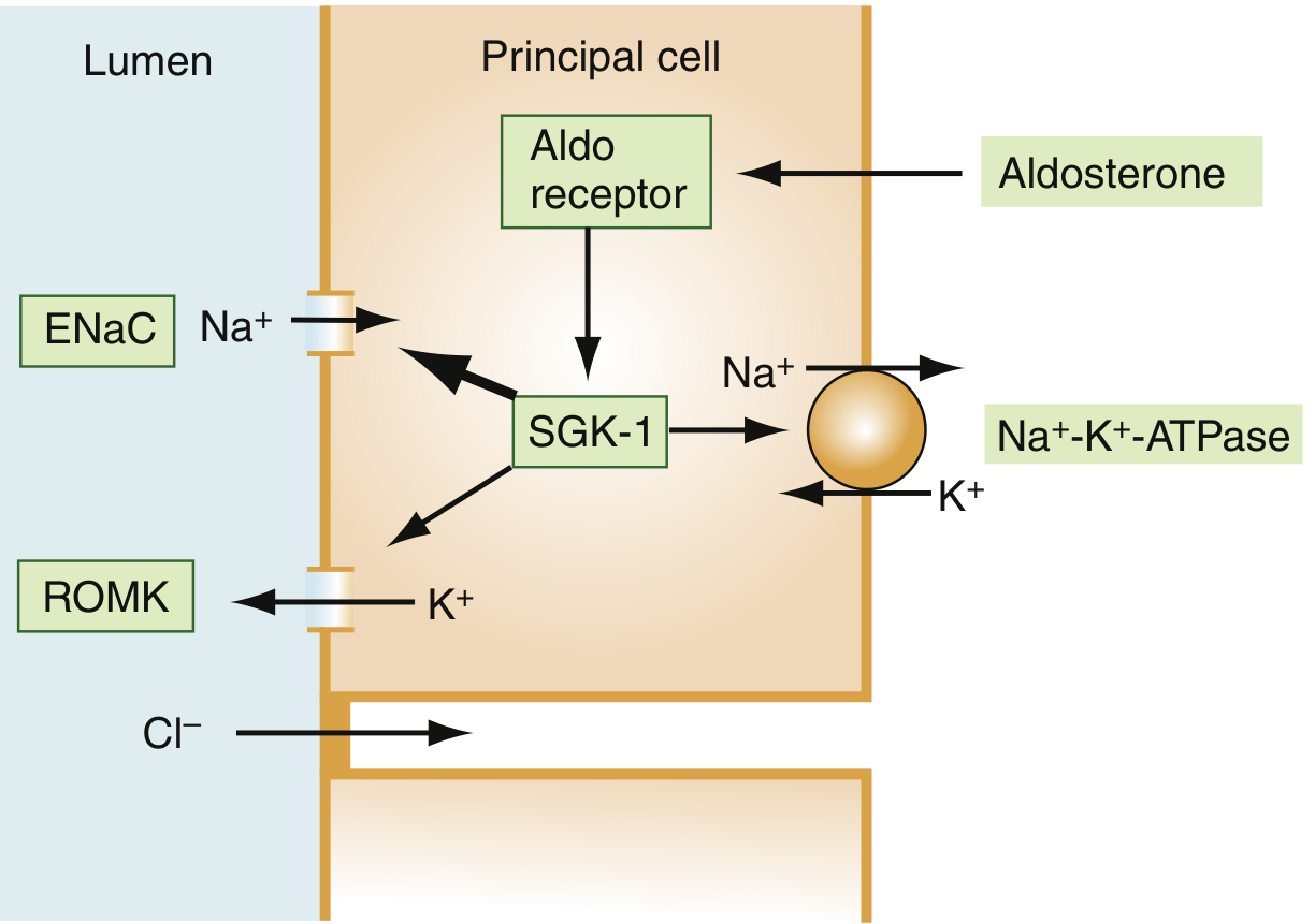
The primary site of action is the principal cells of the distal nephron (cortical collecting duct). The three main gene targets are:
| Gene | Protein | Function |
|---|---|---|
| α-ENaC | Epithelial Na+ Channel (luminal) | Increases Na+ entry from tubular lumen into cell |
| SGK1 | Serum- and glucocorticoid-regulated kinase 1 | Phosphorylates and stabilizes ENaC; activates ROMK and Na+/K+-ATPase |
| GILZ | Glucocorticoid-induced leucine zipper | Modulates sodium transport regulation |
The net renal effect:
- Na+ reabsorption from the lumen via ENaC (apical membrane)
- Na+ pumped out of cell into blood via Na+/K+-ATPase (basolateral)
- K+ enters the cell via Na+/K+-ATPase, then exits into the lumen via ROMK
- The lumen becomes electrically negative, driving paracellular Cl- reabsorption
Result: Na+ and water retention, K+ and H+ secretion - expansion of extracellular fluid volume and blood pressure regulation.
4. The Role of 11β-Hydroxysteroid Dehydrogenase Type 2 (11β-HSD2)
The MR has equal affinity for both aldosterone and cortisol. Since circulating cortisol concentrations are ~100-1000x higher than aldosterone, selectivity is achieved by the enzyme 11β-HSD2 in renal tubular cells, which converts cortisol to the inactive cortisone - preventing it from activating the MR. This ensures the MR responds only to aldosterone in the kidney.
"Because the mineralocorticoid receptor also binds glucocorticoids with high affinity, an enzyme (11β-hydroxysteroid dehydrogenase) in renal tubular cells inactivates glucocorticoids, allowing selective renal responses to mineralocorticoids such as aldosterone." - Harrison's Principles of Internal Medicine 22E
5. Nongenomic Effects
In addition to the slow genomic mechanism (which takes hours), aldosterone can produce rapid nongenomic effects within seconds to minutes - including decreasing intracellular [Ca2+] - that are too fast to be explained by altered gene transcription. These may be mediated by:
- An alternate non-MR membrane receptor
- The classical MR producing rapid signaling in addition to its genomic role
- Nonspecific interactions with other signaling cascades
This is an area of active investigation. - Medical Physiology (Boron & Boulpaep)
6. Extra-Renal Effects (Nonepithelial Tissues)
MRs are also expressed in the heart, blood vessels, and brain. In these tissues, aldosterone excess mediates:
- Fibrosis and collagen deposition (via upregulation of Ang II receptor responsiveness)
- Vascular smooth muscle hypertrophy
- Sodium influx in vascular smooth muscle
- Inhibition of norepinephrine reuptake in myocardial and vascular cells
These effects explain why aldosterone antagonists (spironolactone, eplerenone, finerenone) reduce mortality in heart failure beyond their diuretic action. - Brenner and Rector's The Kidney
Summary Table
| Feature | Detail |
|---|---|
| Receptor type | Nuclear receptor (MR/NR3C2) |
| Location of MR (unliganded) | Cytoplasm (predominantly) + nucleus |
| Chaperone complex | hsp90, hsp70, hsp56, immunophilins |
| Onset of nuclear translocation | ~30 sec; complete ~10 min |
| DNA binding site | Hormone Response Elements (HREs) |
| Key target genes | α-ENaC, SGK1, GILZ |
| Primary renal site | Distal nephron principal cells |
| Net ion effects | Na+/H2O retention; K+/H+ excretion |
| Selectivity mechanism | 11β-HSD2 inactivates cortisol in kidney |
| Antagonists | Spironolactone, eplerenone, finerenone |
Simpler steps
Mechanism of Action of Mineralocorticoids (Simple Steps)
Step 1 - Aldosterone enters the cell
Aldosterone is fat-soluble, so it diffuses straight through the cell membrane. No receptor needed on the surface.
Step 2 - Binds to its receptor in the cytoplasm
Inside the cell, it binds to the Mineralocorticoid Receptor (MR), which is sitting idle, wrapped in protective "chaperone" proteins (heat shock proteins).
Step 3 - Chaperones fall off
Binding causes a shape change in MR. The chaperone proteins detach, and a "nuclear entry signal" is unmasked.
Step 4 - MR moves into the nucleus
The aldosterone-MR complex travels into the nucleus. This happens within minutes.
Step 5 - Binds to DNA
Inside the nucleus, two MR complexes pair up (dimerize) and attach to specific DNA sequences called Hormone Response Elements (HREs).
Step 6 - Target genes are switched on
Three key genes get activated:
- α-ENaC - opens Na⁺ channels on the luminal (urine-facing) side
- SGK1 - a kinase that stabilizes ENaC and boosts Na⁺/K⁺-ATPase activity
- Na⁺/K⁺-ATPase - pumps Na⁺ out into blood, K⁺ into cell
Step 7 - Net effect in the kidney
| What happens | Result |
|---|---|
| Na⁺ reabsorbed from urine | Water follows → blood volume ↑, BP ↑ |
| K⁺ secreted into urine | Blood K⁺ ↓ |
| H⁺ secreted into urine | Mild alkalosis |
One extra point - Why doesn't cortisol do the same thing?
The MR binds cortisol just as well as aldosterone. But an enzyme called 11β-HSD2 in the kidney destroys cortisol before it can reach the MR - so only aldosterone gets through.
In one line: Aldosterone → enters cell → binds MR → MR enters nucleus → switches on ENaC + SGK1 + Na/K-ATPase genes → Na⁺ retained, K⁺ lost.
Hormonal contraceptives drugs and MEchanism of action
Reading File
Searching sources...
Finding Sources
Finding Sources
Reading File
Reading File
Reading File
Reading File
Finding Sources
Reading File
Finding Sources
Finding Sources
Loading Image
I now have comprehensive content. Here is the full structured answer.
Hormonal Contraceptives - Drugs and Mechanism of Action
How They Work - The Core Mechanism
The normal menstrual cycle depends on a precise hormonal sequence:
Hypothalamus (GnRH) → Pituitary (FSH + LH) → Ovary (estrogen + ovulation)
Hormonal contraceptives disrupt this sequence through negative feedback:
| Component | What it suppresses | Effect |
|---|---|---|
| Estrogen | FSH from pituitary | No follicle development |
| Progestin | LH surge from pituitary | No ovulation trigger |
| Progestin | Cervical mucus | Thickens it - sperm can't penetrate |
| Progestin | Endometrium | Atrophies - implantation blocked |
"Exogenously administered estrogen in contraceptives provides negative feedback that blunts release of FSH by the pituitary gland, and progestin inhibits LH secretion, thus preventing ovulation." - Lippincott Illustrated Reviews: Pharmacology
Categories of Hormonal Contraceptives
1. Combined Oral Contraceptives (COCs) - Estrogen + Progestin
Estrogen component: Almost always ethinyl estradiol (EE)
Progestin component - classified by generation:
| Generation | Progestins | Androgen activity |
|---|---|---|
| 1st | Norethindrone, ethynodiol diacetate | Moderate |
| 2nd | Levonorgestrel, medroxyprogesterone | Lower |
| 3rd | Desogestrel, norgestimate, gestodene | Very low |
| 4th | Drospirenone, dienogest, cyproterone acetate | Anti-androgenic |
Types of COC packs:
- Monophasic - same dose of estrogen + progestin every day (21 active + 7 placebo)
- Biphasic/Triphasic - dose of progestin increases progressively (mimics natural cycle)
- Extended cycle - 84 active days + 7 placebo (only 4 periods/year)
- Continuous - active pills every day (no periods)
Mechanism (COC):
- Estrogen suppresses FSH → no follicle growth
- Progestin suppresses LH → no ovulation
- Progestin thickens cervical mucus → blocks sperm
- Endometrium becomes thin and atrophic → implantation unlikely
- Tubal motility is altered → slows sperm/egg transport
2. Transdermal Patch
- Contains ethinyl estradiol + norelgestromin (or levonorgestrel)
- One patch/week for 3 weeks → no patch in week 4 → withdrawal bleed
- Same mechanism as COC
- Less effective in women >90 kg
- Higher total estrogen exposure → increased VTE risk vs. oral pills
3. Vaginal Ring (NuvaRing)
- Contains ethinyl estradiol + etonogestrel
- Inserted vaginally for 3 weeks, removed for 1 week
- Same COC mechanism - local absorption through vaginal mucosa
- Avoids first-pass metabolism
4. Progestin-Only Pills (Mini-Pill)
- Contains norethindrone (or drospirenone in newer formulations)
- Taken every single day without a break
Mechanism (different from COC):
- Does not always suppress ovulation (variable)
- Main effect: thickens cervical mucus making it hostile to sperm
- Thins endometrium
- Safe for breastfeeding mothers (no effect on milk production unlike estrogen)
5. Injectable Progestin (Depot)
- Medroxyprogesterone acetate (DMPA / Depo-Provera)
- IM or SC injection every 3 months
- Provides high, sustained progestin levels
- Suppresses ovulation reliably (unlike mini-pill)
- Common side effects: amenorrhea, weight gain, delayed return of fertility (up to 12 months), bone loss (avoid >2 years)
6. Subdermal Implant
- Etonogestrel implant (Nexplanon)
- Placed under skin of upper arm - lasts up to 3 years
- Most effective hormonal method (failure rate <0.05%)
- Mechanism: suppresses ovulation + thickens cervical mucus
- Not dependent on patient adherence - hence highest efficacy
7. Hormonal Intrauterine Device (IUD)
- Levonorgestrel-releasing IUD (Mirena, Kyleena) - lasts 3-7 years
- Mechanism: mainly local - thickens cervical mucus + thins endometrium
- Minimal systemic progestin absorption
- Also treats heavy menstrual bleeding
- Avoid in: PID, history of ectopic pregnancy
8. Emergency Contraception (Postcoital)
| Drug | Timing | Mechanism |
|---|---|---|
| Levonorgestrel (Plan B) | Within 72 hours | Delays/inhibits ovulation; thickens mucus |
| Ulipristal acetate (ella) | Within 120 hours (5 days) | Progesterone receptor agonist/antagonist; delays ovulation more effectively near LH surge |
| Copper IUD | Within 5 days | Toxic to sperm; also prevents implantation |
Comparison of Failure Rates (Typical Use)
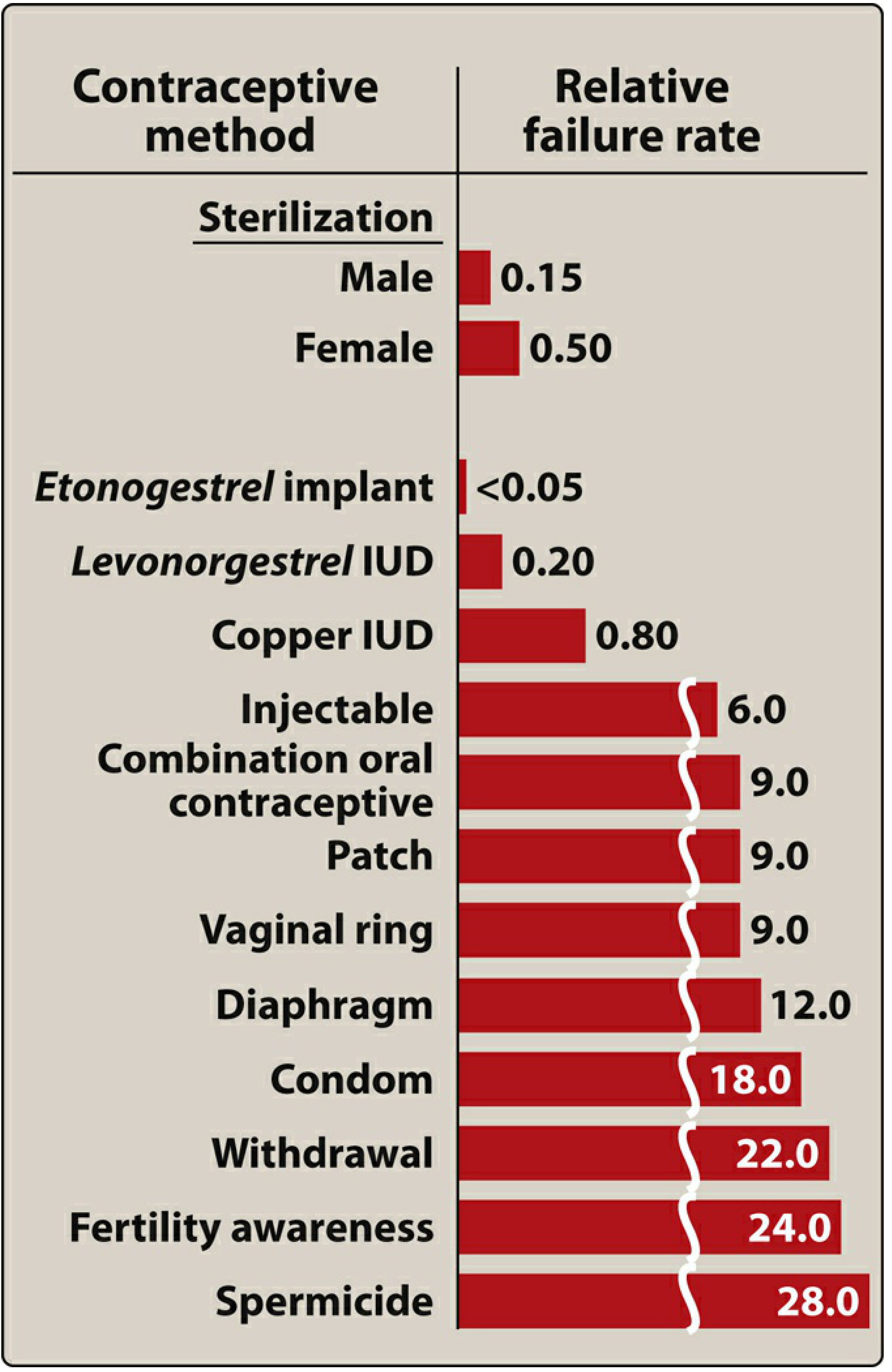
The etonogestrel implant and levonorgestrel IUD have the lowest failure rates among all reversible methods - because they eliminate user error entirely.
Quick Summary Table
| Method | Drugs | Primary Mechanism |
|---|---|---|
| COC | EE + progestin | Suppress FSH + LH → no ovulation |
| Patch | EE + norelgestromin | Same as COC |
| Vaginal ring | EE + etonogestrel | Same as COC |
| Mini-pill | Norethindrone | Cervical mucus thickening |
| Injectable | Medroxyprogesterone | Suppresses ovulation + mucus |
| Implant | Etonogestrel | Suppresses ovulation + mucus |
| Hormonal IUD | Levonorgestrel | Local: mucus + endometrium |
| Emergency (72h) | Levonorgestrel | Delays ovulation |
| Emergency (120h) | Ulipristal | Delays ovulation (near LH surge) |
Sources: Lippincott Illustrated Reviews: Pharmacology; Katzung's Basic and Clinical Pharmacology, 16th Ed.
Hormonal contraceptives drugs and mechanism of action
I just covered this in detail in my previous response! Here's a clean summary version for quick reference:
Hormonal Contraceptives - Drugs & Mechanism of Action
The Core Concept
All hormonal contraceptives work by mimicking the hormonal state of pregnancy - when estrogen and progesterone are high, the body does not ovulate. They exploit the negative feedback axis:
High estrogen → pituitary stops releasing FSH → no follicle grows High progestin → pituitary stops releasing LH → no ovulation trigger
4 Ways They Prevent Pregnancy
| Mechanism | Hormone responsible |
|---|---|
| Suppress FSH → no follicle development | Estrogen |
| Suppress LH surge → no ovulation | Progestin |
| Thicken cervical mucus → sperm can't pass | Progestin |
| Thin endometrium → implantation blocked | Progestin |
All Drug Categories
A. Combined (Estrogen + Progestin)
Estrogen used: Ethinyl estradiol (in almost all products)
Progestins by generation:
| Gen | Drug examples | Special property |
|---|---|---|
| 1st | Norethindrone, ethynodiol | Moderate androgenic |
| 2nd | Levonorgestrel, MDPA | Lower androgenic |
| 3rd | Desogestrel, norgestimate, gestodene | Minimal androgenic |
| 4th | Drospirenone, dienogest | Anti-androgenic (useful in PCOS, acne, hirsutism) |
Delivery forms:
- Oral pill (COC) - daily pill, 21 active + 7 placebo days
- Transdermal patch - weekly patch x3 weeks (ethinyl estradiol + norelgestromin)
- Vaginal ring - monthly ring (ethinyl estradiol + etonogestrel)
All three share the same mechanism: suppress FSH + LH + thicken mucus + thin endometrium
B. Progestin-Only
| Drug / Route | Duration | Primary Mechanism |
|---|---|---|
| Norethindrone (mini-pill, oral) | Daily | Thickens mucus (ovulation not always suppressed) |
| Medroxyprogesterone acetate (Depo-Provera, IM/SC) | Every 3 months | Suppresses ovulation reliably |
| Etonogestrel (subdermal implant, Nexplanon) | Up to 3 years | Suppresses ovulation + thickens mucus |
| Levonorgestrel IUD (Mirena) | 3-7 years | Local effect: mucus + endometrium |
Progestin-only options are preferred when estrogen is contraindicated - e.g., breastfeeding, smokers over 35, history of VTE, migraines with aura.
C. Emergency Contraception
| Drug | Window | Mechanism |
|---|---|---|
| Levonorgestrel (Plan B) | Within 72 hours | Delays/inhibits ovulation |
| Ulipristal acetate (ella) | Within 120 hours | Progesterone receptor modulator - delays ovulation even near LH surge (more effective than levonorgestrel if taken late) |
| Copper IUD | Within 5 days | Spermicidal + prevents implantation (most effective EC) |
Key Points to Remember
| Point | Detail |
|---|---|
| Most potent reversible method | Etonogestrel implant (failure rate <0.05%) |
| Safest in breastfeeding | Progestin-only (estrogen reduces milk) |
| Anti-androgenic progestin | Drospirenone, dienogest, cyproterone |
| Bone loss risk | Depo-Provera (avoid >2 years) |
| VTE risk highest | Patch > COC |
| Progestin-only pill - must take | Same time every day (narrow 3-hour window) |
One-liner: Estrogen kills FSH (no egg grows), progestin kills LH (no egg releases) and makes cervical mucus hostile to sperm. Together = highly effective contraception.
Osteoporosis drugs and mechanism of action
Finding Sources
Finding Sources
Searching sources...
Finding Sources
Reading File
Finding Sources
Finding Sources
Finding Sources
Finding Sources
Finding Sources
Reading File
Finding Sources
Reading File
Reading File
Reading File
Reading File
Reading File
Finding Sources
I now have comprehensive material from multiple authoritative sources. Here is the complete answer.
Osteoporosis Drugs and Mechanism of Action
Understanding the Target: Bone Remodeling
Normal bone is constantly remodeled by two opposing cell types:
- Osteoclasts - break down (resorb) old bone
- Osteoblasts - build new bone
The key signaling axis controlling this balance:
Osteoblasts produce RANKL → binds RANK on osteoclast precursors → osteoclasts mature and resorb bone OPG (osteoprotegerin) - a natural decoy produced by osteoblasts that blocks RANKL
In osteoporosis, osteoclast activity exceeds osteoblast activity - net bone loss occurs. Drugs either suppress osteoclasts (antiresorptive) or stimulate osteoblasts (anabolic).
Classification of Drugs
| Class | Action | Examples |
|---|---|---|
| Bisphosphonates | Antiresorptive | Alendronate, risedronate, zoledronic acid |
| RANKL inhibitor | Antiresorptive | Denosumab |
| SERMs | Antiresorptive | Raloxifene |
| Calcitonin | Antiresorptive | Salmon calcitonin |
| PTH analogues | Anabolic | Teriparatide, abaloparatide |
| Sclerostin inhibitor | Dual (anabolic + antiresorptive) | Romosozumab |
| Estrogen/HRT | Antiresorptive | Conjugated equine estrogen |
1. Bisphosphonates
Drugs: Alendronate (weekly oral), Risedronate (weekly/monthly oral), Ibandronate (monthly oral), Zoledronic acid (yearly IV), Pamidronate (IV)
Mechanism:
- Bisphosphonates are pyrophosphate analogues - they bind tightly to hydroxyapatite (bone mineral) at sites of active remodeling
- Osteoclasts engulf the drug during bone resorption
- Inside the osteoclast: amino-bisphosphonates (alendronate, zoledronate) inhibit farnesyl pyrophosphate synthase in the mevalonate pathway → blocks prenylation of signaling proteins (Ras, Rho) → osteoclast cytoskeletal disruption and apoptosis
- Net result: fewer and less active osteoclasts → reduced bone resorption
Key points:
- Must be taken on empty stomach with full glass of water, stay upright 30 min (oral forms) - prevents esophageal irritation
- "Drug holiday" recommended after 5 years (reduce risk of atypical femur fracture and jaw osteonecrosis)
- Not cleared by kidney - caution in CKD (except zoledronate with care)
2. Denosumab (RANKL Inhibitor)
Drug: Denosumab (Prolia) - SC injection every 6 months
Mechanism:
- Fully humanized monoclonal antibody that mimics OPG (the natural RANKL decoy)
- Binds to RANKL with high affinity → prevents RANKL from binding RANK on osteoclast precursors
- Blocks osteoclast formation, maturation, and survival → reduces bone resorption
- At least as potent as bisphosphonates in inhibiting bone resorption
Key points:
- Can be used in renal failure (unlike bisphosphonates) - not renally cleared
- Do NOT stop abruptly - rebound bone resorption surge causes multiple vertebral fractures
- Watch for hypocalcemia, especially in vitamin D deficiency or CKD
- Slight increased risk of infection (RANKL is also expressed on immune cells)
3. SERMs - Selective Estrogen Receptor Modulators
Drug: Raloxifene (oral daily)
Mechanism:
- Acts as an estrogen receptor agonist in bone but antagonist in breast and uterus
- In bone: mimics estrogen → suppresses osteoclast activity → reduces bone resorption
- Estrogen normally suppresses RANKL production by osteoblasts - raloxifene replicates this effect in bone tissue only
Key points:
- Does NOT increase risk of breast cancer or endometrial cancer (unlike estrogen)
- Reduces risk of vertebral fractures (but weaker hip fracture protection vs bisphosphonates)
- Increases VTE risk
- Used in postmenopausal women with osteoporosis who also have breast cancer risk
4. Calcitonin
Drug: Salmon calcitonin (intranasal or SC)
Mechanism:
- Binds calcitonin receptors on osteoclasts → directly inhibits osteoclast activity
- Reduces bone resorption
- Also has a central analgesic effect (useful in acute vertebral fracture pain)
Key points:
- Weak antiresorptive effect - rarely first-line now
- Main current use: acute pain relief in vertebral compression fractures
- Tachyphylaxis develops with prolonged use (effectiveness decreases)
- Long-term use may be associated with slightly increased cancer risk - use limited to short courses
5. PTH Analogues (Anabolic Agents)
Drugs: Teriparatide (PTH 1-34, daily SC), Abaloparatide (PTHrP analogue, daily SC)
Mechanism:
- Intermittent low-dose PTH (given as a daily injection) has a paradoxical anabolic effect on bone
- Acts on osteoblasts via PTH receptor → increases osteoblast proliferation and survival
- Also inhibits sclerostin secretion by osteocytes → further promotes osteoblast activity via Wnt/β-catenin pathway
- Stimulates both bone formation AND resorption, but net effect is formation > resorption at these doses
- Abaloparatide selectively binds the RG conformation of PTH receptor → more bone formation, less resorption than teriparatide → greater BMD gains
Key points:
- Only agents that actually build new bone (others just slow loss)
- Reserved for severe osteoporosis or failure of antiresorptive therapy
- Limited to 2 years (black box warning for osteosarcoma risk in animal studies)
- Must be followed by antiresorptive (bisphosphonate or denosumab) to preserve gains
6. Romosozumab (Sclerostin Inhibitor) - Dual Action
Drug: Romosozumab (SC injection monthly for 12 months)
Mechanism:
- Monoclonal antibody that binds and neutralizes sclerostin
- Sclerostin is produced by osteocytes and blocks the Wnt receptor on osteoblasts → normally suppresses bone formation
- By blocking sclerostin → Wnt/β-catenin signaling is activated → osteoblast proliferation increases → bone formation goes up
- Simultaneously reduces bone resorption (mechanism not fully understood)
- Dual effect: anabolic + antiresorptive - unique among all osteoporosis drugs
Key points:
- Causes rapid early rise in bone formation marker (P1NP) and sustained fall in resorption marker (CTX)
- Significant BMD gains within 12 months
- Limited to 1 year of treatment, then switch to antiresorptive
- Black box warning: contraindicated if MI or stroke within past year (cardiovascular safety signal in trials)
7. Estrogen / HRT
Mechanism:
- Estrogen suppresses RANKL production by osteoblasts → less osteoclast activation → less bone resorption
- Also promotes osteoblast survival
- Protects bone throughout reproductive years - loss of estrogen at menopause is the main driver of postmenopausal osteoporosis
Key points:
- Effective at preventing and treating osteoporosis
- No longer first-line due to increased risk of breast cancer, VTE, stroke, MI with long-term use
- Still considered in younger postmenopausal women (<60 years, <10 years since menopause) with vasomotor symptoms
Summary: Mechanism at a Glance
OSTEOCLAST SIDE (Antiresorptive)          OSTEOBLAST SIDE (Anabolic)
─────────────────────────────             ──────────────────────────
Bisphosphonates → osteoclast apoptosis    Teriparatide → ↑ osteoblast proliferation
Denosumab → blocks RANKL → no             Abaloparatide → same, more selective
             osteoclast formation
Raloxifene → ER agonist in bone →         Romosozumab → blocks sclerostin →
             ↓ RANKL                                   ↑ Wnt → ↑ osteoblasts
Calcitonin → directly inhibits                         (ALSO reduces resorption)
              osteoclast activity
Estrogen → ↓ RANKL production
Sources: Katzung's Basic and Clinical Pharmacology 16th Ed; Harrison's Principles of Internal Medicine 22E; Goodman & Gilman's Pharmacological Basis of Therapeutics; Rheumatology 2-Vol Set (Elsevier 2022)
This is a shared conversation. Sign in to Orris to start your own chat.