

What is this small water filled bump and how to cure
dyshidrotic eczema pompholyx small vesicles fingers
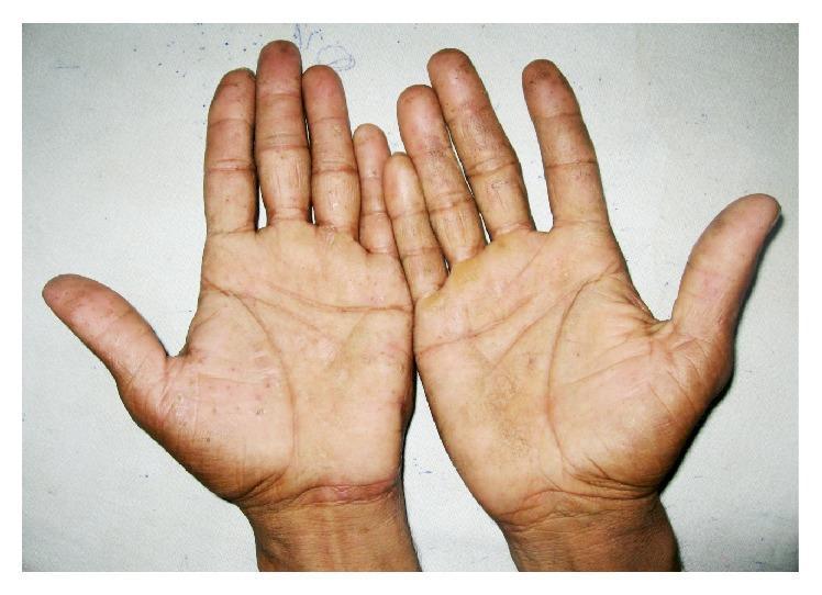
Clinical photograph of both palms demonstrating multiple, small, deep-seated vesicles symmetrically distributed across the palmar surfaces and digits. The lesions are characteristic of chronic vesiculobullous hand dermatitis, also known as dyshidrotic eczema or pompholyx. The vesicles appear as translucent, firm, 'tapioca-like' elevations primarily concentrated on the palms and the lateral aspects of the fingers. The surrounding skin exhibits mild xerosis (dryness) and accentuation of skin markings, with focal areas of post-inflammatory hyperpigmentation. There is no evidence of secondary bacterial infection or prominent erythema. The bilateral and symmetrical involvement is a key clinical sign used to differentiate this condition from contact dermatitis or fungal infections (tinea manuum). This image serves as a diagnostic reference for primary inflammatory hand eczemas in a clinical dermatology setting.
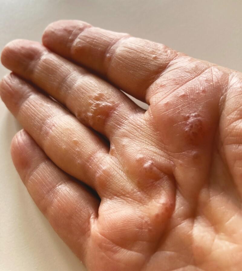
Clinical photograph of a right hand, specifically the palmar surface and digits, displaying dyshidrotic eczema (pompholyx). The image shows multiple deep-seated, small, tense vesicles and papules distributed across the palm, thenar eminence, and lateral aspects of the fingers. The lesions exhibit varying morphology, including clear fluid-filled vesicles and larger inflammatory papular clusters with a background of diffuse erythema. There is a notable absence of significant scaling or crusting, indicating an acute to subacute presentation. The skin appears mildly edematous with accentuated skin markings. Clinically, this visual represents a Grade 3 cutaneous adverse event (CAE) related to ribociclib therapy, illustrating a drug-induced eczematous dermatitis. Key educational concepts include the recognition of pompholyx-like distributions in the context of oncology-related dermatological toxicity and the identification of inflammatory vesiculobullous eruptions on acral surfaces.
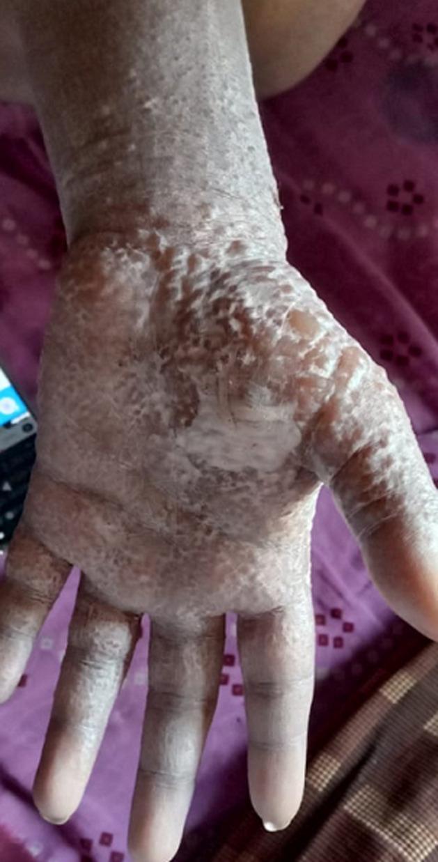
Clinical photograph of the palmar aspect of a human hand and wrist demonstrating a case of dyshidrotic eczema, also known as pompholyx. The image shows a dense, symmetric distribution of numerous small, deep-seated, ‘tapioca-like’ vesicles and bullae. These lesions are primarily concentrated on the palm and extend across the fingers and onto the volar wrist. The affected skin exhibits prominent morphological changes including whitish, translucent blisters, areas of scaling, and desquamation (skin peeling), which give the surface a rough, cobblestone-like texture. There is underlying mild erythema. The clinical presentation is characteristic of an acute pompholyx flare, often associated with severe pruritus (itching) or burning sensations, and serves as a classic educational example of vesicular hand dermatitis within the field of dermatology.
What Is That Small Water-Filled Bump?
What Is Dyshidrotic Eczema (Pompholyx)?
Other Possible Causes (Less Likely)
| Condition | Key Feature |
|---|---|
| Friction blister | Caused by rubbing or tool use; painless |
| Contact dermatitis | Reaction to soap, cleaning chemicals, metal (nickel), or plants |
| Insect bite | Single bump, may be itchy or mildly swollen |
| Herpetic whitlow | Caused by herpes virus - typically painful, often clustered |
How to Treat It
- Do NOT pop or squeeze the vesicle. The intact blister roof acts as a natural barrier against infection.
- Replace soap-and-water handwashing with an alcohol-based hand sanitizer to reduce skin irritation.
- Wash gently with mild, fragrance-free soap and pat dry.
- Apply a greasy emollient (petrolatum/Vaseline, Aquaphor, or similar bland moisturizer) liberally. This moisturizes, protects, and prevents skin cracking (fissures).
- A high-potency topical corticosteroid ointment (e.g., fluocinolone 0.05% or clobetasol propionate) applied twice daily for 1-2 weeks is the mainstay of treatment.
- Cold compresses on the area can relieve the burning/itching sensation.
- A short oral steroid course (prednisone starting at 40 mg/day, tapering over 2 weeks) for acute flares.
- In chronic/recurrent cases, immunosuppressants like cyclosporine may be used under specialist supervision.
- If the skin cracks (fissures), a tiny drop of cyanoacrylate (superglue) can seal the crack and reduce pain.
Triggers to Avoid
- Frequent hand washing with soap and water
- Harsh soaps, detergents, cleaning chemicals
- Contact with nickel (jewelry, coins, metal tools)
- Stress (a known trigger for flares)
- Hot water
When to See a Doctor
- The bump becomes painful, red, or warm (possible infection)
- New blisters keep appearing across multiple fingers or palms
- It does not resolve within 2-3 weeks
- You develop a fever