

IMG_7480.jpg
Answer these questions for a physiotherapy exam add images and tables and flow charts wherever possible
Reading File
The exam has 6 questions. Given the budget constraints, I'll search the medical library for all topics in parallel.
Finding Sources
Finding Sources
Finding Sources
Finding Sources
Reading File
Reading File
Finding Sources
Finding Sources
Reading File
Reading File
I now have sufficient textbook content on shock and gangrene. Let me get the remaining content for management of haemorrhagic shock and the classification sections, then compile the full answer.
Reading File
I now have rich content. Let me compile the comprehensive answer for all 6 questions.Here are comprehensive exam answers for all 6 questions, with tables, flowcharts, and images where applicable.
General Surgery - Physiotherapy Exam Answers
Q1. What is Shock? Classify Types of Shock. Describe Clinical Features and Management of Haemorrhagic Shock. (15 marks)
Definition of Shock
Shock is a state of systemic tissue hypoperfusion - inadequate delivery of oxygen and nutrients to cells relative to metabolic demand, resulting in cellular hypoxia, anaerobic metabolism, and if untreated, organ failure and death.
Classification of Shock
                          SHOCK
                            |
    ┌───────────┬───────────┼───────────┬───────────┐
    ▼           ▼           ▼           ▼           ▼
Haemorrhagic/ Cardiogenic  Obstructive Distributive Endocrine
Hypovolaemic              Shock       Shock        Shock
Shock
    |               |           |           |
- Haemorrhage   - MI        - PE        - Septic
- Burns         - Arrhythmia - Cardiac   - Anaphylactic
- Vomiting      - Valvular     tamponade - Neurogenic
- Diarrhoea       disease   - Tension
- Third-spacing              pneumothorax
| Type | Mechanism | Example Causes |
|---|---|---|
| Haemorrhagic/Hypovolaemic | Reduced circulating volume | Haemorrhage, burns, vomiting, diarrhoea, pancreatitis |
| Cardiogenic | Heart pump failure | MI, arrhythmia, valvular disease, cardiomyopathy |
| Obstructive | Outflow obstruction | PE, cardiac tamponade, tension pneumothorax |
| Distributive | Vasodilation/maldistribution | Sepsis, anaphylaxis, spinal injury |
| Endocrine | Hormonal failure | Addison's disease, adrenal insufficiency |
Source: Bailey and Love's Short Practice of Surgery, 28th Ed
Haemorrhagic Shock - Pathophysiology
Blood Loss
    ↓
↓ Circulating Volume
    ↓
↓ Cardiac Output & ↓ BP
    ↓
Baroreceptor activation
    ↓
Sympathoadrenal response
    ↓
↑ Heart rate, Vasoconstriction, ↑ ADH, ↑ Renin-Angiotensin
    ↓
Compensated Shock (maintained BP)
    ↓ (if loss continues)
Decompensated Shock (↓ BP, ↓ urine output)
    ↓
Organ hypoperfusion → Anaerobic metabolism → Lactic acidosis
    ↓
Multiple Organ Failure → Death
Clinical Features of Haemorrhagic Shock
| Feature | Mild (Compensated) | Moderate | Severe |
|---|---|---|---|
| Lactic acidosis | + | ++ | +++ |
| Urine output | Normal | Reduced (<0.5 mL/kg/hr) | Anuric |
| Conscious level | Mild anxiety | Drowsy, confused | Comatose |
| Respiratory rate | Increased | Increased | Labouring |
| Pulse rate | Increased | Increased | Markedly increased |
| Blood pressure | Normal | Mild hypotension | Severe hypotension |
| Peripheries | Cool, sweaty | Very cool, pale | Mottled |
Blood pressure is usually well maintained until 30-40% of circulating volume is lost. A 15% loss is within normal compensatory limits.
Classes of Haemorrhagic Shock (ATLS Classification)
| Class | Blood Loss | Blood Loss (70 kg adult) | HR | BP | RR | GCS |
|---|---|---|---|---|---|---|
| I | <15% | <750 mL | <100 | Normal | 14-20 | Normal |
| II | 15-30% | 750-1500 mL | 100-120 | Normal/slightly ↓ | 20-30 | Anxious |
| III | 30-40% | 1500-2000 mL | 120-140 | Decreased | 30-40 | Confused |
| IV | >40% | >2000 mL | >140 | Severely decreased | >35 | Lethargic |
Management of Haemorrhagic Shock
MANAGEMENT FLOWCHART
        ↓
    1. STOP THE BLEEDING
       - Direct pressure / tourniquet
       - Surgical haemostasis
        ↓
    2. AIRWAY & BREATHING
       - High-flow O₂ (15 L/min via non-rebreather mask)
       - Intubate if GCS < 8
        ↓
    3. IV ACCESS & FLUID RESUSCITATION
       - 2 large-bore peripheral IV lines (16G or larger)
       - Warm IV crystalloid (Hartmann's / Normal saline)
       - Permissive hypotension in penetrating trauma (SBP ~80-90 mmHg)
        ↓
    4. BLOOD PRODUCTS (if Class III/IV)
       - Packed Red Blood Cells
       - Fresh Frozen Plasma (FFP)
       - Platelets (1:1:1 ratio)
        ↓
    5. MONITORING
       - Urine output (target ≥0.5 mL/kg/hr)
       - BP, HR, SpO₂, ABG, Lactate
        ↓
    6. DEFINITIVE CARE
       - Surgical / interventional radiology to stop bleeding
       - ICU admission
        ↓
    7. PREVENT COMPLICATIONS
       - Hypothermia prevention (warm fluids, blankets)
       - Correct coagulopathy (avoid "lethal triad":
         hypothermia + acidosis + coagulopathy)
Q2. What is Gangrene? Classify Types. Clinical Features and Management of Gas Gangrene. (15 marks)
Definition of Gangrene
Gangrene is the death (necrosis) of body tissue, usually as a result of loss of blood supply, bacterial infection, or both. It may be localised to one area or spread systemically.
Classification of Gangrene
                        GANGRENE
                           |
           ┌───────────────┼───────────────┐
           ▼               ▼               ▼
         DRY             WET             GAS
       Gangrene         Gangrene        Gangrene
           |               |               |
    (Ischaemia,      (Infection +     (Clostridium
    no infection)    Ischaemia)       perfringens)
                           |
                    ┌──────┴──────┐
                    ▼             ▼
              Moist/Wet     Gas Gangrene
              Gangrene    (Clostridial)
                                  |
                           ┌──────┴──────┐
                           ▼             ▼
                    Necrotising     Fournier's
                    Fasciitis       Gangrene
| Type | Cause | Key Feature |
|---|---|---|
| Dry Gangrene | Arterial occlusion (atherosclerosis) | Dry, shrivelled, mummified; no smell; clear line of demarcation |
| Wet Gangrene | Infection + ischaemia | Moist, swollen, foul-smelling; rapid spread |
| Gas Gangrene | Clostridium perfringens | Gas in tissues, crepitus, sweet smell, rapidly fatal |
| Necrotising Fasciitis | Mixed organisms (synergistic) | Fascia involvement, rapid spread, high mortality |
| Fournier's Gangrene | Mixed infection | Perineum/scrotum involvement |
| Diabetic Gangrene | Neuropathy + ischaemia + infection | Painless, foot/toes most common |
Gas Gangrene
Causative Organism
- Clostridium perfringens - Gram-positive, anaerobic, spore-bearing bacillus
- Found widely in soil and faeces
- Produces: collagenase, hyaluronidase, alpha toxin (lecithinase)
Predisposing Factors
- Immunocompromised state
- Diabetes mellitus
- Malignant disease
- Wounds with necrotic tissue, foreign material, or devascularised tissue
- Military wounds (high-velocity missile injuries cause extensive tissue damage)
- Amputation for peripheral vascular disease with open necrotic ulceration
Clinical Features of Gas Gangrene
TIMELINE OF GAS GANGRENE PROGRESSION

Hour 0-6:         Contaminated wound (trauma, surgery)
                           ↓
Hour 6-24:        Severe, disproportionate LOCAL PAIN
                  Wound oedema, pallor, tense skin
                           ↓
Hour 24-48:       CREPITUS (gas in tissues - palpable crackle)
                  Thin, brown, sweet-smelling discharge
                  Gas visible on plain X-ray
                  Skin becomes bronze/purple → black
                           ↓
Hour 48-72:       SYSTEMIC FEATURES:
                  - High fever, tachycardia
                  - Circulatory collapse
                  - Jaundice (haemolysis)
                  - Renal failure
                  - Multi-organ failure → DEATH if untreated
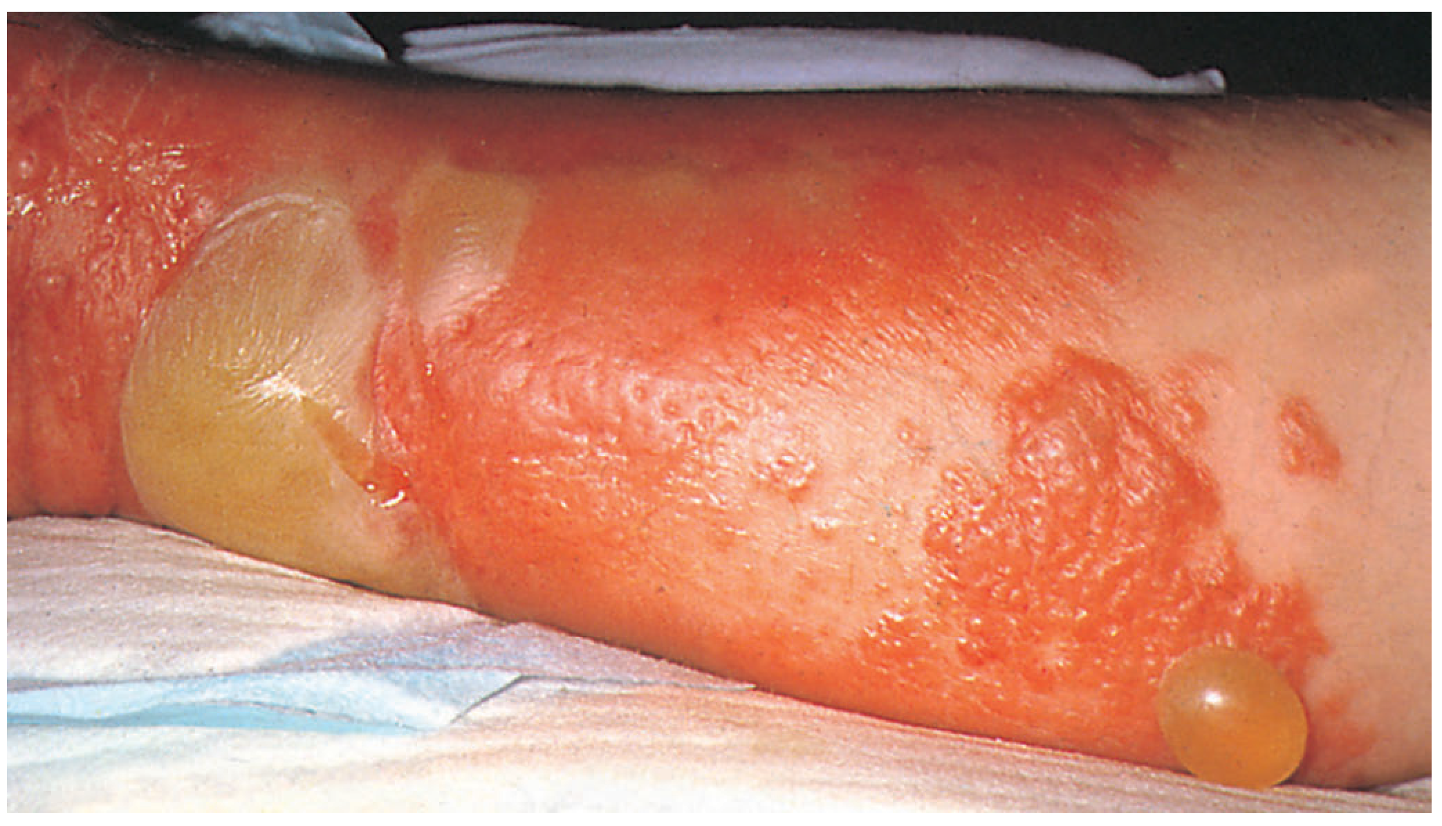
| Feature | Description |
|---|---|
| Pain | Severe, out of proportion to wound appearance - earliest sign |
| Skin appearance | Initially pale, then bronze, then purple-black; haemorrhagic bullae |
| Gas | Crepitus on palpation; gas shadows on X-ray between muscle planes |
| Discharge | Thin, brown, watery, sweet (like rotten apples) - Gram stain shows rods |
| Smell | Characteristic sweet/putrid odour |
| Oedema | Rapid, massive oedema |
| Systemic | Fever, tachycardia, hypotension, jaundice, confusion |
Management of Gas Gangrene
MANAGEMENT ALGORITHM
         ↓
  1. IMMEDIATE RESUSCITATION
     - IV access, fluids, O₂
     - Blood cultures, FBC, coagulation, renal function
         ↓
  2. HIGH-DOSE ANTIBIOTICS (IMMEDIATELY)
     - IV Penicillin G (large doses) - FIRST LINE
     - + Clindamycin (stops toxin production)
     - + Metronidazole (for anaerobes)
         ↓
  3. URGENT SURGICAL DEBRIDEMENT
     - Radical excision of ALL necrotic tissue
     - Fasciotomy if compartment syndrome
     - Amputation may be necessary (limb sacrifice to save life)
         ↓
  4. HYPERBARIC OXYGEN THERAPY (if available)
     - Creates unfavourable anaerobic environment
     - Reduces toxin production
     - Adjunct - NOT a substitute for surgery
         ↓
  5. SUPPORTIVE ICU CARE
     - Mechanical ventilation if needed
     - Vasopressors for haemodynamic support
     - Renal replacement therapy
     - Nutritional support
         ↓
  6. PREVENTION
     - Antibiotic prophylaxis before amputations
     - Adequate wound debridement after trauma
     - Tetanus prophylaxis
Q3. Causes of Cancer + Warburg Effect + Benign vs Malignant Tumour (15 marks)
Causes of Cancer
Cancer arises from uncontrolled cell proliferation due to genetic mutations. The causes can be classified as:
CAUSES OF CANCER

┌─────────────────────────────────────────────────────┐
│                                                     │
│   GENETIC          ENVIRONMENTAL      BIOLOGICAL    │
│   FACTORS          FACTORS            FACTORS       │
│                                                     │
│ - Inherited gene   - Radiation        - Viruses     │
│   mutations        - Chemical         - Bacteria    │
│ - Proto-oncogene     carcinogens      - Parasites   │
│   activation       - Tobacco smoke    - Chronic     │
│ - Tumour           - Alcohol            inflammation│
│   suppressor gene  - UV radiation                   │
│   loss (p53, Rb)   - Asbestos                       │
│ - DNA repair gene  - Aflatoxin                      │
│   mutations        - Diet (processed                │
│ - Chromosomal        foods, red meat)               │
│   instability      - Obesity                        │
└─────────────────────────────────────────────────────┘
| Category | Examples |
|---|---|
| Chemical carcinogens | Tobacco (lung, bladder, oral ca), asbestos (mesothelioma), aflatoxin (hepatocellular ca), benzene (leukaemia) |
| Physical carcinogens | UV radiation (skin melanoma), ionising radiation (thyroid, leukaemia) |
| Viral | HPV (cervical, oropharyngeal ca), HBV/HCV (hepatocellular ca), EBV (Burkitt's lymphoma, NPC), HTLV-1 (T-cell leukaemia) |
| Bacterial | H. pylori (gastric ca, MALT lymphoma) |
| Inherited mutations | BRCA1/2 (breast/ovarian), APC (colorectal), RET (MEN2, medullary thyroid ca), p53 (Li-Fraumeni syndrome) |
| Chronic inflammation | Barrett's oesophagus → oesophageal ca; chronic ulcerative colitis → colorectal ca |
| Hormonal | Oestrogen (endometrial, breast ca) |
The Warburg Effect
Named after Otto Warburg (Nobel Prize 1931).
NORMAL CELL METABOLISM vs WARBURG EFFECT (Cancer Cells)

NORMAL CELLS (Aerobic):
Glucose → Pyruvate → Krebs Cycle → Oxidative Phosphorylation → 36-38 ATP
                                          (requires O₂)

CANCER CELLS (Warburg Effect):
Glucose → Pyruvate → LACTATE
(Even in presence of O₂ - "aerobic glycolysis")
→ Only 2 ATP per glucose
→ BUT rapid glucose uptake compensates

WHY do cancer cells do this?
↓
1. Rapid ATP production (speed > efficiency)
2. Lactate creates acidic tumour microenvironment (promotes invasion)
3. Glycolytic intermediates used for biosynthesis of:
   - Nucleotides (DNA/RNA for cell division)
   - Lipids (cell membranes)
   - Amino acids (proteins)
4. Supports rapid, uncontrolled proliferation
Clinical significance of Warburg Effect:
- PET scan (FDG-PET) exploits it - cancer cells take up more radiolabelled glucose (¹⁸F-FDG) than normal cells, lighting up on scan
- Target for cancer therapy (glycolysis inhibitors under research)
- Associated with poor prognosis (high metabolic activity = aggressive tumour)
Differences Between Benign and Malignant Tumours
| Feature | Benign Tumour | Malignant Tumour |
|---|---|---|
| Growth rate | Slow | Rapid |
| Growth pattern | Expansile, pushing | Infiltrative, invasive |
| Capsule | Usually encapsulated | Usually no capsule |
| Border | Well-defined, smooth | Irregular, ill-defined |
| Cell differentiation | Well differentiated (resembles parent tissue) | Poorly differentiated (anaplastic) |
| Mitoses | Rare, normal | Frequent, abnormal (atypical mitoses) |
| Metastasis | NEVER metastasises | HALLMARK - spreads to distant sites |
| Local invasion | Does not invade | Invades adjacent structures |
| Recurrence | Rare after removal | Common |
| Effect on host | Usually localised pressure only | Systemic effects: cachexia, paraneoplastic syndromes |
| Necrosis | Rare | Common (tumour outgrows blood supply) |
| Vascularity | Normal/scanty | Abnormal, abundant (tumour angiogenesis) |
| Prognosis | Excellent | Variable; may be fatal |
| Example | Lipoma, fibroadenoma, leiomyoma | Liposarcoma, breast carcinoma, leiomyosarcoma |
Q4. Stages of Pregnancy, Management of Full-Term Pregnancy, Complications (15 marks)
Stages of Pregnancy
| Stage/Trimester | Duration | Key Events |
|---|---|---|
| First Trimester | 0-12 weeks | Fertilisation, implantation, organogenesis (teratogen-sensitive), hCG rises, morning sickness |
| Second Trimester | 13-27 weeks | Fetal growth, fetal movement felt (quickening ~18-20 wks), anatomy scan at 18-20 wks |
| Third Trimester | 28-40 weeks | Rapid fetal weight gain, lung maturation, engagement of presenting part |
| Full Term | 37-42 weeks | Ready for delivery; post-term = >42 weeks |
Stages of Labour (Full-Term Pregnancy Management)
STAGES OF LABOUR

STAGE 1: LATENT PHASE
  - Onset of regular contractions → Cervix fully dilated (10 cm)
  - Primigravida: up to 12-14 hrs | Multigravida: up to 6-8 hrs
  - Early: 0-6 cm dilation (slow)
  - Active: 6-10 cm dilation (faster, ~1 cm/hr)

        ↓

STAGE 2: EXPULSION
  - Full dilation → Delivery of baby
  - Pushing (maternal effort)
  - Primigravida: up to 2 hrs (3 hrs with epidural)
  - Crowning → Delivery of head → Body

        ↓

STAGE 3: PLACENTAL
  - Delivery of baby → Delivery of placenta + membranes
  - Active management: oxytocin 10 IU IM + cord traction
  - Normally within 30 minutes

        ↓

STAGE 4: RECOVERY
  - First 1-2 hours post-delivery
  - Monitor for PPH, vital signs stabilisation
Antenatal Management Visits
| Visit | Gestation | Purpose |
|---|---|---|
| Booking visit | 8-12 weeks | History, BP, blood tests (FBC, blood group, rubella, STIs, MSU), USS |
| Dating scan | 11-13+6 weeks | Crown-rump length, nuchal translucency for Down's screening |
| Anomaly scan | 18-21 weeks | Structural survey of fetus |
| 28 weeks | 28 wks | FBC, GDM screen, anti-D if Rh-negative |
| 36 weeks | 36 wks | Position check, GBS screen |
| 40 weeks | 40 wks | Discuss induction if overdue |
Complications During Management of Full-Term Pregnancy
COMPLICATIONS

MATERNAL                           FETAL/NEONATAL
    |                                    |
┌───────────────────┐         ┌──────────────────────┐
│ Antepartum:       │         │ - Fetal distress      │
│ - Placenta praevia│         │ - Cord prolapse       │
│ - Abruption       │         │ - IUGR                │
│ - Pre-eclampsia   │         │ - Macrosomia          │
│ - GDM             │         │ - Premature delivery  │
│ - PROM            │         │ - Shoulder dystocia   │
│                   │         │ - Birth asphyxia      │
│ Intrapartum:      │         └──────────────────────┘
│ - PPH             │
│ - Obstructed      │
│   labour          │
│ - Uterine rupture │
│ - Eclampsia       │
│                   │
│ Postpartum:       │
│ - PPH (>500 mL)   │
│ - Infection       │
│ - DVT/PE          │
│ - Retained        │
│   placenta        │
└───────────────────┘
| Complication | Definition | Management |
|---|---|---|
| PPH | Blood loss >500 mL (vaginal) or >1000 mL (CS) within 24 hours | Oxytocin, uterine massage, bimanual compression, surgical (B-Lynch suture, hysterectomy) |
| Pre-eclampsia | HTN + proteinuria after 20 wks | MgSO₄ (seizure prevention), antihypertensives, deliver baby |
| Eclampsia | Seizures in pre-eclampsia | MgSO₄, airway protection, deliver baby |
| Cord prolapse | Cord before presenting part after membranes rupture | Manual elevation of presenting part, emergency CS |
| Shoulder dystocia | Head delivered but shoulder impacted | McRoberts manoeuvre, suprapubic pressure, Zavanelli, episiotomy |
| Placenta praevia | Placenta covering cervical os | Elective CS at 38 weeks |
Q5. Short Notes (3 × 5 marks)
(a) Common Causes of Blindness and Prevention
| Cause | Mechanism | Prevention |
|---|---|---|
| Cataract (most common worldwide) | Lens opacification | Surgical removal + IOL |
| Glaucoma | ↑ Intraocular pressure → optic nerve damage | Tonometry screening, eye drops, surgery |
| Diabetic Retinopathy | Microangiopathy of retinal vessels | Tight glycaemic control, annual retinal screening |
| Age-related Macular Degeneration (ARMD) | Central vision loss | Anti-VEGF injections, lifestyle changes |
| Trachoma | Chlamydia trachomatis - eyelid scarring | SAFE strategy: Surgery, Antibiotics, Facial hygiene, Environmental improvement |
| Onchocerciasis (River Blindness) | Onchocerca volvulus | Ivermectin treatment, vector control |
| Vitamin A deficiency | Xerophthalmia → corneal ulceration | Vitamin A supplementation |
| Corneal scarring | Trauma, infection | Eye protection, early treatment |
Prevention Strategies:
PRIMARY:    - Vitamin A supplementation (children)
            - Eye protection (workplace, UV protection)
            - Vaccination (measles, which can cause corneal scarring)
            - Vector control (trachoma, onchocerciasis)

SECONDARY:  - Screening (diabetics, elderly for glaucoma/ARMD)
            - Early treatment of infections
            - Refractive error correction

TERTIARY:   - Surgical rehabilitation (cataract, corneal transplant)
            - Low vision aids
            - Rehabilitation services
(b) Keloid
Definition: A keloid is an abnormal, exuberant scar that extends beyond the boundaries of the original wound, invades surrounding normal tissue, and does NOT regress spontaneously.
COMPARISON: NORMAL SCAR vs HYPERTROPHIC SCAR vs KELOID

                    Normal      Hypertrophic    Keloid
                    Scar        Scar
                      |              |             |
Boundaries:       Within       Within           BEYOND
                  wound        wound            wound
Regression:       Yes          Yes (often)      NO
Time to appear:   Months       Early            Late (months-years)
Symptoms:         None         Itch/pain        Itch, pain, tenderness
Recurrence:       No           Low              HIGH after excision
Histology:        Organised    Nodular          Thick, hyalinised
                  collagen     collagen         collagen bundles
Sites affected:   Any          Any              Ear lobes, sternum,
                                               shoulder, upper back
Pathophysiology:
- Overactive fibroblasts → Excess collagen (Type I > Type III) production
- Defective apoptosis of fibroblasts
- ↑ TGF-β signalling
- More common in dark-skinned individuals
Management:
1. PREVENTION:    - Avoid unnecessary surgery in prone individuals
                  - Tension-free wound closure
                  - Silicone gel sheets (prophylactic)

2. TREATMENT (least to most invasive):
   - Silicone gel/sheets (1st line)
   - Intralesional corticosteroids (triamcinolone) - most common
   - Compression therapy (pressure garments)
   - Cryotherapy
   - Laser therapy
   - Surgical excision (ONLY combined with adjuvant - radiation or steroids; high recurrence if excision alone)
   - Radiotherapy (post-excision)
(c) Types of Anaesthesia
                    ANAESTHESIA
                        |
          ┌─────────────┼─────────────┐
          ▼             ▼             ▼
      GENERAL        REGIONAL       LOCAL
    ANAESTHESIA     ANAESTHESIA   ANAESTHESIA
          |               |             |
   Unconscious,    Block of           Small
   complete loss   nerve pathway      area
   of sensation              |
                    ┌────────┴────────┐
                    ▼                 ▼
                CENTRAL           PERIPHERAL
                NEURAXIAL         NERVE BLOCKS
                    |                 |
              ┌─────┴─────┐     - Brachial plexus
              ▼           ▼     - Femoral nerve
           Spinal      Epidural  - Sciatic nerve
          (subarachnoid)          - Intercostal
| Type | Mechanism | Examples/Uses | Agents Used |
|---|---|---|---|
| General (GA) | CNS depression - unconscious + analgesic + muscle relaxation | Major surgeries, airway procedures | Propofol/thiopentone (induction), sevoflurane/isoflurane (maintenance), opioids, NMBDs |
| Spinal | Local anaesthetic into subarachnoid space (L3-L4) | Lower limb, perineal, obstetric surgery | Bupivacaine, lidocaine |
| Epidural | Local anaesthetic into epidural space | Labour analgesia, post-op pain | Bupivacaine + fentanyl |
| Peripheral nerve block | Injection around specific nerve/plexus | Upper/lower limb surgery | Ropivacaine, bupivacaine |
| Local infiltration | Direct tissue injection | Minor procedures, wound closure | Lidocaine (+/- adrenaline) |
| Topical | Surface application | Eye drops, skin, mucosa | Lidocaine, EMLA cream |
Stages of General Anaesthesia (Guedel's Stages):
| Stage | Name | Features |
|---|---|---|
| I | Analgesia | Conscious, amnesia begins |
| II | Excitement/Delirium | Irregular breathing, danger of laryngospasm, vomiting |
| III | Surgical anaesthesia | Regular breathing, safe for surgery (4 planes) |
| IV | Medullary depression | Respiratory/cardiovascular failure - OVERDOSE |
(d) Causes of Sudden Hearing Loss (Sudden Sensorineural Hearing Loss - SSNHL)
Definition: Sudden loss of ≥30 dB in at least 3 consecutive frequencies over ≤72 hours.
CAUSES OF SUDDEN HEARING LOSS

CONDUCTIVE                    SENSORINEURAL
     |                              |
- Foreign body              Vascular:
- Cerumen impaction          - Cochlear infarction
- Otitis media (acute)       - Microvascular disease
- Otitis media with          - Vertebrobasilar insufficiency
  effusion ("glue ear")
- Perforated TM             Infectious:
- Otosclerosis               - Viral (mumps, measles,
- Trauma                       herpes zoster = Ramsay Hunt)
                             - Bacterial (meningitis)

                            Autoimmune:
                             - Autoimmune inner ear disease
                             - SLE, Wegener's

                            Neoplastic:
                             - Acoustic neuroma (CN VIII)
                             - Cerebellopontine angle tumour

                            Trauma:
                             - Temporal bone fracture
                             - Barotrauma (perilymph fistula)
                             - Noise-induced

                            Drugs/Ototoxicity:
                             - Aminoglycosides (gentamicin)
                             - Loop diuretics (furosemide)
                             - Cisplatin, quinine

                            Idiopathic (majority, ~85%):
                             - Presumed viral or vascular
Management of SSNHL:
- Oral corticosteroids (prednisolone) within 2 weeks - mainstay
- Intratympanic steroids if systemic not possible
- MRI brain/internal auditory meatus - exclude acoustic neuroma
- Audiogram
Q6. Blood Groups, Transfusion Indications, Haemolytic Disease of the Newborn (15 marks)
Blood Group Systems
ABO System:
| Blood Group | Antigen on RBC | Antibody in Plasma | Can Donate To | Can Receive From |
|---|---|---|---|---|
| A | A antigen | Anti-B | A, AB | A, O |
| B | B antigen | Anti-A | B, AB | B, O |
| AB | A + B antigens | None | AB only | A, B, AB, O (Universal Recipient) |
| O | None | Anti-A + Anti-B | A, B, AB, O (Universal Donor) | O only |
Rhesus (Rh) System:
- Based on presence/absence of D antigen
- Rh positive (Rh+): has D antigen (~85% of population)
- Rh negative (Rh-): lacks D antigen
Why classified? - Based on naturally occurring surface glycoproteins/glycolipids (ABO antigens) encoded by the ABO gene on chromosome 9, and Rh antigens encoded by genes on chromosome 1. Classification prevents haemolytic transfusion reactions.
BLOOD GROUP COMPATIBILITY

                O-
                ↓ (can donate to all)
        O+   A-   B-   AB-
         ↓    ↓    ↓    ↓
        A+   B+        AB+
                        ↓ (can only receive from all)
                       AB+
Indications for Blood Transfusion
| Indication | Details |
|---|---|
| Acute haemorrhage | Class III/IV haemorrhagic shock; Hb <7 g/dL with symptoms |
| Symptomatic anaemia | Hb <7 g/dL (general); <8 g/dL in cardiac patients |
| Surgical blood loss | Major surgery with significant anticipated/actual blood loss |
| Haematological disorders | Thalassaemia major, sickle cell disease crises |
| Bone marrow failure | Aplastic anaemia, leukaemia |
| Haemolytic anaemia | Autoimmune haemolytic anaemia |
Transfusion Trigger:
- General guideline: Hb <7 g/dL - restrictive strategy (transfuse 1 unit, re-check)
- Cardiac/older patients: Hb <8 g/dL
- Symptomatic patients: Regardless of absolute Hb number - treat symptoms
- "One unit at a time, then reassess" - current best practice
TRANSFUSION DECISION FLOWCHART

         Patient has anaemia/blood loss
                    ↓
         Is patient SYMPTOMATIC?
         (breathlessness, chest pain,
          syncope, tachycardia, hypotension)
                    ↓
          YES                    NO
           ↓                      ↓
     Transfuse                  Check Hb
     regardless                    ↓
     of Hb           Hb <7 g/dL?  (8 g/dL in cardiac)
                        ↓
              YES               NO
               ↓                 ↓
           Transfuse         Conservative management
           1 unit             Iron supplementation
           reassess           Treat underlying cause
Haemolytic Disease of the Newborn (HDN)
Definition: HDN (erythroblastosis fetalis) occurs when maternal IgG antibodies cross the placenta and destroy fetal/neonatal red blood cells.
PATHOPHYSIOLOGY OF HDN

1st PREGNANCY (Rh- mother + Rh+ father):
    Fetal RBCs (Rh+) enter maternal circulation
    (especially at delivery - "fetomaternal haemorrhage")
                    ↓
    Maternal immune system SENSITISED → produces Anti-D IgG antibodies
    (usually NO disease in 1st pregnancy as antibodies form after delivery)

2nd PREGNANCY (with Rh+ fetus):
    Maternal Anti-D IgG crosses placenta (IgG crosses; IgM does not)
                    ↓
    Binds to fetal Rh+ RBCs → opsonisation
                    ↓
    Fetal RBC destruction (extravascular haemolysis in spleen)
                    ↓
         ┌─────────────────────────────┐
         ▼                             ▼
   FETAL ANAEMIA                 Excess BILIRUBIN
         ↓                             ↓
  Extramedullary                Kernicterus (if
  haemopoiesis                  bilirubin crosses
  (liver, spleen,               blood-brain barrier)
  "erythroblastosis")
         ↓
  Hydrops fetalis
  (severe: oedema,
   ascites, death)
| Feature | Details |
|---|---|
| Commonest cause | Rh (D antigen) incompatibility; also ABO |
| ABO HDN | Usually mild; mother O, baby A or B (most common overall) |
| Rh HDN | More severe; requires sensitisation |
| Diagnosis (fetal) | Maternal antibody screen, fetal Doppler (MCA peak velocity) |
| Diagnosis (neonatal) | Jaundice within 24 hours of birth, positive DAT (Direct Antiglobulin/Coombs Test), anaemia |
Management:
| Stage | Intervention |
|---|---|
| Antenatal - Prevention | Anti-D immunoglobulin to Rh-negative mothers at 28 weeks, after any sensitising event (delivery, miscarriage, amniocentesis), and within 72 hours of delivery |
| Antenatal - Established | Serial maternal antibody titres, fetal MCA Doppler (if >1.5 MoM → fetal anaemia), intrauterine transfusion (IUT) |
| Neonatal - Mild | Phototherapy (converts unconjugated bilirubin to water-soluble form) |
| Neonatal - Severe | Exchange transfusion (removes sensitised RBCs + excess bilirubin, replaces with compatible blood) |
Sources: Bailey and Love's Short Practice of Surgery (28th Ed); Pye's Surgical Handicraft (22nd Ed) | Bailey and Love's Short Practice of Surgery, pp. 33-36, 79
This is a shared conversation. Sign in to Orris to start your own chat.