
Anatomical marking and location if cocckrts , boys , adductor and doyd perforators
I'll look up the anatomical markings and locations of these perforators in the medical library. These appear to be vascular perforators used in reconstructive surgery (Cockett's, Boyd's, Adductor canal, and Dodd's perforators).
Searching sources...
Reading File
Now let me search the medical library for these perforators in parallel:
Finding Sources
Finding Sources
Finding Sources
Good results. Now let me read the detailed content from the most relevant sources and fetch the key anatomical images in parallel:
Reading File
Reading File
Reading File
Reading File
Excellent - rich content found. Now let me fetch the key anatomical diagram and read the detailed perforator table:
Loading Image
Reading File
Loading Image
Now let me get the specific measurements/distances for Cockett perforators and also the Dermatology book's detailed perforator table:
Reading File
Finding Sources
I now have comprehensive information. Let me compile the full answer.
Perforating Veins of the Lower Limb: Anatomical Markings and Locations
Perforating veins traverse the deep fascia to connect the superficial venous system (great saphenous vein, GSV; small saphenous vein, SSV) with the deep venous system. The four classically named perforators are the Cockett's, Boyd's, Adductor canal (Dodd's), and Hunterian perforators. Note that modern nomenclature has renamed most of these based on topography rather than eponyms.
Anatomical Diagram
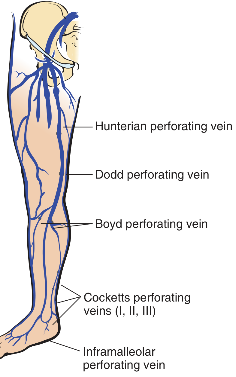
Fig. Locations of the most important perforating veins associated with the great saphenous system. Note the Cockett's and inframalleolar perforating veins are actually separate from the great saphenous system. - Pfenninger and Fowler's Procedures for Primary Care
1. Cockett's Perforators (Posterior Tibial Perforators)
| Feature | Detail |
|---|---|
| Modern name | Posterior tibial perforators |
| Location | Mid-to-distal third of the medial leg, posteromedial aspect |
| Number | Three in number: Cockett I, II, and III |
| Heights above medial malleolus | Cockett I: ~7 cm; Cockett II: ~12 cm; Cockett III: ~18 cm |
| Connection | Posterior accessory GSV of the leg (posterior arch vein / vein of Leonardo) --> posterior tibial veins |
| Key point | These are NOT direct perforators of the GSV itself - they connect the posterior arch vein to the deep system |
| Clinical significance | Most common site of incompetent perforators causing venous stasis ulcers, typically just above the medial malleolus. Stasis ulcers tend to occur at Cockett's perforator site. |
2. Boyd's Perforator (Paratibial Perforator)
| Feature | Detail |
|---|---|
| Modern name | Paratibial perforator |
| Location | Upper third of the medial leg, just below the knee - approximately 10 cm below the tibial plateau / at the level of the tibial tuberosity |
| Connection | GSV (or its tributaries) --> posterior tibial veins |
| Key point | Constantly present, but may drain the saphenous vein itself OR its tributaries. It is the most proximal leg perforator |
| Clinical significance | Important cause of medial calf varicosities and incompetence below the knee |
3. Adductor Canal / Dodd's Perforators (Femoral Canal Perforators - Distal Thigh)
| Feature | Detail |
|---|---|
| Modern name | Perforators of the femoral canal (distal group) |
| Location | Distal third of the medial thigh, at the level of the adductor canal (Hunter's canal), approximately 8-12 cm above the knee |
| Connection | GSV --> femoral vein (through the roof of the adductor canal) |
| Key point | Pass through the adductor magnus aponeurosis. These are in the distal thigh |
| Clinical significance | Incompetence causes thigh varicosities; important to identify pre-operatively |
4. Hunterian Perforators (Femoral Canal Perforators - Mid Thigh)
| Feature | Detail |
|---|---|
| Modern name | Perforators of the femoral canal (proximal/mid group) |
| Location | Middle third of the medial thigh |
| Connection | GSV --> femoral vein in the mid-thigh |
| Key point | Dodd and Hunterian are sometimes grouped together as "femoral canal perforators" - Dodd = distal thigh, Hunterian = mid thigh |
| Clinical significance | Can be the highest point of saphenous reflux when the saphenofemoral junction is competent |
Summary Table
| Perforator | Old Name | Modern Name | Location | Connection |
|---|---|---|---|---|
| Cockett I, II, III | Cockett perforators | Posterior tibial perforators | Mid-distal medial leg (7, 12, 18 cm above medial malleolus) | Posterior arch vein --> posterior tibial veins |
| Boyd | Boyd perforator | Paratibial perforator | Upper medial leg (just below knee, ~10 cm below tibial plateau) | GSV/tributaries --> posterior tibial veins |
| Dodd | Adductor canal perforator | Femoral canal perforator (distal) | Distal third of medial thigh (~8-12 cm above knee) | GSV --> femoral vein |
| Hunterian | Hunterian perforator | Femoral canal perforator (mid) | Middle third of medial thigh | GSV --> femoral vein |
Physiology and Clinical Relevance
- All perforators normally carry flow superficial to deep (outward flow = incompetent/pathological)
- During calf muscle contraction, perforator valves close, preventing flow into the superficial system
- During calf muscle relaxation, perforator valves open, allowing flow from superficial into deep
- Incompetent perforators transmit high ambulatory venous pressure to the superficial system, causing venous hypertension, lipodermatosclerosis, and ulceration
- The medial malleolar region (Cockett's zone) is the most common site for venous stasis ulcers due to high perforator incompetence
Sources:
- Schwartz's Principles of Surgery, 11th Ed., "Lower Extremity Veins"
- Fitzpatrick's Dermatology, Table 212-2, "Perforating Veins by Location"
- Pfenninger and Fowler's Procedures for Primary Care, Fig. 92-4, "Cockett's, Boyd, Dodd, Hunterian Perforating Veins"
- Dermatology 2-Volume Set 5e, Fig. 155.4, "Major sites of perforator veins"
Differenciate btw hydrocele and inguinal hernia
Searching sources...
Finding Sources
Finding Sources
Reading File
Reading File
Reading File
Reading File
Finding Sources
Finding Sources
Loading Image
Finding Sources
Reading File
I now have comprehensive information from multiple authoritative surgical textbooks. Here is the full differentiation:
Hydrocele vs. Inguinal Hernia - Differential Diagnosis
Both conditions present as groin/scrotal swellings and share a common embryological basis (patent processus vaginalis), making their differentiation clinically important.
Types of Hydrocele (Quick Review)
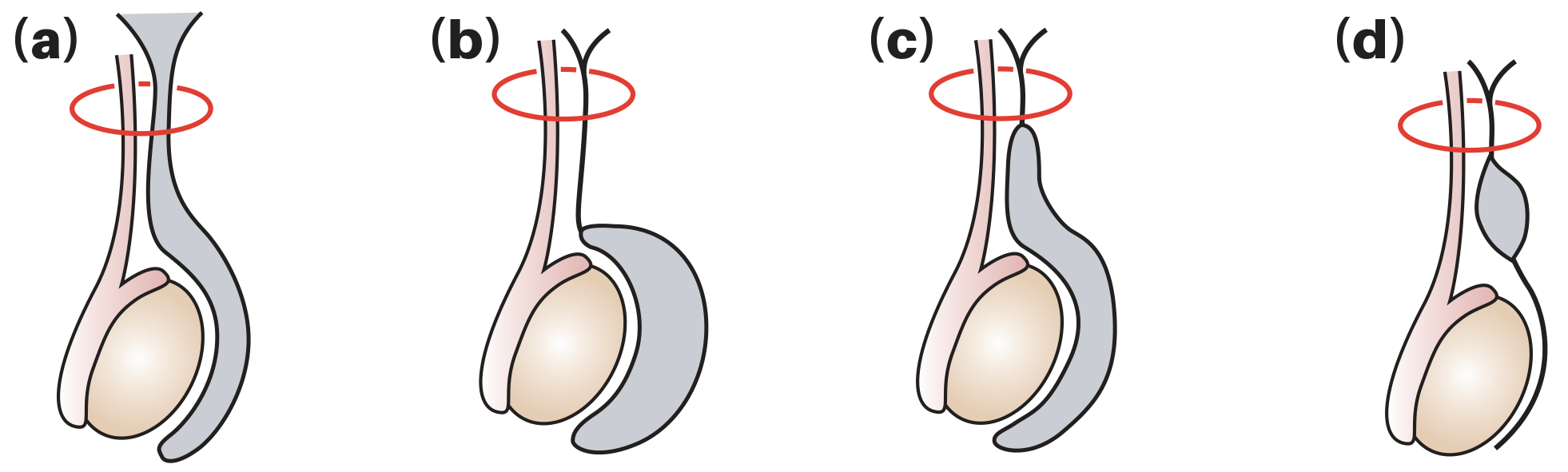
(a) Normal - (b) Vaginal hydrocele (very common) - (c) Congenital/communicating hydrocele - (d) Hydrocele of the cord. Red ring = internal inguinal ring. - Bailey & Love's Surgery, 28th Ed.)
Comparison Table
| Feature | Hydrocele | Inguinal Hernia |
|---|---|---|
| Definition | Abnormal collection of serous fluid in the tunica vaginalis or along the processus vaginalis | Protrusion of abdominal contents (bowel, omentum, ovary) through the inguinal canal |
| Contents | Fluid only | Bowel, omentum, or other viscera |
| Onset | Usually gradual; congenital type may be intermittent | May be sudden (on straining) or gradual |
| Pain | Usually painless | Usually painless unless incarcerated/strangulated |
| Shape | Smooth, oval, tense cystic | Elongated, follows the inguinal canal; may extend into scrotum |
Key Clinical Examination Signs
1. "Getting Above the Swelling" (Most Important Sign)
| Hydrocele | Inguinal Hernia | |
|---|---|---|
| Can you get above the swelling? | YES - you can palpate a normal spermatic cord above the swelling | NO - the swelling continues upward into the inguinal canal; you cannot get above it |
"If it is possible to get above the swelling to palpate a normal cord - it is NOT an inguinal hernia that has entered the scrotum." - Bailey & Love's, 28th Ed.
2. Transillumination
| Hydrocele | Inguinal Hernia | |
|---|---|---|
| Result | Brilliantly transilluminates (fluid-filled, translucent) | Does NOT transilluminate (solid/gas-filled bowel) |
| Caveat | Caution in infants: A thin-walled herniated loop of bowel in infants/children CAN transilluminate, making this sign unreliable in the pediatric age group |
3. Testis and Epididymis Palpability
| Hydrocele | Inguinal Hernia | |
|---|---|---|
| Testis palpable? | Impalpable - the hydrocele encloses both testis and epididymis so they cannot be felt separately | Palpable separately from the swelling |
4. Reducibility
| Hydrocele | Inguinal Hernia | |
|---|---|---|
| Reducible? | Non-reducible (non-communicating type) | Reducible - contents can be pushed back into abdomen (unless incarcerated) |
| Communicating hydrocele | May reduce when the child lies flat (fluid drains into peritoneum) | Reduces on lying down or manual pressure |
5. Impulse on Coughing
| Hydrocele | Inguinal Hernia | |
|---|---|---|
| Cough impulse | Absent (fluid does not respond to increased intra-abdominal pressure) | Present - a visible/palpable expansile impulse is felt on coughing |
6. Percussion Note
| Hydrocele | Inguinal Hernia | |
|---|---|---|
| Percussion | Dull (fluid-filled) | Resonant (if enterocele - bowel) or Dull (if epiplocele - omentum) |
7. Bowel Sounds
| Hydrocele | Inguinal Hernia | |
|---|---|---|
| Auscultation | Absent | May be present (bowel sounds audible over the swelling if intestine is the content) |
8. Effect of Posture / Size Change
| Hydrocele | Inguinal Hernia | |
|---|---|---|
| Change with posture | No change (non-communicating); may decrease on lying down (communicating) | Disappears or reduces in size on lying down (reducible hernia) |
9. Traction Test (for Hydrocele of the Cord)
- Pull the testis downward - a hydrocele of the cord descends slightly and becomes fixed (moves with the cord)
- An inguinal hernia does not behave this way
Special Situations
| Scenario | Points |
|---|---|
| Congenital hydrocele vs. indirect inguinal hernia | Both arise from a patent processus vaginalis. In a hernia, the processus is wide enough for bowel to enter; in communicating hydrocele, only fluid passes through the small opening |
| Acute hydrocele | Can be associated with epididymitis, torsion, or testicular tumor - ultrasound is mandatory. Do NOT aspirate if tumor is suspected (risk of malignant needle-track implantation) |
| Strangulated hernia | Presents with an irreducible, painful, tender scrotal/inguinal swelling with absent cough impulse, vomiting, and bowel obstruction - a surgical emergency |
| Silk glove sign | In children with inguinal hernia (even without visible swelling), rubbing the empty hernial sac between fingers produces a "silk rubbing on silk" sensation |
Investigation
| Hydrocele | Inguinal Hernia | |
|---|---|---|
| Ultrasound | Confirms anechoic fluid around testis; excludes testicular pathology | Identifies bowel/omentum in canal; Doppler shows peristalsis |
| Transillumination | Positive (with caveats above) | Negative |
| Role | Ultrasound is "almost always a useful adjunct" to clarify scrotal swellings | Diagnosis is primarily clinical; US confirms in equivocal cases |
Management Summary
| Hydrocele | Inguinal Hernia | |
|---|---|---|
| Congenital/pediatric | Herniotomy (ligation of patent processus vaginalis); observe until 18 months as most resolve spontaneously | Herniotomy (repair as soon as diagnosed due to incarceration risk) |
| Adult - primary | Small ones: observe. Large/bothersome: Lord's operation (plication), eversion (Jaboulay), or excision | Herniorrhaphy (open or laparoscopic mesh repair) |
| Secondary hydrocele | Treat underlying cause first | - |
Sources:
- Bailey & Love's Short Practice of Surgery, 28th Ed., "Hydrocele," p. 1586-1587
- Sabiston Textbook of Surgery, "Hernias," p. 2696
- S Das Manual on Clinical Surgery, 13th Ed., Inguinal Hernia examination
- Pfenninger & Fowler's Procedures for Primary Care, "Venous Anatomy and Pathology"
This is a shared conversation. Sign in to Orris to start your own chat.