
Events occurring at the bare area of liver
bare area of liver anatomy diagram peritoneum
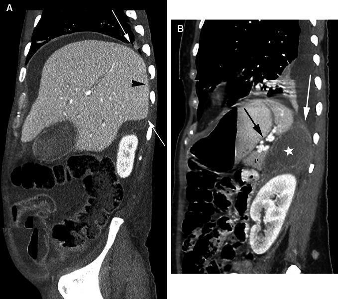
Two sagittal CT images demonstrating the anatomy and pathways of the extraperitoneal spaces in the upper abdomen. Image A shows the right upper quadrant where peritoneal fluid surrounds most of the liver. White arrows indicate the superior and inferior peritoneal reflections. The area posterior and medial to these reflections, known as the 'bare area' of the liver (black arrowhead), is devoid of peritoneum and directly abuts the diaphragm. Image B illustrates the left side, showing a hypodense fluid collection (asterisk) within the left perinephric space. This collection extends superiorly to contact the diaphragm (white arrow) and is positioned posterior to the pancreas (P) and the splenorenal ligament (black arrow). These images are pedagogically used to highlight how anatomical boundaries like the bare area and renal fascia determine the spread of intraperitoneal versus extraperitoneal fluid or disease.
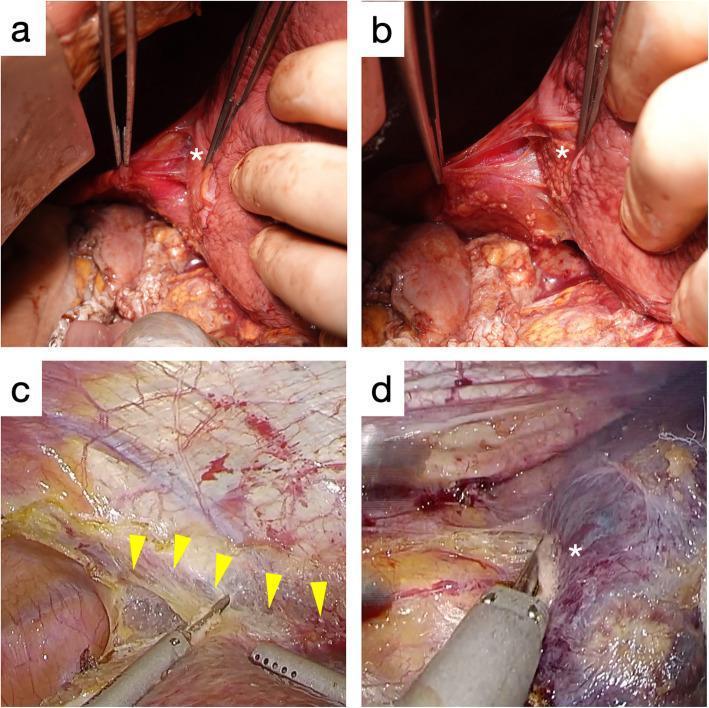
This composite figure presents clinical photographs and intraoperative images comparing dissection layers of the liver's bare area. Panels (a) and (b) show cadaveric anatomy; (a) demonstrates the liver surface (*) covered in dense, glossy white connective tissue when dissected at the outer layer of the fused peritoneum (near the diaphragm), while (b) reveals a 'naked' or exposed liver parenchyma (*) when dissected at the inner layer (near the liver). Panels (c) and (d) provide intraoperative laparoscopic views. Panel (c) highlights the transitional line between the outer and inner peritoneal layers (yellow arrowheads) during a partial hepatectomy for hepatocellular carcinoma. Panel (d) shows the exposure of the liver surface (*) covered only by thin connective tissue during dissection of the inner layer for colorectal liver metastasis. These images illustrate the surgical importance of recognizing the multilayered structure of the fused peritoneum to ensure appropriate oncological margins and safe dissection planes during laparoscopic hepatic surgery.
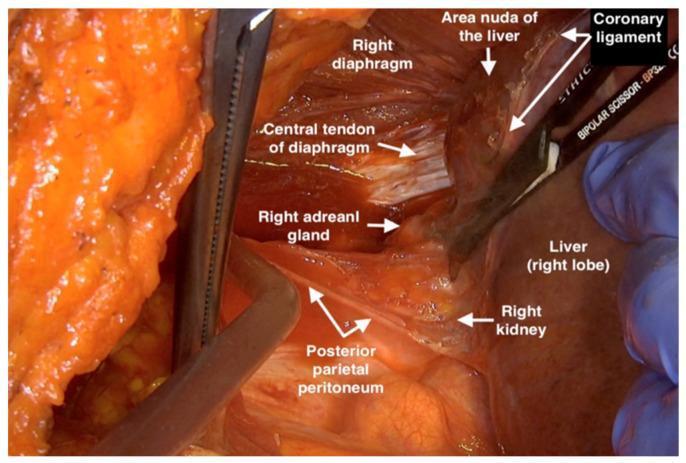
This clinical photograph captures an intraoperative view of the right upper abdominal quadrant during a surgical dissection, specifically showing the mobilization of the liver. The image demonstrates the anatomical relationship between the right diaphragm, liver, and retroperitoneal structures. Key labeled landmarks include the striated right diaphragm and its whitish, fibrous central tendon. The liver's right lobe is retracted, exposing the coronary ligament and the 'area nuda' (bare area) of the liver. In the retroperitoneal space, the yellowish right adrenal gland and the superior aspect of the right kidney are visible. The posterior parietal peritoneum is seen as a thin, translucent membrane being dissected. Surgical instruments, including bipolar scissors and retractors, are positioned to provide exposure of the diaphragmatic area. This visual serves as an educational resource for understanding surgical anatomy and the planes of dissection required for liver mobilization and posterior parietal peritonectomy.
bare area of liver anatomy clinical significance events
Bare Area of the Liver - Events and Clinical Significance
Anatomy of the Bare Area
- Formed embryologically because the liver contacts the diaphragm before peritoneum can invest that surface (Langman's Medical Embryology)
- Enclosed between the anterior (superior) and posterior (inferior) leaves of the coronary ligament
- The two leaves of the coronary ligament converge at the right triangular ligament on the right, and at the left triangular ligament on the left
- The inferior vena cava (IVC) runs in a groove at the medial end of the bare area - Sleisenger & Fordtran's, p. 959
Events Occurring at the Bare Area
1. Portacaval (Portosystemic) Anastomosis
"Other regions where portal and caval systems interconnect include: where the liver is in direct contact with the diaphragm (the bare area of the liver)..." - Gray's Anatomy for Students, p. 964
2. Lymphatic Drainage - Direct Route to Thorax
- Phrenic (inferior diaphragmatic) lymph nodes, OR
- Deep lymphatics that accompany the hepatic veins converging on the IVC, passing with the IVC through the diaphragm into the posterior mediastinal lymph nodes
"Superficial lymphatics from the posterior aspects of the diaphragmatic and visceral surfaces of the liver drain toward the bare area of the liver into the phrenic lymph nodes or join deep lymphatics that have accompanied the hepatic veins converging on the inferior vena cava and pass with the inferior vena cava through the diaphragm to drain into the posterior mediastinal lymph nodes." - Fischer's Mastery of Surgery, p. 4784
3. Spread of Infection / Inflammation
- Subphrenic or hepatic abscesses at the bare area can spread directly into the thoracic cavity (empyema, pleuropulmonary involvement)
- Conversely, diaphragmatic infections or right-sided pleural disease can spread downward to the liver through this zone
- This explains why amoebic liver abscess of the right lobe can rupture into the right pleural cavity (the right lobe's bare area directly abuts the right diaphragm)
- Right subphrenic space is divided by the bare area into right anterior subphrenic space and right posterior subphrenic space (Morrison's pouch)
4. Traumatic / Surgical Hemorrhage
- Bleeding is harder to detect on FAST ultrasound (which looks for intraperitoneal fluid)
- Mobilization of the liver during surgery requires careful dissection of the bare area to avoid injury to the hepatic veins and suprahepatic IVC - Mulholland & Greenfield's Surgery, p. 3187; Fischer's Mastery, p. 4489
5. Relationship to the Right Adrenal Gland and IVC
- The right adrenal gland is directly posterior to the bare area - its superior part contacts the bare area of the liver anteriorly, which is why right adrenalectomy requires careful dissection here (Fischer's Mastery, p. 2611)
- The IVC lies in a groove at the medial border of the bare area and receives the major hepatic veins (right, middle, left) here - this is the zone of hepatic venous outflow
- Multiple small retrohepatic veins also enter the IVC along this course, creating surgical hazard during hepatic resections
6. Embryological Significance
Summary Table
| Event | Mechanism | Clinical Relevance |
|---|---|---|
| Portacaval anastomosis | Portal veins communicate with phrenic/diaphragmatic systemic veins | Collateral formation in portal hypertension |
| Lymphatic drainage to mediastinum | Lymphatics pass through diaphragm with IVC | Metastatic spread of liver cancer to thorax; pleural effusions in cirrhosis |
| Spread of infection to thorax | No peritoneal barrier between abdomen and thorax | Amoebic abscess → empyema; hepatic abscess → pleuropulmonary fistula |
| Retroperitoneal hemorrhage | No peritoneum covering bare area | Trauma/surgery here causes retroperitoneal, not intraperitoneal bleed |
| IVC and hepatic vein relations | IVC runs in groove at medial end; hepatic veins drain here | Major surgical hazard during hepatectomy/liver mobilization |
| Right adrenal contact | Right adrenal gland lies directly posterior | Involved in right adrenalectomy; secondary spread in hepatic/adrenal disease |
- Gray's Anatomy for Students - Portacaval anastomoses section
- Fischer's Mastery of Surgery (8th ed.) - Lymphatic Drainage; Peritoneal Reflections of Diaphragm to Liver
- Sleisenger & Fordtran's Gastrointestinal and Liver Disease - Anatomy
- Mulholland & Greenfield's Surgery (7th ed.) - Topographic and Surface Anatomy
- Langman's Medical Embryology - Liver and Gallbladder development
- Wikipedia - Bare area of the liver