
PENG block and QL block Anesthesia md final exam structured question
PENG block QL block anesthesia MD final exam structured question answer 2025
PENG block pericapsular nerve group hip ultrasound anatomy
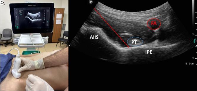
This composite educational image illustrates the Pericapsular Nerve Group (PENG) block, a regional anesthesia technique for hip analgesia. Panel A shows the clinical setup, including the patient’s supine position and the transverse orientation of a curved-array ultrasound probe at the level of the inguinal crease. Panel B presents the corresponding sonographic anatomy. Key landmarks include the hyperechoic bony contours of the anterior inferior iliac spine (AIIS) and the iliopubic eminence (IPE). The psoas tendon (PT) is identified as a hypoechoic oval structure resting between these bony prominences. The femoral artery (FA) is visualized medially as an anechoic, pulsatile vessel. A dashed red arrow demonstrates the in-plane needle trajectory, targeting the fascial plane between the psoas tendon and the pubic ramus. This block specifically targets the articular branches of the femoral nerve, obturator nerve, and accessory obturator nerve to provide sensory blockade of the anterior hip capsule without significant motor impairment.
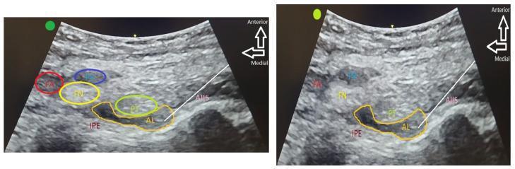
Diagnostic ultrasound images illustrating the Pericapsular Nerve Group (PENG) block for regional anesthesia. The composite contains two panels showing the cross-sectional anatomy of the anterior hip region. The left panel identifies key landmarks: the femoral artery (FA), femoral vein (FV), and femoral nerve (FN) located medially and superiorly. Deep to these lie the psoas tendon (PT), iliopubic eminence (IPE), and anterior inferior iliac spine (AIIS). The right panel demonstrates the clinical implementation of the block, where hypoechoic local anesthetic (AL) is seen spreading within the fascial plane between the psoas tendon and the underlying bony cortex (IPE and AIIS). The images highlight the anatomical plane containing sensory branches of the femoral, obturator, and accessory obturator nerves targeted for hip fracture analgesia. Orientation arrows indicate anterior and medial directions, emphasizing the deep approach required to reach the periarticular space while avoiding superficial neurovascular structures.
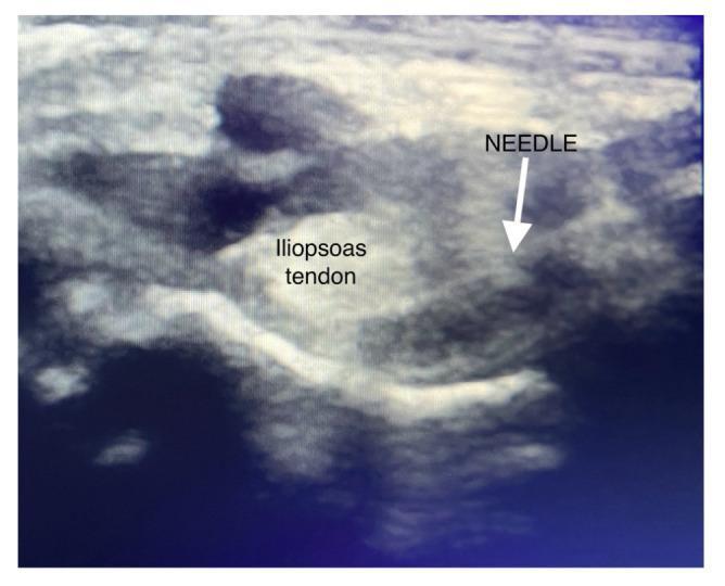
This diagnostic ultrasound image demonstrates the technique for a PENG (Pericapsular Nerve Group) block in the hip region. The image shows a transverse view of the pelvic anatomy, focusing on the space between the anterior inferior iliac spine (AIIS) and the iliopubic eminence. The iliopsoas tendon is clearly identified as a prominent, hyperechoic (bright), oval-shaped structure with a homogeneous internal texture. A needle tip is visualized and labeled, positioned laterally to the iliopsoas tendon. The needle path enters from a lateromedial direction to target the fascial plane beneath the tendon, adjacent to the bone surface of the ilium. This modified technique is intended for regional anesthesia to provide analgesia for hip fractures or surgeries while minimizing quadriceps motor weakness by depositing local anesthetic away from the femoral nerve.
quadratus lumborum block QL ultrasound shamrock view anatomy
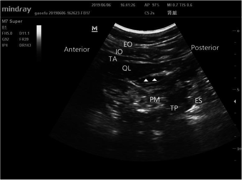
This diagnostic ultrasound image demonstrates the 'Shamrock sign' anatomy used for a transmuscular quadratus lumborum (TQL) block. The sonogram shows a cross-sectional view of the posterior abdominal wall with clearly labeled anatomical structures. From anterior to posterior (left to right), the external oblique (EO), internal oblique (IO), and transversus abdominis (TA) muscles are visible as distinct superficial layers. Deep to these, the quadratus lumborum (QL) muscle is positioned adjacent to the psoas major (PM) muscle. The transverse process (TP) of the lumbar vertebra and the erector spinae (ES) muscle are situated posteriorly. White arrowheads point to a hypoechoic region between the QL and PM muscles, representing the interfascial plane where local anesthetic injectate spreads during the procedure. The image illustrates the 'Shamrock' configuration formed by the QL, PM, and ES muscles surrounding the transverse process, providing a critical landmark for anesthesiologists to perform regional anesthesia for postoperative pain management.
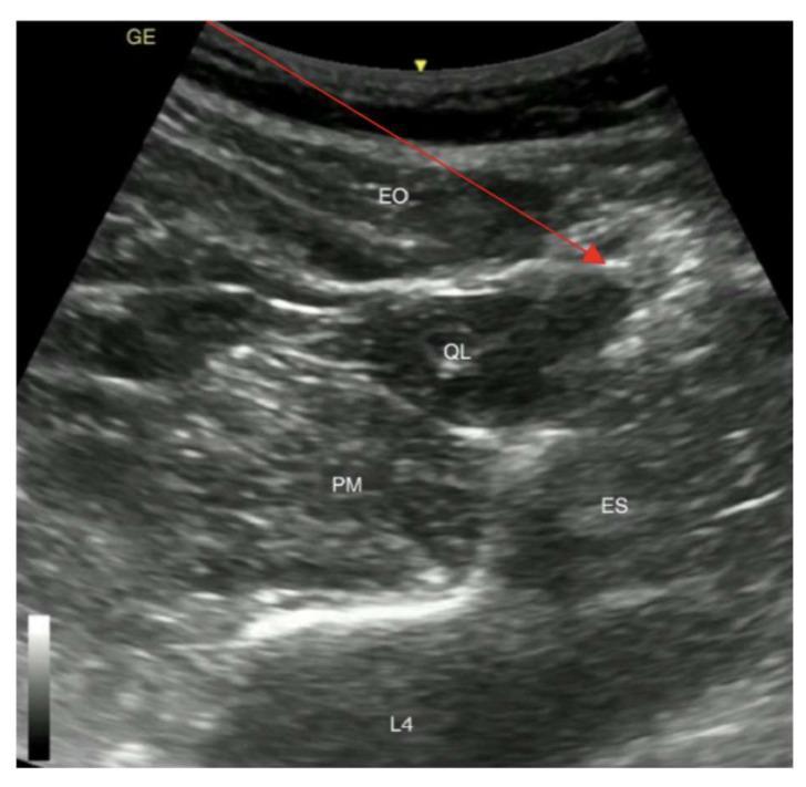
This diagnostic ultrasound image demonstrates the regional anatomy for a posterior Quadratus Lumborum Block (QLB type 2). The sonogram shows a transverse view of the abdominal wall and paraspinal musculature at the level of the fourth lumbar vertebra (L4). Key anatomical structures are labeled, including the superficial external oblique (EO) muscle, the quadratus lumborum (QL) muscle, the psoas major (PM) muscle situated anteriorly, and the erector spinae (ES) muscle group located posteriorly. The hyperechoic shadow of the L4 vertebral body serves as a deep posterior landmark. A yellow arrow identifies the patient's skin surface. A red arrow illustrates the planned needle trajectory for a posterior QLB, originating from the skin, traversing the EO, and targeting the interfacial plane on the posterior aspect of the QL muscle (the 'shamrock sign' area). This image is an essential educational resource for regional anesthesia, illustrating the sonographic landmarks required for safe and effective truncal nerve blocks.
PENG Block and QL Block - MD Final Exam Structured Answer
PENG (PERICAPSULAR NERVE GROUP) BLOCK
Definition / Introduction
- Miller's Anesthesia, 10e
- Morgan & Mikhail's Clinical Anesthesiology, 7e
Anatomy and Nerve Targets
| Nerve | Contribution |
|---|---|
| Femoral nerve (articular branches) | Primary target |
| Obturator nerve (articular branches) | Primary target |
| Accessory obturator nerve | Primary target |
Indications
- Hip fracture analgesia (most evidence - emergency and preoperative)
- Total hip arthroplasty (THA) - perioperative analgesia
- Hip arthroscopy
- Patients where motor preservation is important (early mobilization, falls risk)
- Multimodal opioid-sparing analgesia
Technique
- Anterior inferior iliac spine (AIIS) - laterally
- Iliopubic eminence (IPE) - medially/deep
- Iliopsoas muscle and its tendon (between AIIS and IPE)
- Femoral artery (medially, as safety landmark)
- Superior pubic ramus (deep)
Advantages over Femoral Nerve Block / FIC Block
| Feature | PENG Block | Femoral Nerve Block |
|---|---|---|
| Motor sparing | Yes (motor-sparing) | No (quadriceps weakness) |
| Mobility post-op | Preserved | Impaired |
| Target | Articular branches only | Entire femoral nerve trunk |
| Falls risk | Lower | Higher |
| Coverage | Anterior capsule | Anterior thigh + anterior capsule |
Limitations and Complications
- Does not cover the posterior hip capsule or lateral thigh (LFCN often needs to be blocked separately)
- Intramuscular injection or large volume (>20 mL) can cause femoral nerve blockade and anterior thigh / quadriceps weakness
- Proximity to the LFCN during skin infiltration can injure it
- Standard block risks: infection, hematoma, local anesthetic systemic toxicity (LAST)
- Block failure
- Evidence still evolving - initially only case series; RCTs now emerging
Clinical Pearl
QL (QUADRATUS LUMBORUM) BLOCK
Definition / Introduction
- Miller's Anesthesia, 10e
- Barash Clinical Anesthesia, 9e
- Morgan & Mikhail's Clinical Anesthesiology, 7e
Anatomy
- Mechanoreceptors
- Nociceptors
- Sympathetic fibers
- Psoas major (anterior leaf)
- Quadratus lumborum (lateral leaf)
- Erector spinae (posterior leaf)
- Transverse process = the "stem"
Types of QL Block
| Type | Injection Plane | Approach |
|---|---|---|
| QL1 (Lateral) | Between transversus abdominis muscle and QL (lateral aspect of QL) | Anterior-to-posterior or lateral |
| QL2 (Posterior) | Between QL and erector spinae (posterior aspect of QL), at lumbar interfascial triangle (LIFT) | Posterior-to-anterior |
| QL3 (Anterior / Transmuscular) | Between QL and psoas major (anterior QL plane) | Posterior-to-anterior through QL |
- QL3 (anterior/transmuscular) provides the widest spread and is most relevant for hip surgery; however, it has highest risk of LA spreading to fascia iliaca causing quadriceps weakness.
Indications
- Abdominal surgery - laparoscopic, open abdominal procedures (renal surgery, cesarean section, bowel surgery, herniorrhaphy)
- Hip surgery - total hip arthroplasty, hip fracture surgery (especially via anterior QL block)
- Flank incisions
- Alternative to TAP block when visceral analgesia is needed (TAP block does not provide visceral analgesia; QL block does via paravertebral spread)
- Opioid-sparing multimodal analgesia
Technique
- Place probe transversely on the midaxillary line, between the costal margin and iliac crest - identify the 3 muscle layers of the TAP
- Slide the probe posteriorly above the iliac crest until the transversus abdominis tapers and QL becomes visible
- Continue sliding posteriorly until the transverse process comes into view - this gives the shamrock sign
Dermatomal Coverage
Advantages over TAP Block
| Feature | QL Block | TAP Block |
|---|---|---|
| Dermatomal coverage | T8-L1 | T10-L1 (variable) |
| Visceral analgesia | Yes (via paravertebral spread) | No |
| Hip analgesia | Yes (anterior QL3) | Limited |
| Technical difficulty | Higher (deeper) | Moderate |
Complications and Safety
- Kidney puncture - most significant risk; IP needle technique and US guidance are mandatory
- Bowel and peritoneal puncture
- Fourth lumbar artery injury → hematoma (especially with anticoagulation)
- Quadriceps weakness with anterior QL block (LA spreads to fascia iliaca)
- LAST
- Block failure
PENG vs QL BLOCK - COMPARATIVE SUMMARY (High-Yield for Exam)
| Feature | PENG Block | QL Block |
|---|---|---|
| Primary target | Articular branches of femoral, obturator, accessory obturator nerves (anterior hip capsule) | Thoracolumbar fascia around QL muscle |
| Coverage | Anterior hip capsule | T8-L1 somatic + visceral |
| Motor sparing | Yes (goal) | Yes, but QL3 can cause quadriceps weakness |
| Main indication | Hip fracture, THA, hip arthroscopy | Abdominal/flank/hip surgery |
| Probe | Curvilinear (along inguinal crease) | Curvilinear (transverse, midaxillary) |
| Needle endpoint | Deep to psoas tendon, on pubic ramus (AIIS-IPE plane) | QL1: lateral to QL / QL2: posterior to QL / QL3: QL-psoas plane |
| LA volume | 20 mL (max) | 15-20 mL per side |
| Visceral analgesia | No | Yes |
| Guidance modality | Ultrasound only | Ultrasound only (no NS) |
| Key risk | Femoral nerve spread if intramuscular / volume >20 mL | Kidney/peritoneal puncture; quadriceps weakness (QL3) |
| Mnemonic / sign | AIIS + IPE + psoas tendon = target plane | Shamrock sign / PQRST |
| Evidence in THA | PENG superior postop analgesia vs anterior QL in hip fracture surgery (2025 RCT) | Broader dermatomal coverage than PENG; useful in THA |
Recent Evidence (2025)
- PENG block provided superior postoperative analgesia vs anterior QL block
- Tramadol consumption was significantly lower in the PENG group
- Time to first rescue analgesia was significantly longer with PENG
Exam Tips (Structured Answer Format)
- Definition (1 mark)
- Anatomy / nerve targets (2 marks)
- Indications (1 mark)
- Technique - position, probe, landmarks, needle, drug (3 marks)
- Advantages / clinical significance (2 marks)
- Complications (1 mark)