
I have my university exam of 1 st year of subject biochemistry These are the important question give me answer of this "Alkaptonuria" and remeber it's a university examination The answer should be easy to memorize and follow the pattern of answer writing as in University paper
Reading File
Now let me search the medical library for Alkaptonuria content.
Finding Sources
Finding Sources
Excellent! Let me read the most relevant sources - Lippincott Biochemistry (the standard 1st year text) and Harper's Biochemistry, plus Harrison's for clinical details.
Reading File
Reading File
Reading File
Let me read the rest of the Harrison's section on Alkaptonuria and also fetch the relevant biochemical pathway image.
Reading File
Loading Image
I now have all the information needed to write a comprehensive university-exam-style answer on Alkaptonuria. Let me also quickly get the tyrosine metabolic pathway image from Lippincott.
Finding Sources
I have all the content needed. Now let me write the full university-style answer.
ALKAPTONURIA
Definition
Alkaptonuria is a rare, autosomal recessive inborn error of metabolism characterized by deficiency of the enzyme homogentisate 1,2-dioxygenase (homogentisic acid oxidase), leading to accumulation and excretion of homogentisic acid (HGA) in urine and tissues.
- Frequency: approximately 1 in 250,000 births
- It was one of the first diseases described by Sir Archibald Garrod (1902) as an "inborn error of metabolism"
Biochemical Basis / Enzyme Defect
Alkaptonuria occurs in the tyrosine degradation pathway:
Phenylalanine → Tyrosine → p-Hydroxyphenylpyruvate → Homogentisic Acid (HGA)
                                                              ↓ ← ENZYME DEFECT HERE
                                                 Homogentisate 1,2-dioxygenase (BLOCKED)
                                                              ↓
                                                   Maleylacetoacetate
                                                              ↓
                                                  Fumarylacetoacetate
                                                              ↓
                                               Fumarate + Acetoacetate
Deficient enzyme: Homogentisate 1,2-dioxygenase (homogentisic acid oxidase)
- Because this enzyme is blocked, HGA accumulates in the body and is excreted in large amounts in urine
- HGA is oxidized by atmospheric oxygen to form a dark brown-black pigment (alkapton bodies)
Clinical Features (The Classic Triad)
| Feature | Description |
|---|---|
| 1. Homogentisic Aciduria | Dark urine on standing or on addition of alkali; diapers may appear dark in infants |
| 2. Ochronosis | Deposition of black/bluish-black pigment in cartilage, ear cartilage, sclera, tendons, and other collagenous tissues |
| 3. Arthritis | Degenerative arthritis of large joints (spine, hips, knees), usually appears in the 3rd-4th decade |
Additional features:
- Dark staining of diapers may be the first sign in infants
- Gray-brown pigmentation of the sclera and ear (concha, anthelix)
- Cardiovascular involvement in severe cases (pigment deposition in heart valves and coronary arteries)
- Usually asymptomatic until ~40 years of age; men affected twice as often as women (2:1)
Pathogenesis of Ochronosis
- Excess HGA accumulates in blood and tissues
- HGA is oxidized to benzoquinone acetate, which polymerizes
- This polymer binds irreversibly to collagen and connective tissue, producing the characteristic dark pigment
- Pigment deposition in cartilage causes brittleness and leads to severe degenerative joint disease
Diagnosis
| Test | Finding |
|---|---|
| Urine examination | Dark urine on standing; turns black on addition of NaOH (alkali test) |
| Ferric chloride test | Urine turns dark green/black |
| Benedict's test | Positive (HGA is a reducing substance) |
| Urine HGA levels | Elevated (confirmed by chromatography/mass spectrometry) |
| X-ray | Calcification and narrowing of intervertebral discs ("ochronotic spondylosis") |
Key point for exam: The urine of an alkaptonuric patient does NOT appear dark when freshly voided - it darkens only on exposure to air (due to oxidation of HGA).
Inheritance
- Autosomal Recessive
- Gene: HGD gene (encoding homogentisate 1,2-dioxygenase)
Treatment
| Approach | Details |
|---|---|
| Dietary restriction | Low phenylalanine and tyrosine diet (reduces substrate load) |
| Ascorbic acid (Vitamin C) | Acts as antioxidant; may reduce ochronosis (inhibits oxidation of HGA to pigment) |
| Nitisinone (NTBC) | Inhibits 4-hydroxyphenylpyruvate dioxygenase (upstream enzyme), reduces HGA production - emerging treatment |
| Symptomatic | Analgesics and joint replacement for arthritis |
Historical Significance
Alkaptonuria holds a special place in the history of medicine:
- Described as early as the 16th century (dark urine on standing)
- Sir Archibald Garrod (1902) used alkaptonuria to establish the concept of "inborn errors of metabolism"
- Earliest known case was detected in an Egyptian mummy dating from ~1500 B.C.!
Key Points to Remember (Memory Aid)
"HAD" = 3 cardinal features:
- Homogentisic aciduria (dark urine)
- Arthritis (degenerative, large joints)
- Dark pigment deposition = Ochronosis
Enzyme blocked = Homogentisate 1,2-dioxygenase (Think: the enzyme that "oxidizes" HGA is absent, so HGA accumulates and "oxidizes itself" in the urine and tissues)
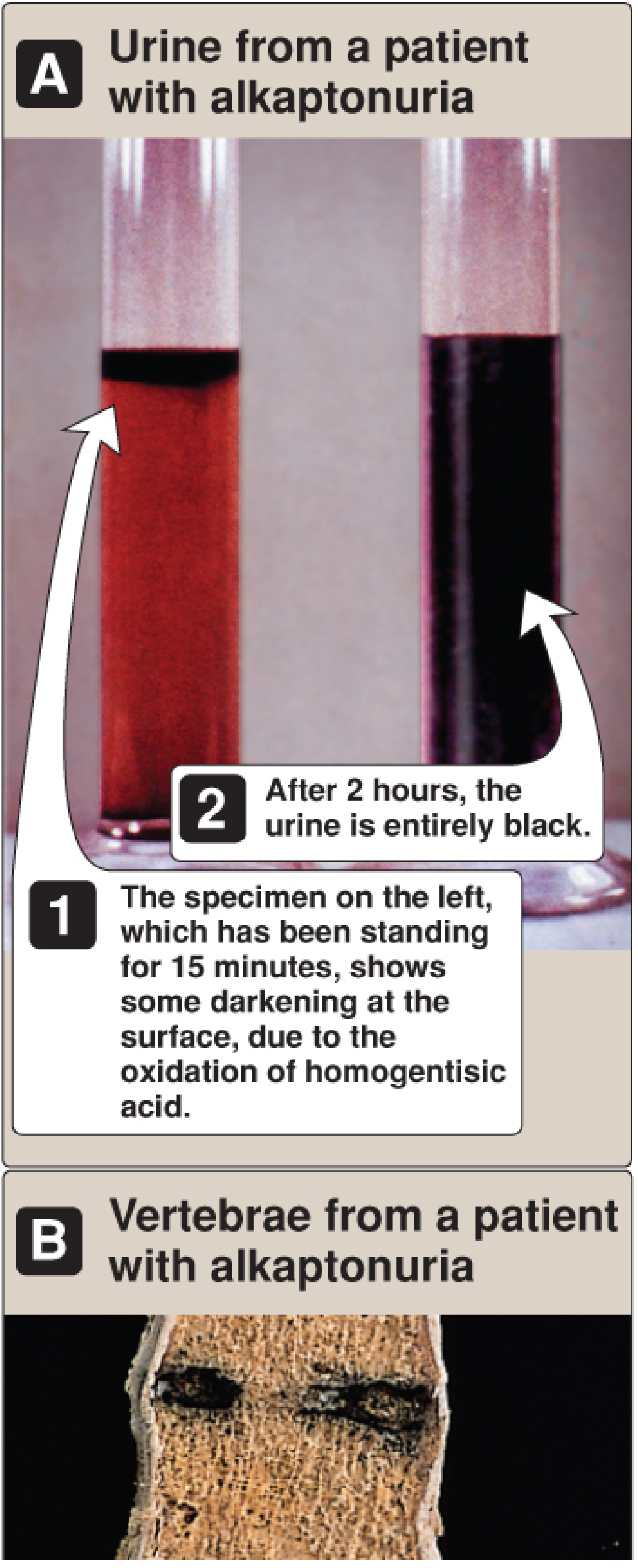
Fig: (A) Urine from a patient with alkaptonuria - darkens on standing due to oxidation of homogentisic acid. (B) Black ochronotic pigment deposited in vertebrae.
Sources: Lippincott's Illustrated Reviews: Biochemistry 8th Ed., p. 771 | Harper's Illustrated Biochemistry 32nd Ed. | Harrison's Principles of Internal Medicine 22nd Ed.
Exam Writing Tips for This Topic:
- Always start with a clear definition + enzyme defect
- Draw or describe the tyrosine metabolic pathway showing where the block occurs
- List the classic triad (the examiner always looks for all three)
- Mention autosomal recessive inheritance
- Include diagnosis (the urine darkening test is a favorite exam question)
- End with treatment (dietary restriction + nitisinone)
- The historical connection to Garrod earns bonus marks in university exams
This is a shared conversation. Sign in to Orris to start your own chat.