
Periodic paralysis
periodic paralysis channelopathy
periodic paralysis hypokalemia ECG muscle weakness
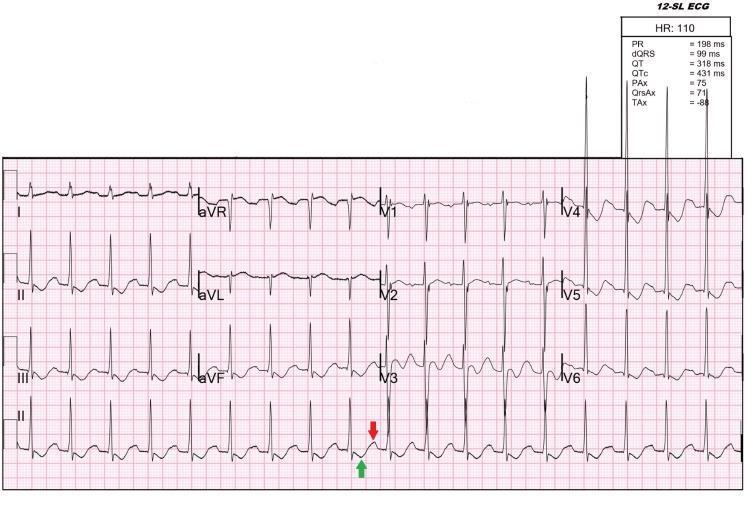
This diagnostic image is a 12-lead electrocardiogram (ECG) demonstrating classic features of severe hypokalemia in a patient with thyrotoxic periodic paralysis. The tracing shows sinus tachycardia with a heart rate of approximately 110 beats per minute. Key visible abnormalities include prominent U waves, most notably in leads V2 through V6 (indicated by a red arrow in lead V3), and T-wave inversion (marked with a green arrow in lead V3). The U waves are positive deflections that appear after the T waves, separated by a brief isoelectric period. Additionally, there are inverted T waves in the inferior leads (III and aVF) and mid-precordial leads. Automated measurements indicate a PR interval of 198 ms, QRS duration of 99 ms, and a QTc of 431 ms. The combination of tachycardia, T-wave inversion, and prominent U waves is clinically significant for identifying severe electrolyte disturbances like hypokalemia (1.7 mEq/L in this case) which can occur during thyrotoxic crisis or periodic paralysis.
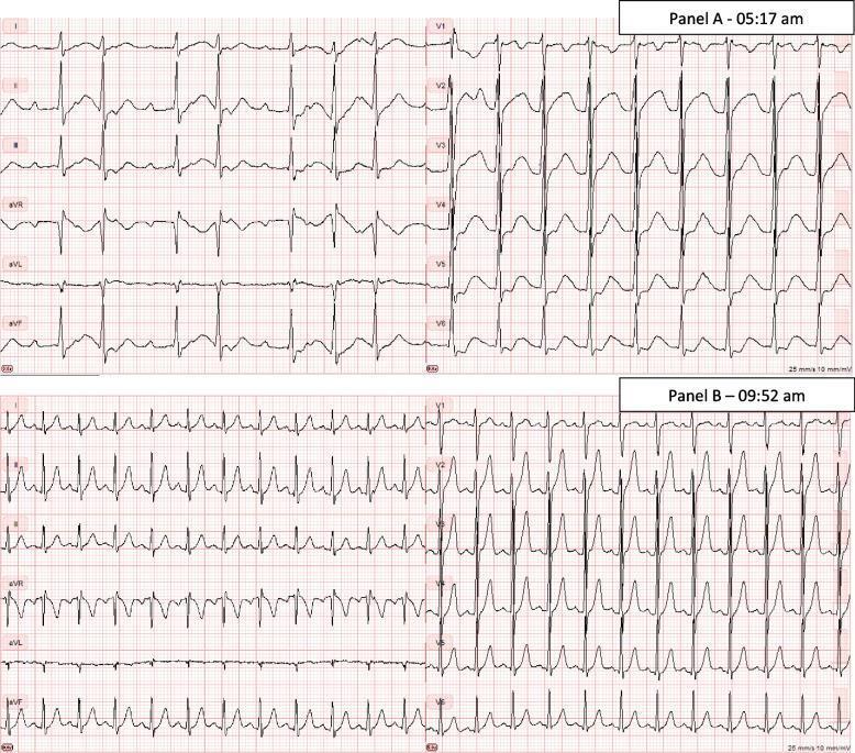
A comparison of two 12-lead electrocardiograms (ECG) labeled Panel A and Panel B, documenting the resolution of rhythm and morphological abnormalities in a patient with thyrotoxic periodic paralysis and severe hypokalemia. Panel A (on admission) demonstrates an irregular rhythm consistent with atrial flutter featuring variable conduction (1:2 and 1:3 block). Morphological markers of hypokalemia are visible, including prominent U-waves fused with T-waves and a flattened or pseudo-prolonged QT interval. Panel B, taken approximately four hours later following oral potassium chloride treatment, shows a transition to a regular sinus tachycardia. The atrial flutter waves have disappeared, replaced by distinct P-waves preceding each QRS complex. The ST-segments and T-waves have largely normalized compared to the baseline hypokalemic tracing. This side-by-side comparison illustrates the dynamic cardiac manifestations of electrolyte derangement (potassium 1.4 mM) and the rapid electrocardiographic improvement upon potassium replacement therapy.
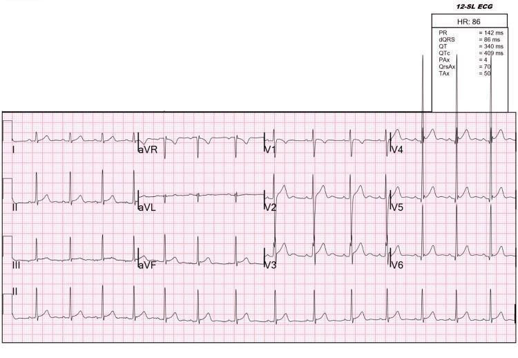
A 12-lead electrocardiogram (ECG) recorded on standard pink grid paper, demonstrating a normal sinus rhythm with a heart rate of 86 beats per minute. The tracing shows a consistent 1:1 relationship between P waves and QRS complexes. Key intervals reported in the data summary include a PR interval of 142 ms, QRS duration of 86 ms, and a corrected QT (QTc) of 409 ms. The ECG specifically illustrates the near-complete resolution of pathological U waves, which were previously associated with severe hypokalemia in a case of thyrotoxic periodic paralysis. Residual, faint U waves are still identifiable in the precordial leads, specifically V2 through V5, appearing as small positive deflections following the T waves. There are no significant ST-segment deviations or pathological Q waves. This visual is representative of cardiac repolarization changes during the recovery phase of metabolic and electrolyte disturbances related to hyperthyroidism.
Periodic Paralysis
Definition and Overview
- Muscle strength and serum potassium are usually normal between attacks
- Respiratory muscles are typically spared (though severe HypoPP can affect swallowing/breathing)
- Frequent attacks can lead to progressive, permanent weakness over time
- Hypothermia exacerbates both frequency and severity
Classification
1. Hypokalemic Periodic Paralysis (HypoPP)
| Feature | Details |
|---|---|
| Gene | CACNA1S (HypoPP Type 1, L-type Ca²⁺ channel Cav1.1) or SCN4A (HypoPP Type 2, Na⁺ channel Nav1.4) |
| Inheritance | Autosomal dominant; higher penetrance in males |
| Mechanism | ~90% of mutations cause loss of positively charged arginine in S4 voltage-sensor domains, creating an abnormal "gating pore current" that leads to paradoxical depolarization during hypokalemia. Reduced KATP channel activity + insulin-mediated Na+/K+-ATPase stimulation shifts K⁺ intracellularly |
| Serum K⁺ during attack | < 3.0 mEq/L (often markedly low) |
| No myotonia | Unlike hyperkalemic form |
| Triggers | High-carbohydrate meals, insulin, rest after exercise, high-salt meals, stress, hypothermia, pregnancy, menstruation |
| Classic presentation | Weakness on waking after a large carbohydrate-rich dinner |
- Acute: Oral KCl 40 mEq preferred; IV potassium sparingly (10 mEq over 1 hour, 1-2 doses) - caution against overtreatment since total body K⁺ is NOT depleted, only shifted
- Prophylaxis: Acetazolamide (first-line for Type 1); spironolactone for Type 2 (acetazolamide may worsen Type 2)
- Dichlorphenamide is the only FDA-approved treatment (potent carbonic anhydrase inhibitor)
- Avoid triggers
2. Hyperkalemic Periodic Paralysis (HyperPP)
| Feature | Details |
|---|---|
| Gene | SCN4A (Nav1.4 sodium channel gain-of-function) |
| Inheritance | Autosomal dominant; prevalence ~0.17 per 100,000 |
| Mechanism | Sustained abnormal inward Na⁺ current → prolonged membrane depolarization → myotonia → membrane desensitization → paralysis. Simultaneous K⁺ efflux causes hyperkalemia |
| Serum K⁺ during attack | Elevated (>5 mEq/L); >7 mEq/L between episodes suggests secondary cause |
| Associated features | Mild myotonia (often subclinical), elevated CK (5-10x normal) |
| Triggers | Potassium-rich foods, rest after exercise, cold, fasting, glucocorticoids, stress, pregnancy |
| Attack duration | Typically 15 minutes to 1 hour |
- Acute: Glucose + insulin, albuterol (promotes intracellular K⁺ shift)
- Prophylaxis: Acetazolamide (125-500 mg), mexiletine (for myotonia)
- Avoid K⁺-rich foods, strenuous exercise, fasting, cold exposure
3. Normokalemic Periodic Paralysis (NormoKPP)
- Now considered a variant of HyperPP, not a distinct disease
- Same gene (SCN4A mutation), similar clinical and laboratory features
- Paroxysmal weakness without detectable change in serum potassium
4. Thyrotoxic Periodic Paralysis (TPP)
- Classic in Asian and Hispanic men (genetic variation in KCNJ18, encoding Kir2.6 inward-rectifying K⁺ channel)
- Pathophysiology: Excess thyroid hormones overstimulate Na⁺/K⁺-ATPase (via β-adrenergic/insulin activation) → marked intracellular K⁺ shift → hypokalemia → paralysis
- Clinical picture almost identical to hereditary HypoPP, but occurs later in life
- Associated with signs/symptoms of hyperthyroidism
- Severity correlates with degree of hypokalemia, not with thyroid hormone levels
- Non-selective β-blockers (propranolol) = first-line (blocks adrenergic overstimulation of Na⁺/K⁺-ATPase)
- Antithyroid drugs (methimazole) for underlying hyperthyroidism
- Caution: large IV K⁺ supplementation risks rebound hyperkalemia and dysrhythmias
- All patients with a first hypokalemic PP episode should have thyroid function tests
5. Andersen-Tawil Syndrome
- Mutations in KCNJ2 gene (Kir2.1 inward-rectifying K⁺ channel)
- Triad: periodic paralysis + cardiac arrhythmias + dysmorphic features
- K⁺ level during attacks can be normo-, hypo-, or hyperkalemic (but consistent within families)
6. Secondary Forms
| Cause | Notes |
|---|---|
| GI/renal K⁺ loss (Gitelman, Fanconi, dRTA) | Weakness may be episodic; K⁺ levels much lower |
| Barium salt ingestion | Barium blocks K⁺ channels; treat with oral KCl + stopping exposure |
| Secondary hyperkalemia (renal disease) | K⁺ > 7 mEq/L between attacks; target primary disease |
Key Diagnostic Approach
- Serum K⁺ during attack - essential for classification
- ECG during attack:
- Hypokalemia: flat/inverted T waves, prominent U waves, prolonged QT, ventricular arrhythmias
- Hyperkalemia: peaked T waves, prolonged PR/QRS, loss of P waves, sine wave pattern
- Magnesium and glucose levels
- Thyroid function tests (all first-episode hypokalemic PP)
- EMG: HyperPP shows myotonic discharges; HypoPP usually does not
- Genetic testing: SCN4A (HyperPP/NormoKPP/HypoPP-2), CACNA1S (HypoPP-1), KCNJ2 (Andersen)
- Muscle biopsy: small peripheral sarcoplasmic vacuoles in HyperPP
Quick Comparison Table
| Feature | HypoPP | HyperPP | TPP |
|---|---|---|---|
| K⁺ during attack | Low (<3.0) | High | Low |
| Gene | CACNA1S / SCN4A | SCN4A | KCNJ18 (Kir2.6) |
| Myotonia | No | Yes (mild) | No |
| Trigger | Carbs, rest, insulin | K⁺-rich food, exercise, cold | Hyperthyroidism, carbs |
| Acute Rx | Oral/IV KCl | Glucose + insulin, albuterol | Propranolol + oral KCl |
| Prophylaxis | Acetazolamide (Type 1) / Spironolactone (Type 2) | Acetazolamide | Treat hyperthyroidism |
Anesthetic Considerations
- Succinylcholine is contraindicated in HyperPP (and probably all forms) - risk of dangerous hyperkalemia and prolonged paralysis
- Avoid glucose-containing IV fluids in HypoPP (worsens hypokalemia)
- Avoid hyperventilation (alkalosis lowers K⁺) in HypoPP
- Avoid anticholinesterases (neostigmine) in HyperPP - aggravates myotonia
- Nondepolarizing NMBs: response is unpredictable; increased sensitivity in HypoPP; monitor neuromuscular function carefully
- Maintain normothermia - hypothermia triggers attacks
- Frequent intraoperative K⁺ monitoring + ECG surveillance is mandatory
stuttering paralysis)
stuttering hemiplegia lacunar infarction
stuttering hemiplegia lacunar infarct internal capsule
stuttering hemiplegia capsular warning syndrome neurology
capsular warning syndrome treatment outcome
Stuttering Paralysis
What Is It?
Pathophysiology
- Lipohyalinosis or microatheroma of a single small penetrating vessel causes fluctuating perfusion
- Partial occlusion or thrombosis forms, dissolves, and re-forms
- Each episode produces transient ischemia in the posterior limb of the internal capsule (most commonly), corona radiata, or basis pontis
- Final complete occlusion results in a lacunar infarct
Capsular Warning Syndrome (CWS)
Three or more stereotyped, transient, lacunar-type ischemic attacks within 24-72 hours, affecting the contralateral face, arm, and leg, WITHOUT cortical signs (no aphasia, no visual field defect, no cortical sensory loss)
Classic Features:
| Feature | Details |
|---|---|
| Location of ischemia | Internal capsule (posterior limb most common), pons, or thalamus |
| Deficit type | Pure motor hemiparesis (face + arm + leg = full corticospinal involvement) |
| Cortical signs | Absent - no aphasia, agnosia, or neglect |
| Episode duration | Minutes; fully reversible between attacks initially |
| Progression | Escalating - attacks increase in frequency/severity culminating in a fixed capsular lacunar stroke |
| Natural history | Very high risk of completing stroke - 40-80% progress to infarction |
Why No Cortical Signs?
Clinical Context: Where "Stuttering" Fits
"Strokes caused by occlusion of small penetrating vessels of the brain - lacunes - may have a propensity to be intermittent ('stuttering') at their onset and occasionally allow virtually complete restitution of function between discrete episodes."
"Crescendo episodes of cerebral ischemia that increase in frequency, severity, or duration must be treated as neurological emergencies. The capsular warning syndrome is characterized by restricted, stereotyped, repeated episodes of capsular ischemia causing contralateral symptoms involving the face, arm, and leg. Occasionally, 'stuttering TIAs' may be confused with epileptic events."
Differential Diagnosis of Stuttering Weakness
| Condition | Key Differentiating Feature |
|---|---|
| Capsular warning syndrome | Stereotyped, pure motor, face+arm+leg, no cortical signs |
| Partial seizures (Todd's paralysis) | Preceded by convulsion or jerking; EEG abnormal |
| Migraine with aura | Headache, visual aura, may march through an extremity |
| Lacunar TIA with large-vessel disease | Cervical bruit, carotid stenosis on imaging |
| MS relapse | Younger patient, other CNS lesions, MRI white matter lesions |
| Metabolic (hypoglycemia) | Blood glucose resolves symptoms; history of diabetes |
| Conversion disorder/malingering | Inconsistent examination; non-anatomical pattern |
Lacunar Syndromes Associated with Stuttering Onset
- Pure Motor Hemiparesis (PMH) - Most common; internal capsule posterior limb or basis pontis; face + arm + leg weakness; most likely to present with stuttering/capsular warning
- Pure Sensory Stroke - Thalamic (VPL nucleus); numbness/paresthesias in face, arm, trunk, leg
- Sensorimotor Stroke - Internal capsule + thalamus combined
- Ataxic Hemiparesis - Ipsilateral ataxia + contralateral leg weakness; basis pontis or internal capsule
- Dysarthria-Clumsy Hand Syndrome - Pons or genu of internal capsule
Management
Acute:
- Immediate hospitalization - treat as high-risk TIA/pre-stroke
- Urgent MRI with DWI - detect early restricted diffusion; CT often negative early
- Dual antiplatelet therapy: Aspirin + Clopidogrel (POINT/CHANCE trial evidence) for short-term dual antiplatelet in minor stroke/high-risk TIA
- Blood pressure control: avoid over-lowering acutely (perfusion pressure dependent)
- IV heparin or anticoagulation: controversial in CWS - risk of hemorrhagic conversion; some centers use it for crescendo lacunar TIA
- Intra-arterial nimodipine infusion into lenticulostriate arteries: reported in case series but experimental
Long-term secondary prevention:
- Antiplatelet therapy (aspirin ± clopidogrel)
- Strict blood pressure control (target <130/80 mmHg)
- Statin therapy
- Lifestyle modification; treat modifiable risk factors (diabetes, smoking, dyslipidemia)
Prognosis
- CWS has a high rate of completing infarction despite treatment (reported 40-80% in various series)
- Even after stabilization, patients with lacunar disease have ongoing risk of recurrent lacunar strokes and vascular cognitive impairment
- A 2025 retrospective study (PMID 40791913) examined predictors of infarction and outcomes in CWS
- A 2022 systematic review (PMID 35074686) reviewed the "stroke warning syndrome" across 190 cases (1993-2022), highlighting heterogeneity in diagnostic criteria and management approaches
Key Takeaway
Seizures types and explanation
seizure classification ILAE 2017
seizure types classification ILAE focal generalized EEG diagram
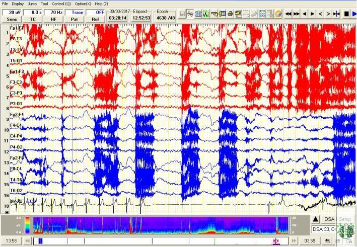
This diagnostic image displays a 30-second compressed video-EEG segment captured during a focal seizure evolving into a generalized event. The EEG layout uses a longitudinal bipolar montage, with upper channels (1-7) highlighted in red and lower channels (9-16) in blue. The tracing demonstrates high-amplitude, rhythmic ictal discharges consisting of spiky theta and alpha frequencies, particularly prominent in the left temporal regions (F7-T3, T3-T5). These discharges are accompanied by intermittent bursts of generalized high-voltage activity. Channel 17 shows a simultaneous single-lead ECG recording. Clinical significance is highlighted by the temporal correlation between the ictal EEG activity and a period of cardiac asystole, evidenced by a prolonged flatline on the ECG following initial bradycardia. This visual illustrates the rare but critical phenomenon of ictal asystole, an autonomic complication of epilepsy. A Density Spectral Array (DSA) is provided at the bottom of the interface, visualizing power distribution over time.
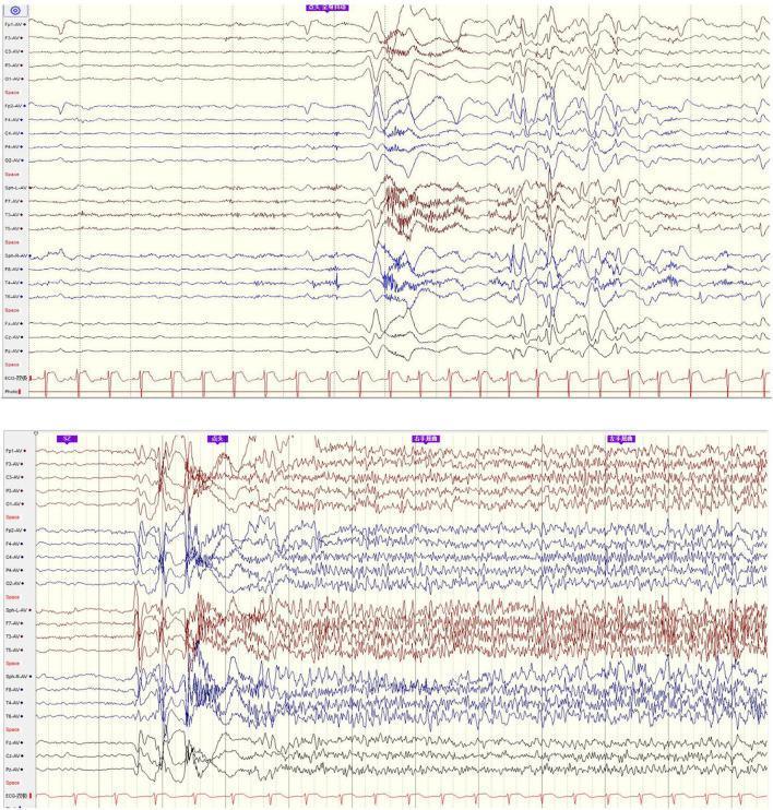
Two multi-channel electroencephalogram (EEG) tracings demonstrating the onset of different seizure types in a patient with Lennox-Gastaut syndrome (LGS). The top segment depicts an epileptic spasm, characterized by a sudden burst of high-amplitude, generalized slow waves followed by relative background suppression and lower-amplitude activity. The bottom segment shows a tonic seizure onset, featuring a transition from interictal slow spike-wave (SSW) complexes to generalized paroxysmal fast activity (GPFA). This generalized activity consists of sustained, high-frequency, rhythmic discharges appearing across all recording channels with minimal localization value. Both segments include an ECG rhythm strip at the bottom for monitoring cardiac synchrony. The images serve as diagnostic examples of the complex ictal patterns associated with refractory epilepsy and encephalopathy, highlighting the transition from intermittent focal-like events to generalized, persistent abnormal cortical discharges.
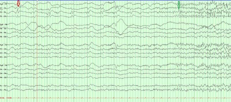
This diagnostic image shows a multi-channel continuous electroencephalogram (EEG) tracing used to monitor brain electrical activity. The tracing demonstrates the evolution of focal epileptiform activity into a generalized seizure. A red arrow on the left side of the tracing identifies interictal epileptiform discharges, specifically sharp waves, localized to the left temporal region (prominent in channels F7-T3, T3-T5, and T5-O1). A green arrow toward the right indicates the electrographic onset of a seizure, characterized by a transition from the background rhythm to rhythmic, high-frequency, and high-amplitude discharges. This ictal pattern shows rapid alpha and beta frequency activity that originates in the left temporal channels before demonstrating focal spread. The tracing is displayed on a standard green grid, providing a visual representation of status epilepticus and the transformation from focal sharps to rhythmic seizure activity, essential for neurocritical care and epilepsy diagnostics.
Seizure Types and Classification
What Is a Seizure?
The 2017 ILAE Classification Framework
SEIZURES
├── FOCAL ONSET
│   ├── Aware (simple partial)
│   └── Impaired awareness (complex partial)
│       └── ± Focal-to-bilateral tonic-clonic (secondary generalized)
├── GENERALIZED ONSET
│   ├── Motor: Tonic-clonic, Tonic, Clonic, Myoclonic, Atonic, Epileptic spasms
│   └── Non-motor: Absence (typical, atypical, myoclonic absence)
└── UNKNOWN ONSET
PART 1: FOCAL ONSET SEIZURES
1a. Focal Aware Seizure
- Consciousness fully preserved - patient remains alert and responsive
- Duration: usually seconds to 1-2 minutes
- May be a warning (aura) before a larger seizure
- Features depend on focus location:
| Cortical Location | Symptom |
|---|---|
| Primary motor cortex | Contralateral jerking of face, arm, or leg (Jacksonian march - spreads along homunculus) |
| Primary sensory cortex | Contralateral numbness, tingling, paresthesias |
| Occipital lobe | Flashing lights, sparks, color, brief blindness |
| Temporal lobe | Rising epigastric sensation (most common aura), déjà vu, jamais vu, fear, olfactory/gustatory hallucinations |
| Parietal lobe | Distortion of body image, vertiginous sensations |
| Frontal (supplementary motor) | Tonic posturing, head/eye deviation |
1b. Focal Impaired Awareness Seizure
- Consciousness is impaired or lost - patient is unresponsive to commands
- Most commonly arise from the temporal lobe or frontal lobe
- Duration: 1-3 minutes
- Key features:
- Automatisms - repetitive, semi-purposeful, involuntary movements: lip-smacking, chewing, swallowing, hand fumbling, picking, walking in circles
- Staring with behavioral arrest
- Patient may appear "out of contact" - can look at examiner but not respond
- Postictal confusion lasting minutes to hours (important distinguishing feature)
- Patient has no memory of the event
"The motor components take the form of automatisms such as lip smacking, chewing or swallowing movements, salivation, fumbling of the hands, or shuffling of the feet... the patients are obviously out of contact with their surroundings." - Adams & Victor
1c. Focal-to-Bilateral Tonic-Clonic Seizure
- Begins as focal (often with an aura or focal motor jerk), then spreads bilaterally to produce a full tonic-clonic convulsion
- The focal onset may be very brief and easily missed - only noticed in retrospect
- Clinically important: implies a focal brain lesion (tumor, AVM, scar, cortical dysplasia) - requires MRI investigation
- Distinguishing from primary generalized TC: focal onset = secondary = structural cause more likely
PART 2: GENERALIZED ONSET SEIZURES
2a. Generalized Tonic-Clonic (GTC) Seizure
- Sudden loss of consciousness + fall
- Sustained muscle stiffness - back, neck, then arms and legs
- Jaw clamps shut - may bite lateral tongue (NOT tip - biting the tip suggests psychogenic)
- Loud cry as air is forced through closed vocal cords
- Apnea + cyanosis
- Pupils dilated and unreactive
- Bladder may empty
- Rhythmic jerking of limbs begins at ~8 Hz, slows to ~4 Hz, then stops
- Grimacing, salivation, profuse sweating, hypertension, tachycardia
- Rapid alternation of contraction and relaxation
- Deep unresponsiveness/coma immediately after
- Then gradually awakens - confused, agitated, exhausted, headache
- May sleep for hours
- No memory of event
- Postictal confusion is the hallmark - distinguishes GTC from syncope (which has rapid full recovery)
2b. Absence Seizure
| Feature | Typical Absence | Atypical Absence |
|---|---|---|
| Duration | 4-20 seconds (usually <10 s) | Longer, more variable |
| Onset/offset | Abrupt - no warning, no postictal | Gradual onset/offset |
| Consciousness | Complete interruption | Less complete loss |
| Motor features | May have mild eyelid flicker, automatisms | More prominent motor signs |
| EEG | Classic 3 Hz spike-and-wave | Slow spike-wave 1-2.5 Hz |
| Age | Childhood (5-7 years), remits by age 12 | Part of epileptic encephalopathies (Lennox-Gastaut) |
| Trigger | Hyperventilation reliably provokes it | - |
"Brief episodes of unconsciousness (4-20 seconds) with no warning and immediate resumption of consciousness - NO postictal abnormality." - Katzung
2c. Myoclonic Seizure
- Sudden, brief (<100 ms), involuntary jerks of muscles or muscle groups
- Can be a single jerk or a rapid salvo of jerks
- Consciousness usually preserved (too brief to lose)
- Often occur on awakening - classic in Juvenile Myoclonic Epilepsy (JME)
- Patient may fling objects (drops coffee cup in the morning)
- EEG: polyspike-and-wave complexes
- If prominent + progressive + with dementia → look for storage diseases, mitochondrial disorders
2d. Tonic Seizure
- Sudden sustained stiffening of axial and/or limb muscles (lasting 2-30 seconds)
- No clonic phase
- Often causes falls + injuries
- Seen in Lennox-Gastaut syndrome and other epileptic encephalopathies
2e. Clonic Seizure
- Rhythmic jerking WITHOUT a preceding tonic phase
- Rare in isolation; more common in infants
- Bilateral, symmetric
2f. Atonic Seizure (Drop Attack)
- Sudden complete loss of muscle tone - head drops, knees buckle, falls
- Duration: 1-2 seconds
- No warning, extremely dangerous for head injuries
- Seen almost exclusively in Lennox-Gastaut syndrome
- EEG: burst of slow spike-wave or generalized paroxysmal fast activity
2g. Epileptic Spasms
- Sudden flexion, extension, or mixed extension-flexion of trunk and proximal limbs
- Lasts 1-3 seconds; occur in clusters, especially on waking
- Onset: 3-12 months of age
- EEG: Hypsarrhythmia - chaotic, high-amplitude disorganized pattern between spasms
- Triad of West Syndrome: infantile spasms + hypsarrhythmia + developmental arrest/regression
- Treatment: ACTH, vigabatrin (tuberous sclerosis specifically), prednisolone
PART 3: SPECIAL SEIZURE TYPES & SYNDROMES
Febrile Seizures
- Age 6 months - 5 years, triggered by fever (>38°C)
- Simple: single, <15 min, generalized, fully resolves
- Complex: >15 min, focal features, or recurs within 24 hours
- Most benign - low risk of developing epilepsy (~2-4%)
Status Epilepticus (SE)
- A single seizure lasting >5 minutes, or two+ seizures without return to baseline
- Medical emergency - risk of permanent neuronal injury after 30 minutes
- Convulsive SE: obvious; Non-convulsive SE (NCSE): subtle - only detected by EEG
Todd's (Postictal) Paralysis
- Focal motor weakness following a focal motor seizure
- Lasts minutes to hours (occasionally up to 24-48 hours)
- Resolves spontaneously
- Mimics stroke - important differential in emergency setting
Key Localizing Features of Focal Seizures
| Feature | Likely Focus |
|---|---|
| Rising epigastric aura | Temporal lobe (mesial) |
| Déjà vu, fear, olfactory hallucination | Temporal lobe (amygdala/hippocampus) |
| Visual sparks/lights | Occipital lobe |
| Jacksonian march (motor march) | Primary motor cortex |
| Head/eye deviation | Frontal (contralateral) |
| Hypermotor behavior (bicycling, thrashing) | Frontal lobe |
| Versive (turning) followed by tonic posturing | Supplementary motor area |
| Gelastic (laughing) seizure | Hypothalamic hamartoma |
EEG Correlates
| Seizure Type | EEG Pattern |
|---|---|
| Typical absence | 3 Hz spike-and-wave (bilaterally synchronous) |
| Atypical absence (LGS) | Slow spike-wave 1-2.5 Hz |
| Myoclonic | Polyspike-and-wave |
| GTC (primary generalized) | Bilateral symmetric spike-wave from onset |
| Focal impaired awareness | Temporal lobe spike/sharp waves; focal ictal pattern |
| Infantile spasms | Hypsarrhythmia |
| Lennox-Gastaut (tonic) | GPFA (generalized paroxysmal fast activity) |
Quick Summary Table
| Seizure Type | Consciousness | Duration | Postictal | Key Feature |
|---|---|---|---|---|
| Focal aware | Preserved | Seconds-2 min | None/minimal | Aura, focal motor/sensory |
| Focal impaired awareness | Impaired | 1-3 min | Confusion (minutes-hours) | Automatisms, staring |
| Generalized tonic-clonic | Lost | 1-3 min | Prolonged confusion + sleep | Tonic then clonic, tongue bite, incontinence |
| Absence (typical) | Completely lost | <10 sec | None | Abrupt stare, no fall |
| Myoclonic | Preserved | <100 ms | Minimal | Brief jerk, morning clustering |
| Atonic | Brief loss | 1-2 sec | Minimal | Sudden fall, dangerous |
| Epileptic spasms | Variable | 1-3 sec (clusters) | Variable | Clusters on waking, infant |