
hey orris i have exam pf pathology practical i name you some slide name give me microscopic features and in short defination and imp point which is related to viva so slide list is leiomyoma serous cystadenoma serous cystadenocarcinoma mucius cystadenoma ssc of cervix teratoma hyadatid mole
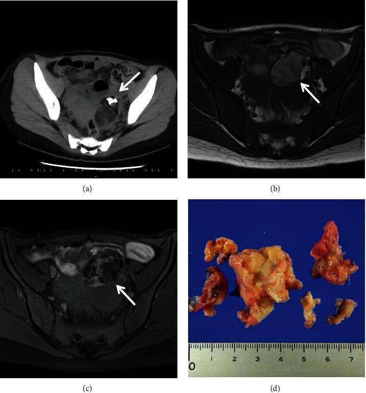
Pathology Practical Exam - Slide Guide 🔬
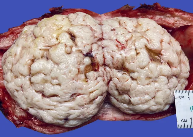
1. LEIOMYOMA (Uterine Fibroid)
- Interlacing/whorled bundles of smooth muscle cells arranged in fascicles (in different planes - longitudinal and transverse sections seen together)
- Cells are elongated, spindle-shaped with cigar-shaped (blunt-ended) nuclei
- Abundant pale pink fibrillar cytoplasm
- No nuclear atypia, no pleomorphism
- Mitotic figures absent or very rare (< 5 per 10 HPF)
- Fibrous stroma between muscle bundles
- Well-circumscribed with a pseudo-capsule
- Most common tumor in females overall
- Estrogen-dependent (enlarges in pregnancy, regresses post-menopause)
- Three locations: submucosal (causes menorrhagia), intramural (most common), subserosal
- Malignant transformation to leiomyosarcoma is VERY RARE (<1%)
- Leiomyosarcoma diagnosis: >10 mitoses/10 HPF + necrosis + atypia
- Whorled appearance on gross - key distinguishing feature
- Hyaline degeneration is the most common degeneration
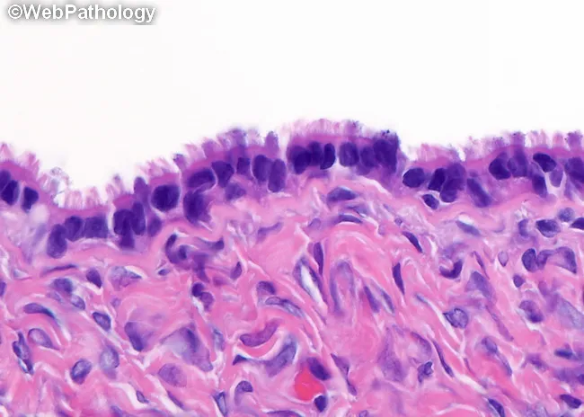
2. SEROUS CYSTADENOMA (Ovary)
- Thin-walled cyst lined by a single layer of flat to cuboidal to tall columnar ciliated epithelium (resembles fallopian tube lining)
- Cells have uniform nuclei, no atypia
- Papillary projections may be present - lined by the same simple epithelium
- No stromal invasion
- Psammoma bodies (concentrically laminated calcified bodies) may be seen (~30% of cases)
- Fibrous stroma in the wall
- Most common ovarian tumor overall
- 20% are bilateral
- Psammoma bodies are characteristic (also seen in papillary thyroid carcinoma and meningioma)
- Spectrum: Benign (cystadenoma) → Borderline → Malignant (cystadenocarcinoma)
- Borderline = increased papillary complexity + epithelial stratification but NO invasion
- Derives from surface (coelomic) epithelium of ovary
3. SEROUS CYSTADENOCARCINOMA (Ovary)
- Complex papillary and solid growth patterns
- Multilayered/stratified pleomorphic epithelium with marked nuclear atypia
- Stromal invasion - key feature distinguishing it from borderline tumor
- High mitotic activity (>12 mitoses per 10 HPF)
- Nuclear pleomorphism (>3-fold variation in nuclear size)
- Atypical mitotic figures, multinucleation
- Psammoma bodies may be present
- Necrosis and hemorrhage in high-grade tumors
- Two types: Low-grade (KRAS/BRAF mutations) and High-grade (TP53 mutations)
- Most common malignant ovarian tumor
- ~66% are bilateral
- BRCA1/BRCA2 mutations - almost all are high-grade serous type
- Spreads via transcoelomic route (peritoneal seeding), then lymphatics
- CA-125 is the tumor marker
- Most tumors are HIGH-grade at diagnosis (poor prognosis)
- Many high-grade serous carcinomas actually originate from fallopian tube (STIC - Serous Tubal Intraepithelial Carcinoma)
- TP53 mutation present in >96% of high-grade serous carcinoma
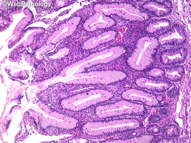
4. MUCINOUS CYSTADENOMA (Ovary)
- Cyst wall lined by tall columnar cells with basal nuclei and apical mucin (goblet cells abundant)
- Resembles endocervical or intestinal epithelium
- Single layer of cells - no stratification, no atypia
- No stromal invasion
- Multiple locules (multilocular cyst) separated by fibrous septa
- Epithelium may show intestinal-type goblet cells
- If cyst ruptures: mucin extravasation into stroma, with inflammatory reaction
- Largest ovarian tumors (can be enormous - up to 100+ lbs reported in old literature)
- Usually UNILATERAL (important - bilateral mucinous should think metastasis from GI)
- KRAS mutations common
- Pseudomyxoma peritonei - massive gelatinous deposits in peritoneum; usually from appendiceal/GI mucinous tumor, not primary ovarian
- ~10% associated with dermoid cysts or Brenner tumors
- Endocervical vs intestinal type - intestinal type has goblet cells
| Feature | Serous | Mucinous |
|---|---|---|
| Lining | Ciliated columnar | Tall columnar with mucin |
| Bilaterality | 20% bilateral | Usually unilateral |
| Psammoma bodies | Present | Absent |
| Size | Smaller | Larger |
| Locules | Unilocular (usually) | Multilocular |
5. SQUAMOUS CELL CARCINOMA (SCC) OF CERVIX
- Keratin pearls (concentric whorls of keratin) - in well-differentiated (keratinizing) type
- Intercellular bridges (prickles) between tumor cells
- Individual cell keratinization (pink glassy cytoplasm)
- Tumor cells form irregular infiltrative nests invading stroma
- Nuclear atypia, pleomorphism, prominent nucleoli
- Mitotic figures (including atypical)
- Desmoplastic stromal reaction
- Three subtypes: Large cell keratinizing, Large cell non-keratinizing, Small cell
- Well differentiated (Grade I): Prominent keratin pearls
- Moderately differentiated (Grade II): Some keratinization, intercellular bridges
- Poorly differentiated (Grade III): Little/no keratinization, high mitoses
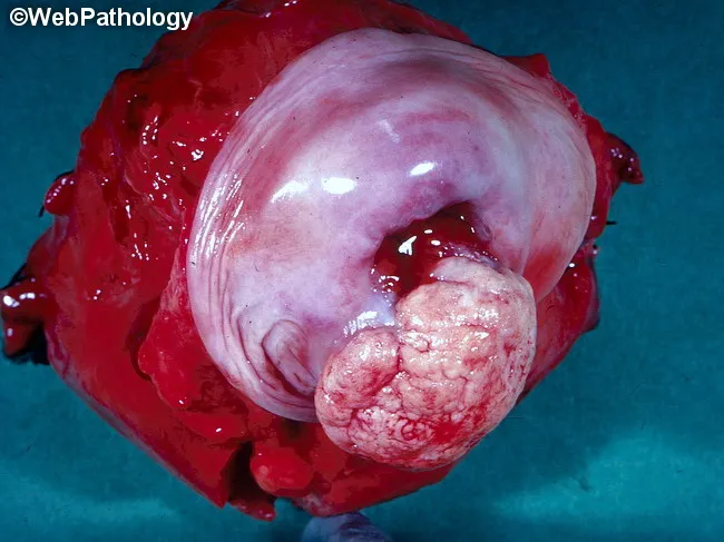
- HPV (Human Papillomavirus) is the most important cause - HPV 16 (SCC), HPV 18 (adenocarcinoma)
- Arises at squamo-columnar junction (transformation zone)
- CIN (Cervical Intraepithelial Neoplasia) is the precursor lesion
- Pap smear is the screening test
- HPV vaccine (Gardasil) protects against HPV 6, 11, 16, 18
- Spreads by direct extension first, then lymphatics
- FIGO staging used
- Koilocytes - hallmark of HPV infection in Pap smear (perinuclear cytoplasmic halo + wrinkled raisin-like nucleus)
6. TERATOMA (Ovary - Mature Cystic / Dermoid Cyst)
- Ectoderm: Skin with hair follicles, sebaceous glands, sweat glands, teeth, neural tissue
- Mesoderm: Cartilage, bone, muscle, fat
- Endoderm: Respiratory/GI epithelium, thyroid tissue
- All elements are well-differentiated (mature)
- Lined by squamous epithelium (skin) - "dermoid" appearance
- Low-power: classic image shows skin on one edge, brain tissue on the other
- No atypia in mature teratoma
- Contains immature neuroepithelium (key diagnostic feature)
- Graded I-III based on amount of immature neuroepithelium
- Seen in young females, mean age 18 years
- Most common ovarian germ cell tumor (and most common ovarian tumor in young girls)
- Mature cystic teratoma = dermoid cyst - BENIGN
- Contains teeth, hair, sebum - classic gross finding
- Rokitansky's protuberance / Dermoid plug - solid area from which most tissues arise
- 1-2% undergo malignant transformation to SCC
- Struma ovarii - teratoma composed entirely of thyroid tissue (can cause hyperthyroidism)
- Carcinoid syndrome can occur with carcinoid type teratoma even without liver mets (ovarian veins drain directly to systemic circulation)
- Teratoma = tumor arising from totipotent germ cells
7. HYDATIDIFORM MOLE (Complete Mole)
- Markedly hydropic (edematous/swollen) chorionic villi - villi are large, balloon-like
- Central cistern formation - central acellular fluid-filled space in villi (key feature)
- Avascular villi - no fetal blood vessels
- Trophoblastic hyperplasia - both cytotrophoblast and syncytiotrophoblast proliferate circumferentially (360° around villi)
- Trophoblastic cells show atypia
- No fetal parts / no embryo (in complete mole)
- Stroma is pale, myxoid, edematous
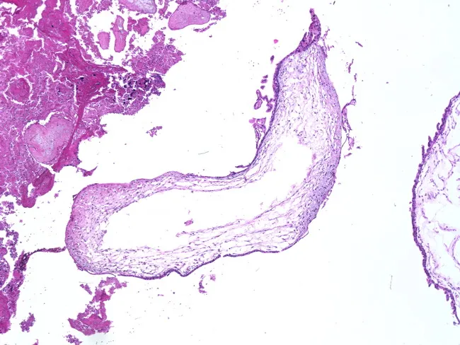
- Complete mole: 46XX (androgenetic) - ALL chromosomes paternal, no fetal tissue, diffuse trophoblastic proliferation
- Partial mole: 69XXX/XXY (triploid) - 2 paternal + 1 maternal set, fetal tissue present, focal trophoblast
- Grape-like clusters appearance on gross
- Beta-hCG markedly elevated - best tumor marker
- "Snowstorm" appearance on ultrasound
- Theca lutein cysts in ovaries (due to high hCG)
- 2% complete moles → choriocarcinoma (most dangerous complication)
- p57/KIP2 immunostaining: NEGATIVE in complete mole (paternally imprinted, maternal gene product), POSITIVE in partial mole and normal placenta
- Follow up with serial beta-hCG levels after evacuation
Quick Viva Summary Table
| Slide | Key Microscopic Feature | Tumor Marker/Special Test | Viva Must-Know |
|---|---|---|---|
| Leiomyoma | Whorled smooth muscle bundles, cigar nuclei | - | Most common female tumor, estrogen-dependent |
| Serous Cystadenoma | Single layer ciliated columnar, psammoma bodies | - | Most common ovarian tumor |
| Serous Cystadenocarcinoma | Papillary + stromal invasion, atypia | CA-125 | Most common ovarian malignancy, BRCA1/2 |
| Mucinous Cystadenoma | Tall columnar + goblet cells, no invasion | - | Largest tumors, usually unilateral |
| SCC Cervix | Keratin pearls, intercellular bridges | Pap smear, HPV | HPV 16, CIN precursor |
| Teratoma (Dermoid) | All 3 germ layers, mature elements, skin/hair | - | Rokitansky's protuberance, Struma ovarii |
| Hydatidiform Mole | Hydropic villi, cisterns, trophoblastic hyperplasia, avascular | Beta-hCG | 46XX androgenetic, snowstorm USG, 2% → choriocarcinoma |

explain the slide of teratoma
Your Teratoma Slide - Detailed Explanation 🔬
What You Can See in This Slide
🔵 TOP EDGE - Stratified Squamous Epithelium (Epidermis-like)
- Multiple cell layers visible
- Basal layer = dark basophilic nuclei
- Surface = thin pink keratin layer (stratum corneum)
- Germ layer: ECTODERM
🔴 LARGE PALE LOBULATED CLUSTERS (lower half of slide) - Sebaceous Glands
- Cells have abundant pale, vacuolated (foamy) cytoplasm - because lipid/fat dissolves during H&E processing
- Small, round, centrally placed nuclei
- Arranged in lobules/clusters
- These are holocrine glands (whole cell secretion = sebum)
- Germ layer: ECTODERM
🟣 CIRCULAR/OVAL STRUCTURES (center and right) - Hair Follicles
- Seen as circular epithelial structures
- Inner keratin/hair shaft appears eosinophilic (pink)
- Outer root sheath = basophilic (blue-purple) stratified epithelium
- Germ layer: ECTODERM
🟤 PINK BACKGROUND TISSUE - Dermis-like Connective Tissue (Fibrous Stroma)
- Abundant collagen fibers = bright eosinophilic (pink)
- Scattered elongated fibroblast nuclei
- Germ layer: MESODERM
Annotated Summary Table
| Structure Seen in Slide | Appearance | Germ Layer |
|---|---|---|
| Stratified squamous epithelium (top) | Layered purple-pink strip | Ectoderm |
| Sebaceous glands (big pale lobules) | Foamy/vacuolated clusters | Ectoderm |
| Hair follicles (circular structures) | Round epithelial rings | Ectoderm |
| Fibrous stroma (background) | Pink collagen bundles | Mesoderm |
How to Describe This Slide in Viva (Exact Wording)
"This is a low-power H&E section showing a mature cystic teratoma (dermoid cyst). The section shows skin-like stratified squamous epithelium lining the top surface, with underlying dermis-like fibrous stroma. In the stroma, there are numerous well-formed sebaceous glands showing characteristic foamy vacuolated cells with round central nuclei, along with hair follicles showing outer root sheath epithelium. All elements are mature and well-differentiated, with no cytological atypia or mitotic activity, confirming the benign nature of this teratoma."
Key Viva Points Specific to This Slide
- Diagnosis: Mature cystic teratoma / Dermoid cyst
- The most dominant element in YOUR slide is ectodermal (skin + skin appendages)
- The key is the combination of features from different germ layers in one tumor
- No atypia = mature = BENIGN
- If asked "what is the classic low-power appearance?" = skin on one side, other tissue on the other (as in your slide)
- The sebaceous glands are the easiest structure to identify and the most important to mention
can you marke this in the image i sent to you ?
mkdir -p /tmp/workspace/teratoma && curl -s -o /tmp/workspace/teratoma/slide.jpg "https://d361swluvexzqs.cloudfront.net/chats-attachments/photo-1785282498602-1785282499149-hoomrm.jpg" && echo "Downloaded OK" && python3 -c "from PIL import Image; img = Image.open('/tmp/workspace/teratoma/slide.jpg'); print(img.size)"
python3 << 'EOF' from PIL import Image, ImageDraw, ImageFont import math img = Image.open('/tmp/workspace/teratoma/slide.jpg').convert('RGB') draw = ImageDraw.Draw(img) W, H = img.size # 1200 x 1600 # Try to load a font, fallback to default try: font = ImageFont.truetype("/usr/share/fonts/truetype/dejavu/DejaVuSans-Bold.ttf", 28) font_small = ImageFont.truetype("/usr/share/fonts/truetype/dejavu/DejaVuSans-Bold.ttf", 22) except: font = ImageFont.load_default() font_small = font def draw_arrow(draw, start, end, color, width=4): """Draw an arrow from start to end""" draw.line([start, end], fill=color, width=width) # arrowhead dx = end[0] - start[0] dy = end[1] - start[1] length = math.sqrt(dx*dx + dy*dy) if length == 0: return dx, dy = dx/length, dy/length arrow_len = 20 arrow_width = 12 left = (end[0] - arrow_len*dx + arrow_width*dy, end[1] - arrow_len*dy - arrow_width*dx) right = (end[0] - arrow_len*dx - arrow_width*dy, end[1] - arrow_len*dy + arrow_width*dx) draw.polygon([end, left, right], fill=color) def draw_label_box(draw, text, box_xy, text_color, bg_color, font): """Draw a filled label box""" x, y = box_xy bbox = draw.textbbox((x, y), text, font=font) pad = 6 draw.rectangle([bbox[0]-pad, bbox[1]-pad, bbox[2]+pad, bbox[3]+pad], fill=bg_color, outline=text_color, width=2) draw.text((x, y), text, fill=text_color, font=font) # ------------------------------------------------------- # 1. STRATIFIED SQUAMOUS EPITHELIUM - top strip ~y=370-400 # Bracket along top edge of tissue # ------------------------------------------------------- color_ecto = (255, 50, 50) # Red for ectoderm structures color_meso = (50, 180, 255) # Blue for mesoderm color_label_bg = (0, 0, 0) # Draw a horizontal bracket on the squamous epithelium strip # The tissue starts around y=370, epithelium strip is thin ~y=370-400, x=200-1000 epi_y = 400 draw.rectangle([210, 370, 990, 410], outline=(255,50,50), width=4) # Arrow pointing to it from the right side label draw_arrow(draw, (1060, 390), (1000, 390), color_ecto, 4) draw_label_box(draw, "Stratified Squamous\nEpithelium (Ectoderm)", (1060, 370), (255,255,255), (200,0,0), font_small) # ------------------------------------------------------- # 2. SEBACEOUS GLANDS - large pale lobulated clusters # Major ones: lower-left ~(250,1000), center-bottom ~(550,1050), lower-right ~(900,1100) # ------------------------------------------------------- color_seb = (255, 165, 0) # Orange # Lower left sebaceous cluster draw.ellipse([150, 900, 430, 1150], outline=(255,165,0), width=5) draw_arrow(draw, (80, 780), (180, 910), color_seb, 4) draw_label_box(draw, "Sebaceous\nGlands\n(Ectoderm)", (20, 730), (255,255,255), (180,100,0), font_small) # Center-right sebaceous cluster draw.ellipse([500, 950, 780, 1200], outline=(255,165,0), width=5) # Lower right draw.ellipse([820, 1050, 1100, 1300], outline=(255,165,0), width=5) draw_arrow(draw, (1120, 1100), (1090, 1100), color_seb, 4) draw_label_box(draw, "Sebaceous\nGlands", (1070, 1070), (255,255,255), (180,100,0), font_small) # ------------------------------------------------------- # 3. HAIR FOLLICLES - circular structures center/upper right ~(700-800, 500-650) # ------------------------------------------------------- color_hair = (50, 220, 50) # Green # Upper center hair follicle region (round structures near center-top) draw.ellipse([650, 490, 750, 580], outline=(50,220,50), width=4) draw.ellipse([720, 530, 810, 620], outline=(50,220,50), width=4) draw_arrow(draw, (500, 440), (655, 510), color_hair, 4) draw_label_box(draw, "Hair Follicles\n(Ectoderm)", (370, 400), (255,255,255), (0,140,0), font_small) # ------------------------------------------------------- # 4. FIBROUS STROMA (DERMIS) - the pink background areas # Mark with a bracket on the right side mid-area # ------------------------------------------------------- color_stroma = (50, 180, 255) # Blue # Mid-right area with clear stroma draw.rectangle([860, 650, 1050, 850], outline=(50,180,255), width=4) draw_arrow(draw, (1090, 750), (1050, 750), color_stroma, 4) draw_label_box(draw, "Fibrous Stroma /\nDermis (Mesoderm)", (1060, 720), (255,255,255), (0,80,180), font_small) # ------------------------------------------------------- # TITLE BOX at the top # ------------------------------------------------------- draw.rectangle([0, 0, W, 80], fill=(0,0,0)) title_font = font try: title_font = ImageFont.truetype("/usr/share/fonts/truetype/dejavu/DejaVuSans-Bold.ttf", 34) except: pass draw.text((20, 18), "MATURE CYSTIC TERATOMA (DERMOID CYST) - Annotated", fill=(255, 220, 50), font=title_font) # Save img.save('/tmp/workspace/teratoma/teratoma_annotated.jpg', quality=92) print("Saved OK", img.size) EOF
teratoma annotated
Image · JPG
| Color | Label | Structure |
|---|---|---|
| 🔴 Red box | Top edge | Stratified Squamous Epithelium (Ectoderm) |
| 🟠 Orange ovals | Large pale clusters | Sebaceous Glands (Ectoderm) |
| 🟢 Green ovals | Small circular structures, center | Hair Follicles (Ectoderm) |
| 🔵 Blue box | Pink background, mid-right | Fibrous Stroma / Dermis (Mesoderm) |


give ans and imp point related to patho viva
CLINICAL CASE NO. 4
1) Diagnosis: Acute Myocardial Infarction (AMI)
2) Other Cardiac Markers
| Marker | Rises | Peak | Returns to Normal |
|---|---|---|---|
| Troponin I/T (MOST SPECIFIC) | 3-6 hrs | 18-24 hrs | 7-14 days |
| CPK-MB (EARLIEST specific) | 4-6 hrs | 18-24 hrs | 3-4 days |
| LDH (LDH-1 > LDH-2) | 24-48 hrs | 3-6 days | 8-14 days |
| AST | 8-12 hrs | 24-36 hrs | 3-4 days |
| Myoglobin (EARLIEST, non-specific) | 1-3 hrs | 6-8 hrs | 24 hrs |
3) Risk Factors
- Hypercholesterolemia (280 mg/dl - HIGH - in this case)
- Hypertriglyceridemia (220 mg/dl - HIGH - in this case)
- Hypertension, Diabetes, Smoking, Obesity, Sedentary lifestyle
- Age >45 male / >55 female, Male sex, Family history, Post-menopausal women
- Troponin is the GOLD STANDARD cardiac marker today
- CPK-MB is used for reinfarction (returns to normal in 3-4 days, so any new rise = new MI)
- Zone of coagulative necrosis appears at 4-12 hours
- Neutrophil infiltration = 12-24 hours post MI
- Macrophage infiltration = Day 3-7
- Granulation tissue = Day 7-10
CLINICAL CASE NO. 10
1) Diagnosis: Type 1 Diabetes Mellitus (Insulin-Dependent DM)
2) Significance of HbA1C
- HbA1C reflects average blood glucose over the PAST 2-3 MONTHS (life of RBC)
- 8.4% = poorly controlled diabetes (target <7% in diabetics, normal <5.7%)
- It is a glycated hemoglobin (glucose irreversibly binds to Hb)
- Used for monitoring, not screening
- NOT affected by short-term fluctuations in blood sugar
- Falsely low in: hemolytic anemia, blood transfusion (new RBCs)
- Falsely high in: iron deficiency anemia
3) Confirmatory Tests (WHO Criteria - any one sufficient)
- Fasting plasma glucose ≥126 mg/dl (on two occasions)
- PPBS / Random plasma glucose ≥200 mg/dl + symptoms
- OGTT - 2 hr post 75g glucose load ≥200 mg/dl
- HbA1C ≥6.5%
- Green precipitate = trace/0.5%
- Yellow precipitate = 1% (this case)
- Orange precipitate = 1.5%
- Brick red precipitate = 2%+
- Type 1 DM: autoimmune destruction of beta cells, HLA DR3/DR4 association
- Type 2 DM: insulin resistance + relative deficiency
- Classic triad = 3 Ps (polyuria, polydipsia, polyphagia)
- In children with weight loss = always think Type 1
CLINICAL CASE NO. 11
1) Diagnosis: Diabetic Ketoacidosis (DKA) in Type 2 DM
2) Interpretation of Findings
| Finding | Interpretation |
|---|---|
| FBS 260, PPBS 600 | Severe uncontrolled hyperglycemia |
| HbA1C 12% | Prolonged poor glycemic control (past 2-3 months) |
| Brick red precipitate | Urine glucose ≥2% (heavy glycosuria) |
| Ketone bodies in urine | DKA - fat breakdown → ketogenesis due to insulin deficiency |
3) Morphological Lesions of Diabetes
- Kimmelstiel-Wilson nodules (nodular glomerulosclerosis) - PATHOGNOMONIC
- Diffuse glomerulosclerosis
- Armanni-Ebstein lesion (glycogen in tubular cells)
- Pyelonephritis, papillary necrosis
- Atherosclerosis (large vessels)
- Hyaline arteriolosclerosis (small vessels - especially afferent arteriole)
CLINICAL CASE NO. 12
1) Clinical Diagnosis: Acute Post-Streptococcal Glomerulonephritis (APSGN)
2) Causes of Glomerulonephritis
- Post-streptococcal (group A beta-hemolytic strep) - this case
- Post-staphylococcal, post-viral
- IgA nephropathy (Berger's disease) - hematuria during/immediately after URTI
- Lupus nephritis, Goodpasture syndrome, Henoch-Schonlein purpura
- APSGN: 2 weeks after URTI, low complement (C3 low)
- IgA nephropathy: hematuria DURING/same time as URTI, normal complement
3) Significance of Casts
| Cast | Significance |
|---|---|
| RBC casts | PATHOGNOMONIC of glomerulonephritis |
| WBC casts | Pyelonephritis/interstitial nephritis |
| Granular casts | Chronic renal disease, non-specific |
| Waxy casts | Advanced/chronic renal failure |
| Hyaline casts | Normal, concentrated urine (strenuous exercise) |
| Fatty casts | Nephrotic syndrome |
| Epithelial casts | Acute tubular necrosis, glomerulonephritis |
- Benzidine test = for blood/hemoglobin in urine (positive = hematuria)
- Heller's test = for albumin in urine (positive = proteinuria)
- "Smoky/cola-colored urine" = classic of APSGN
- Light microscopy: diffuse proliferative glomerulonephritis, "lumpy bumpy" deposits on EM
- Immunofluorescence: granular IgG + C3 deposits
- Complement C3 is LOW in APSGN (consumed)
CLINICAL CASE NO. 13
1) Clinical Diagnosis: Nephrotic Syndrome
- Massive proteinuria (albumin ++++ in urine)
- Hypoalbuminemia (1.8 gm/dl - very low)
- Generalized edema (face, pedal, ascites)
- Hypercholesterolemia (280 mg/dl)
2) Causes of Nephrotic Syndrome
- Minimal Change Disease - most common in CHILDREN (3-8 yrs) ← THIS CASE
- Membranous GN - most common in ADULTS
- Focal Segmental Glomerulosclerosis (FSGS)
- Membranoproliferative GN
- Diabetes (Kimmelstiel-Wilson)
- Amyloidosis
- Lupus (SLE) - Class V
- Drugs (NSAIDs, gold, penicillamine)
- Infections (malaria, HBV, HCV)
- Nephrotic syndrome = proteinuria >3.5 g/day (adult) / >40 mg/m²/hr (child)
- Hypoalbuminemia → low oncotic pressure → edema
- Hypercholesterolemia: liver synthesizes more lipoproteins to compensate
- Minimal Change Disease: electron microscopy shows effacement of podocyte foot processes - light microscopy is NORMAL (hence "minimal change")
- Waxy/fatty casts = nephrotic syndrome
- MCD responds beautifully to steroids
CLINICAL CASE NO. 14
1) Clinical Diagnosis: Acute Pyelonephritis (Upper UTI)
2) Significance of Urinary Findings
| Finding | Significance |
|---|---|
| Turbid, foul smelling urine | Bacterial infection |
| Albumin ++ | Inflammation/infection-related proteinuria |
| Pus cells (pyuria) | Infection (>5 WBC/HPF = abnormal) |
| Bacteria ++ | Active bacterial infection |
| WBC casts | DIAGNOSTIC of pyelonephritis (upper UTI) |
| Waxy/granular casts | Chronic/severe renal involvement |
| Epithelial cells | Tubular damage |
3) Causative Organisms
- Ascending route (most common) - from bladder via ureter
- Hematogenous - rare (Staph aureus)
- Lower UTI (cystitis): pus cells + bacteria, NO casts
- Upper UTI (pyelonephritis): pus cells + bacteria + WBC casts ← KEY DIFFERENTIATOR
- Women more susceptible: short urethra, close proximity to anus
- Vesicoureteral reflux is main risk factor in children
- Chronic pyelonephritis → coarse asymmetric scarring with cortical thinning
CLINICAL CASE NO. 17
1) Diagnosis: Urinary Tract Infection (Upper UTI / Acute Pyelonephritis)
2) Differentiating Lower vs Upper UTI
| Feature | Lower UTI (Cystitis/Urethritis) | Upper UTI (Pyelonephritis) |
|---|---|---|
| Symptoms | Dysuria, frequency, urgency | + fever, chills, flank pain |
| Fever | Absent/low grade | High grade (>38.5°C) |
| Pus cells | Present | Present |
| WBC casts | ABSENT | PRESENT ← key |
| Bacteria | Present | Present |
| Blood urea/creatinine | Normal | May be elevated |
| TLC | Normal/mild rise | Elevated + neutrophilia |
3) Investigations to Confirm Diagnosis
- Urine culture and sensitivity - GOLD STANDARD (significant bacteriuria = >10⁵ CFU/ml)
- Urine microscopy - pus cells, WBC casts, bacteria
- Complete blood count - leukocytosis, neutrophilia
- Ultrasound KUB - enlarged kidney, obstruction
- Blood culture - if septicemia suspected
- DMSA scan - for renal scarring in chronic cases
4) Complications
- Renal abscess (perinephric abscess)
- Septicemia / Urosepsis
- Chronic pyelonephritis → renal scarring → CKD
- Papillary necrosis (especially in diabetics)
- Hypertension (from chronic scarring)
- Xanthogranulomatous pyelonephritis (rare but important viva point)
- In pregnancy: premature labor, low birth weight
Master Viva Cheat Sheet - All Cases
| Case | Diagnosis | Single Most Important Finding |
|---|---|---|
| Case 4 | Acute MI | Troponin is gold standard; CPK-MB for reinfarction |
| Case 10 | Type 1 DM | HbA1C = 2-3 months glycemic control |
| Case 11 | DKA in Type 2 DM | Kimmelstiel-Wilson nodule = pathognomonic |
| Case 12 | APSGN | RBC casts = pathognomonic of GN |
| Case 13 | Nephrotic Syndrome | MCD in children; effaced foot processes on EM |
| Case 14 | Acute Pyelonephritis | WBC casts = upper UTI |
| Case 17 | UTI/Pyelonephritis | Urine C&S gold standard; WBC casts = upper UTI |


give ans in same manner
CLINICAL CASE NO. 18
1) Diagnosis: Renal Calculus (Urolithiasis / Kidney Stone)
2) Urine Microscopy - Positive Findings
| Finding | Significance |
|---|---|
| RBCs / Hematuria | Stone traumatizing urothelium → red urine |
| RBC casts (occasionally) | Renal parenchymal involvement |
| Crystals | Specific to stone type (see below) |
| Pus cells | If secondary infection present |
| Epithelial cells | Urothelial damage from stone passage |
| Increased specific gravity | Concentrated urine (predisposes to stone) |
| Crystal | Appearance | Stone type |
|---|---|---|
| Calcium oxalate | Envelope/dumbbell shaped | Most common stone (80%) |
| Triple phosphate (struvite) | Coffin lid shaped | Infection stones (Proteus) |
| Uric acid | Rhomboid/needle shaped | Gout/hyperuricemia |
| Cystine | Hexagonal | Cystinuria |
3) Other Investigations to Confirm Diagnosis
- Urine routine & microscopy - hematuria, crystals
- X-ray KUB - radio-opaque stones (calcium oxalate, calcium phosphate, struvite visible; uric acid is RADIOLUCENT)
- Ultrasound KUB - stone location, hydronephrosis
- CT scan KUB (non-contrast) - GOLD STANDARD, detects all stone types
- Serum calcium, phosphate, uric acid - underlying cause
- 24-hr urine analysis - for calcium, oxalate, uric acid, citrate
- Stone analysis (after passing) - definitive stone type
- Most common stone = Calcium oxalate (80%)
- Most common site of stone impaction = Pelvi-ureteric junction (PUJ)
- Proteus produces urease → alkaline urine → struvite (staghorn) calculi
- Staghorn calculus = fills entire renal pelvis + calyces
- Renal colic = pain radiates from loin to groin (follows ureter)
- Risk factors: low fluid intake, hot climate, high oxalate diet (spinach, tomato), hyperparathyroidism, gout
- Lesch-Nyhan syndrome → uric acid stones
CLINICAL CASE NO. 21
1) Diagnosis: Carcinoma of the Rectum / Colorectal Carcinoma
2) Significance of CEA (Carcinoembryonic Antigen)
- A glycoprotein tumor marker normally produced during fetal development, disappears after birth
- Normally < 2.5 ng/ml (non-smoker), < 5 ng/ml (smoker)
| Use | Details |
|---|---|
| Diagnosis support | Raised CEA strongly suggests colorectal carcinoma |
| NOT a screening test | Not specific enough (also raised in other conditions) |
| Prognosis | Higher levels = more advanced disease |
| Post-surgical monitoring | Should fall to normal after tumor resection; if rises again = RECURRENCE |
| Monitoring response to chemotherapy | Falling CEA = good response |
- GI cancers (colon, rectum, stomach, pancreas)
- Lung cancer, breast cancer
- Non-malignant: smokers, cirrhosis, IBD, pancreatitis
- Adenocarcinoma is the most common type of colorectal carcinoma (95%)
- Right-sided colon cancer: occult blood, anemia, dull pain (presents LATE)
- Left-sided/rectal cancer: rectal bleeding, change in bowel habits, obstruction (presents EARLY) ← this case
- Most common predisposing lesion = Adenomatous polyp (villous adenoma has highest malignant potential)
- APC gene mutation (chromosome 5q) - familial adenomatous polyposis
- "Apple core" / "napkin ring" appearance on barium enema
- Spread: direct → lymphatic → portal vein → liver (most common site of metastasis)
- Duke's staging:
- A = confined to mucosa
- B = through muscularis, no nodes
- C = lymph node involvement
- D = distant metastasis
CLINICAL CASE NO. 22
1) Diagnosis: Duodenal Ulcer (Peptic Ulcer Disease)
- Pain RELIEVED by food = Duodenal ulcer (food buffers acid)
- Night pain = acid secretion peaks at night, wakes patient
- Tenderness in right hypochondrium/epigastrium
- Age 46, male
2) Microscopic Features of Peptic Ulcer
- Necrotic debris, fibrin, dead cells
- Pink, amorphous eosinophilic material
- Neutrophils, fibrinous exudate
- Acute inflammatory cells
- Proliferating capillaries, fibroblasts
- Chronic inflammatory cells (lymphocytes, plasma cells, macrophages)
- Dense collagen fibers (fibrosis)
- Obliterative endarteritis of vessels
- Muscularis mucosa is destroyed (distinguishes ulcer from erosion - erosion only involves mucosa)
- Edges may show metaplastic changes (intestinal metaplasia, gastric metaplasia)
- H. pylori organisms may be seen at mucosal surface (curved gram-negative rods)
- H. pylori = most important cause of peptic ulcer (90% DU, 70% GU)
- Duodenal ulcer = ALWAYS benign (never malignant)
- Gastric ulcer = can be malignant (biopsy mandatory)
- Most common site: DU = first part of duodenum (anterior wall); GU = lesser curvature
- Complications: Bleeding (most common), Perforation, Pyloric stenosis (obstruction), Penetration
- Zollinger-Ellison syndrome = multiple recurrent ulcers → think gastrinoma (↑↑ gastrin)
- Curling's ulcer = stress ulcer in burns
- Cushing's ulcer = stress ulcer in CNS injury (↑ vagal tone → ↑ acid)
CLINICAL CASE NO. 23
1) Diagnosis: Gastric Ulcer (Peptic Ulcer Disease - Gastric type)
- Pain AGGRAVATED by food = Gastric ulcer (food stimulates acid → more pain)
- Patient AVOIDS food (unlike DU patient who eats to relieve pain) → leads to weight loss
- Midline epigastric tenderness
- Age 62 (older age group), vomiting, weight loss
2) Differences Between Duodenal and Gastric Ulcer
| Feature | Duodenal Ulcer | Gastric Ulcer |
|---|---|---|
| Age | Younger (30-50 yrs) | Older (50-70 yrs) |
| Sex | M > F (4:1) | M > F (2:1) |
| Site | 1st part duodenum (anterior wall) | Lesser curvature (incisura angularis) |
| Pain | Relieved by food, night pain | Aggravated by food |
| Appetite/Weight | Normal appetite, no weight loss | Anorexia + weight loss ← this case |
| Vomiting | Uncommon | Common |
| Acid secretion | HIGH (hypersecretion) | Normal or LOW |
| H. pylori | 90% | 70% |
| Malignancy risk | NEVER malignant | Can become malignant (1-2%) |
| Blood group | Group O more common | No strong association |
| Gastric emptying | Rapid | Delayed |
| Number | Usually single | Usually single |
- ALWAYS biopsy a gastric ulcer to rule out carcinoma
- Benign GU: punched out edges, flat margins, regular base
- Malignant GU: raised everted edges, irregular base, heaped-up margins
- H. pylori detection: Rapid Urease Test (CLO test), Urea breath test, Histology (Giemsa stain - shows curved bacilli)
- Treatment: Triple therapy = PPI + Amoxicillin + Clarithromycin (14 days)
CASE 6 (Jaundice Case)
Diagnosis: Obstructive Jaundice (Cholestatic Jaundice) due to Cholelithiasis (Gallstones)
| Finding | Interpretation |
|---|---|
| Conjugated bilirubin VERY HIGH | Obstruction - conjugated bili cannot excrete into intestine, regurgitates to blood |
| Unconjugated bilirubin normal | No hemolysis |
| ALP 940 (very high) | HALLMARK of obstructive jaundice (bile duct obstruction → ALP released from bile duct epithelium) |
| AST/ALT near normal | No significant hepatocellular damage |
| Urine bile salts +ve | Conjugated bilirubin is water-soluble → excreted in urine |
| Urine bile pigments +ve | Conjugated bilirubin in urine |
| Urine urobilinogen -ve | Obstruction blocks bilirubin reaching intestine → no urobilinogen formed → none absorbed → ABSENT in urine |
| Itching (pruritus) | Bile salts deposited in skin |
| Pale/clay stools (implied) | No bile reaching intestine |
| Feature | Hemolytic (Pre-hepatic) | Hepatocellular | Obstructive (Post-hepatic) |
|---|---|---|---|
| Unconjugated bili | ↑↑ | ↑ | Normal/slight↑ |
| Conjugated bili | Normal | ↑ | ↑↑ |
| ALP | Normal | Slight↑ | ↑↑↑ |
| AST/ALT | Normal | ↑↑↑ | Normal/slight↑ |
| Urine urobilinogen | ↑↑ | ↑ | ABSENT ← key |
| Urine bile pigments | Absent | Present | Present |
| Stool color | Dark (excess urobilin) | Pale | Clay/pale ← key |
| Pruritus | Absent | Variable | Present ← key |
- Van den Bergh test: Direct (conjugated) = obstructive; Indirect (unconjugated) = hemolytic; Biphasic = hepatocellular
- ALP rises dramatically in obstructive jaundice - most important enzyme here
CASE 7
Diagnosis: Thalassemia Major (Beta-Thalassemia Major) with Hemolytic Jaundice
- Child 8 yrs + severe anemia requiring multiple blood transfusions ← hallmark of Thalassemia major
- Hepatosplenomegaly (extramedullary hematopoiesis + RES hyperplasia)
- Target cells on peripheral smear ← classic of thalassemia
- Unconjugated hyperbilirubinemia = hemolytic jaundice
- Urobilinogen PRESENT (excessive hemolysis → more bilirubin → more urobilinogen)
- Bile salts/pigments ABSENT in urine (unconjugated bilirubin is NOT water-soluble → cannot pass in urine)
| Finding | Significance |
|---|---|
| Hb 7 gm/dl | Severe anemia |
| Unconjugated bilirubin 4.7↑↑ | Hemolysis → RBC breakdown → excess unconjugated bilirubin |
| Conjugated bilirubin 0.3 (normal) | Liver is conjugating normally |
| Target cells | Abnormal Hb (HbF, HbA2 excess) → less Hb in cell → excess membrane → target cell |
| Polychromasia | Reticulocytosis (bone marrow trying to compensate) |
| Microcytic hypochromic | Defective globin synthesis → less Hb |
| Urine urobilinogen +ve | Excess bilirubin → more urobilinogen |
| Bile salts/pigments -ve | UCB is fat-soluble, not filtered by kidney |
| Hepatosplenomegaly | Extramedullary hematopoiesis (liver, spleen take over BM function) + spleen destroying abnormal RBCs |
- Beta-thalassemia major = defect in beta-globin chain synthesis (chromosome 11)
- Blood transfusion dependency starts at 6 months - 2 years
- Complication of multiple transfusions = Hemosiderosis / Secondary hemochromatosis (iron overload)
- Peripheral smear: target cells, microcytic hypochromic, nucleated RBCs, Howell-Jolly bodies
- Diagnosis confirmed by: Hb electrophoresis - shows HbF↑↑, HbA2↑, HbA absent/very low
- Bone: "Hair on end" / "Sun-burst" appearance on X-ray skull (marrow expansion)
- Chipmunk facies due to maxillary overgrowth
- Treatment: Regular blood transfusion + desferrioxamine (iron chelation)
- Cure = Bone marrow transplantation
Master Viva Summary Table
| Case | Diagnosis | Single Most Important Point |
|---|---|---|
| Case 18 | Renal Calculus | Calcium oxalate most common; CT KUB = gold standard |
| Case 21 | Colorectal Carcinoma | CEA for monitoring recurrence, not screening |
| Case 22 | Duodenal Ulcer | Pain relieved by food; DU never malignant |
| Case 23 | Gastric Ulcer | Pain aggravated by food; ALWAYS biopsy GU |
| Case 6 | Obstructive Jaundice (Gallstones) | ALP ↑↑↑ + Urobilinogen absent in urine |
| Case 7 | Thalassemia Major / Hemolytic Jaundice | Target cells + UCB↑ + Urobilinogen↑ in urine |
left and right lung differentiation
stages of atherosclerosis