
Gaucher’s disease Refer all related article and textbooks Mainly refer Nelson genereview and omim and give notes in detail and at last give me how to approach Gaucher’s disease in flowchart format
"Gaucher disease"[MeSH Terms] AND treatment
Gaucher disease OMIM GeneReviews Nelson pathophysiology types genetics treatment 2024
Gaucher disease Gaucher cells bone marrow histology glucocerebrosidase
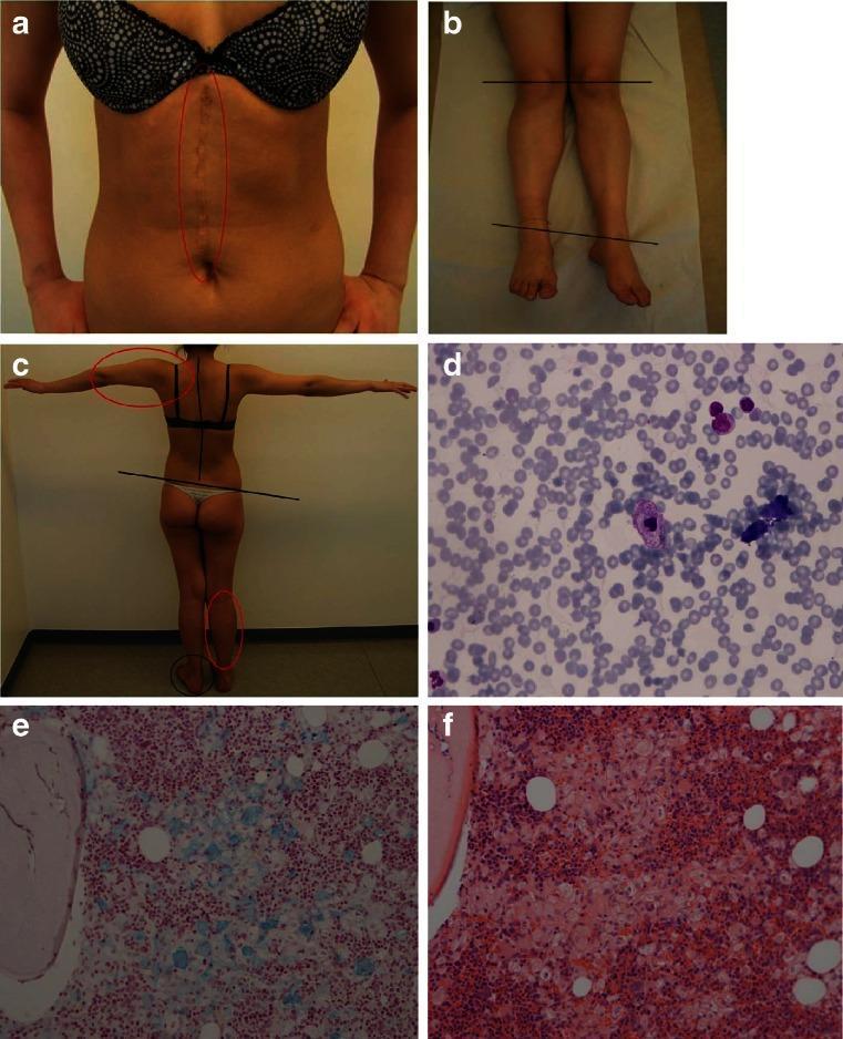
A multi-panel figure illustrating clinical and pathological findings associated with Gaucher disease (GD). Panel a is a clinical photograph of a patient's abdomen showing a large, midline vertical surgical scar (post-splenectomy). Panels b and c show clinical musculoskeletal photographs with annotations highlighting skeletal deformities; red circles indicate primary malformations of the right leg and left arm, while black lines and circles denote secondary postural changes in the spine and feet. Panel d is a bone marrow cytology slide (May-Grünwald Giemsa stain) showing a centrally located histiocyte with classic 'crinkled paper' Gaucher cell morphology. Panels e and f present bone marrow histology sections stained with iron-hematoxylin (e) and hematoxylin and eosin (f), respectively, demonstrating extensive histiocytic infiltration composed of pale-staining Gaucher cells among marrow elements. This visual summary correlates the gross clinical manifestations of lysosomal storage disorders, such as massive organomegaly and skeletal complications, with microscopic cellular evidence of substrate accumulation.
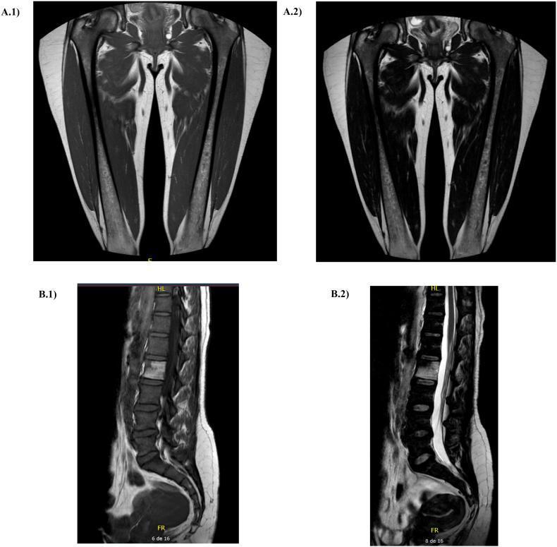
This figure presents a series of Magnetic Resonance Imaging (MRI) scans used for the assessment of bone marrow involvement in Gaucher disease (GD). Panels A.1 and A.2 display coronal views of the bilateral femurs. Panel A.1 is a T1-weighted image showing characteristic low signal intensity (darkening) within the femoral shafts, indicating displacement of normal fatty marrow by Gaucher cells. Panel A.2 is a T2-weighted sequence of the same region, highlighting marrow infiltration patterns. Panels B.1 and B.2 show sagittal views of the lumbar spine and sacrum. Panel B.1 (T1-weighted) reveals diffuse low signal intensity throughout the vertebral bodies, consistent with Gaucher cell infiltration. Panel B.2 (T2-weighted) shows the spinal canal with hyperintense cerebrospinal fluid (CSF) providing contrast against the vertebral bodies. These images are used to calculate the Bone Marrow Burden (BMB) score, a clinical tool for quantifying skeletal involvement and monitoring treatment response in patients with lysosomal storage disorders. The scans demonstrate typical skeletal manifestations including infiltration of the axial and appendicular skeleton without evidence of acute bone crisis or significant avascular necrosis in this specific view.
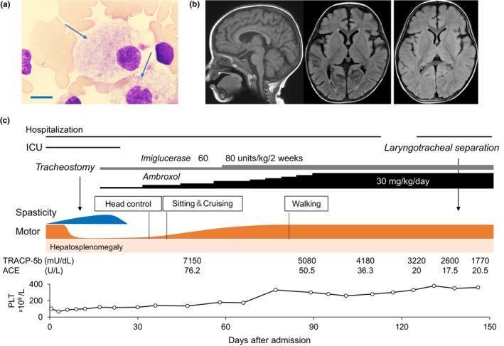
This composite educational graphic details the diagnosis, neuroimaging, and treatment response for a patient with Gaucher disease. (a) A high-power light micrograph of a May-Grünwald-Giemsa stained bone marrow smear reveals characteristic Gaucher cells (arrows), which are large macrophages featuring voluminous, pale, 'crinkled paper' or striated cytoplasm. A 10 μm scale bar is included for reference. (b) Neuroradiological assessment includes a sagittal T1-weighted MRI and axial FLAIR images from day 24 of admission and a follow-up axial FLAIR at 3 months post-discharge, all demonstrating normal brain parenchyma without degenerative or inflammatory lesions. (c) A comprehensive clinical timeline tracks therapeutic interventions, including Imiglucerase and Ambroxol, against clinical milestones. The chart correlates pharmacological dosing with motor development (head control to walking), reduction in hepatosplenomegaly, and improvements in laboratory biomarkers such as Tartrate-Resistant Acid Phosphatase 5b (TRACP-5b), Angiotensin-Converting Enzyme (ACE), and platelet (PLT) counts over a 150-day period.
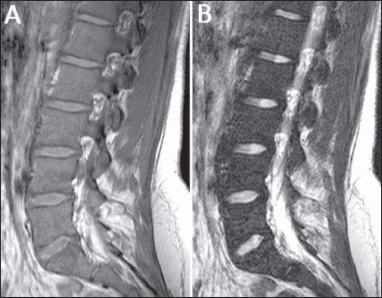
This diagnostic image consists of two sagittal MRI views of the lumbar spine from a 22-year-old patient with Gaucher's disease: a T1-weighted image (A) and a T2-weighted image (B). The primary finding is diffuse bone marrow infiltration, which manifests as a significant decrease in marrow signal intensity across the vertebral bodies. In image A (T1), the vertebral bone marrow appears abnormally hypointense compared to the hyperintense intervertebral discs and presacral fat. In image B (T2), the marrow remains dark, further emphasizing the displacement of normal fatty marrow by Gaucher cells. Additionally, there is a visible loss or replacement of the high-signal fat that normally surrounds the basivertebral veins. This pattern of signal change—marrow becoming darker than the adjacent discs on T1 sequences—is a hallmark of infiltrative marrow disorders. The images serve as an educational example of how systemic metabolic diseases like Gaucher's disease can be staged using MRI-based bone marrow burden (BMB) scoring systems.
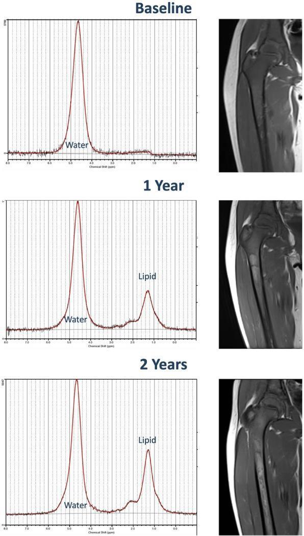
This composite educational image illustrates the longitudinal response of bone marrow to enzyme replacement therapy (ERT) in a pediatric patient with Gaucher disease over a two-year period. The panel is organized into three time points: Baseline, 1 Year, and 2 Years. The left column displays Magnetic Resonance Spectroscopy (MRS) spectra, which quantitatively track the marrow's chemical composition. At Baseline, a single dominant water peak is visible with a negligible lipid peak, representing high cellular infiltration. By 1 Year and 2 Years, a progressive increase in the 'Lipid' peak (fat fraction) is observed relative to the 'Water' peak, indicating therapeutic replacement of Gaucher cells with normal yellow marrow. The right column contains corresponding T1-weighted coronal MRI scans of the right proximal femur. These clinical images show a transition from diffuse, low-signal marrow infiltration at Baseline to a subtle but progressive increase in T1 signal intensity by Year 2. This brightening of the medullary canal correlates with the increasing fat fraction identified on MRS, serving as a visual marker for improved marrow health and treatment efficacy.
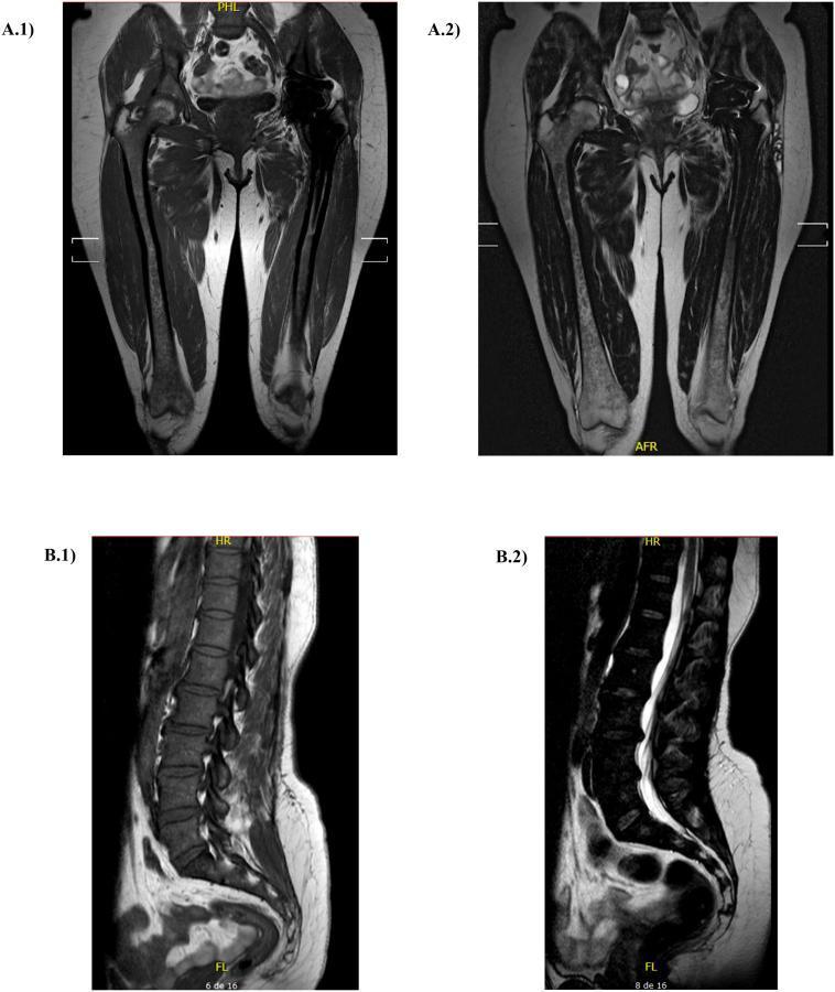
This composite diagnostic image displays magnetic resonance imaging (MRI) of the femurs and lumbar spine, used for assessing Bone Marrow Burden (BMB) in Gaucher disease. A.1 and A.2 provide coronal views of the bilateral femurs. A.1 is a T1-weighted sequence showing fatty bone marrow as high signal intensity (bright), while A.2 is a T2-weighted sequence. Significant signal loss (hypointensity) is visible within the femoral shafts and epiphyses, particularly on T1, suggesting the infiltration of Gaucher cells replacing normal fatty marrow. There is a visible asymmetry due to a left total hip arthroplasty (prosthetic replacement) which appears as a large signal void. B.1 and B.2 provide sagittal views of the lumbar spine. B.1 (T1-weighted) shows dark, heterogeneous signal intensity within the vertebral bodies, indicating diffuse marrow infiltration. B.2 (T2-weighted) illustrates the hyperintense (bright) cerebrospinal fluid (CSF) in the spinal canal, contrasting with the relatively low-signal bone marrow. These images are clinically relevant for scoring skeletal involvement and monitoring treatment response in metabolic storage disorders.
https://cocukmetabolizma.com/wp-content/uploads/2020/08/gauc…
PMID: 40611398
GAUCHER'S DISEASE - Comprehensive Notes
1. DEFINITION AND CLASSIFICATION
| OMIM # | Phenotype |
|---|---|
| 230800 | Gaucher disease, Type I (non-neuronopathic) |
| 230900 | Gaucher disease, Type II (acute neuronopathic) |
| 231000 | Gaucher disease, Type III (subacute/chronic neuronopathic) |
| 231005 | Gaucher disease, Type IIIC (cardiovascular form) |
| 608013 | Gaucher disease, perinatal lethal form |
| 606463 | GBA1 gene (glucosidase, beta, acid) |
- Thompson & Thompson Genetics, p. 493; GeneReviews (Pastores & Hughes 2018)
2. EPIDEMIOLOGY
-
General population prevalence: 1 in 40,000 to 1 in 100,000 live births
-
Ashkenazi Jews: ~1 in 450-855 live births (highest frequency of any ethnic group)
-
Carrier frequency among Ashkenazi Jews: 1 in 12-15 individuals
-
Type 1 accounts for >90% of all GD in Western populations
-
~600 pathogenic GBA1 variants have been described
-
Harrison's 22E, p. 3399; Tietz Laboratory Medicine 7th Ed
3. BIOCHEMISTRY AND PATHOGENESIS
3.1 Enzymatic Defect
- Periphery: Breakdown of senescent blood cells and tissue debris
- CNS: Turnover of neuronal membrane glycolipids
3.2 Gaucher Cells
- Large cell (20-100 μm), eccentric nucleus, condensed chromatin
- Cytoplasm has classic "crumpled/crinkled tissue paper" or "wrinkled silk" appearance
- PAS-positive, CD68-positive
- Mainly infiltrate: bone marrow > spleen > liver > lungs > skin
- Considered the primary instigators of disease pathogenesis
3.3 Mechanism of Organ Damage
| Organ | Mechanism |
|---|---|
| Bone marrow | Marrow packing → infarction, ischemia, cortical destruction |
| Spleen | Infiltration → massive splenomegaly + hypersplenism → cytopenia |
| Liver | Kupffer cell replacement → hepatomegaly; occasionally cirrhosis |
| Bone | Defective bone remodeling + osteoclast/osteoblast dysregulation → osteopenia, avascular necrosis |
| CNS (types 2/3) | GL-1 accumulation + neuronal loss → neurodegeneration |
| Parkinson risk | Defective GCase → compromised lysosomal protein degradation → α-synuclein accumulation in substantia nigra |
- Thompson & Thompson Genetics, p. 494-495; Harrison's 22E, p. 3399-3400
4. CLINICAL FEATURES BY TYPE
Type 1 - Non-neuronopathic (OMIM 230800)
- Younger patients: greater hepatosplenomegaly + cytopenias
- Older patients: greater chronic bone disease
- Hepatosplenomegaly - virtually universal; spleen typically >> liver
- Splenic infarctions (can mimic acute abdomen)
- Severe liver dysfunction uncommon; cirrhosis rare
- Coagulation factor abnormalities → bleeding diathesis (esp. epistaxis)
- Anemia, thrombocytopenia, leukopenia (from hypersplenism + marrow infiltration)
- Pancytopenia severity not directly proportional to organ volumes
- Erlenmeyer flask deformity - undertubulation of distal femur
- Osteopenia/osteoporosis
- Avascular necrosis (esp. femoral head)
- Bone crises - acute excruciating pain, localized erythema, fever, leukocytosis (represent acute bone infarction; absent uptake on pyrophosphate nuclear scan)
- Vertebral compression fractures
- Endosteal scalloping on X-ray
- Fracture of femoral neck
- Pulmonary hypertension
- Alveolar Gaucher cell accumulation
- More common in splenectomized females
- Monoclonal gammopathy, myelodysplasia, myeloma, lymphoma (adult patients)
- Parkinson's disease: GBA mutations in heterozygous or homozygous state = significantly increased lifetime risk (20-30x the general population risk); 5-10% of all Parkinson's patients carry a GBA variant
Type 2 - Acute Neuronopathic (OMIM 230900)
- Onset: 3-6 months of life
- Features: Failure to thrive, hepatosplenomegaly → developmental regression, neurological deterioration, spasticity, seizures
- Rapid brainstem involvement with bulbar palsy, oculomotor abnormalities, opisthotonus
- Recurrent pulmonary infections
- No effective neuroprotective therapy; ERT does not cross blood-brain barrier
Type 3 - Chronic Neuronopathic (OMIM 231000)
- Early childhood: Rapidly progressive massive visceral disease + slowly progressive CNS
- Adolescence: Dementia
- Early adulthood: Rapidly progressive uncontrolled myoclonic seizures + mild visceral disease
- Supranuclear lateral gaze palsy (SNLGP) - earliest and characteristic finding; may be static for decades
- Dementia, ataxia, myoclonic seizures
- Cognitive degeneration (slowly progressive or static)
- Visceral (but NOT CNS) involvement responds to ERT
- More frequent among non-Western world populations
Type IIIC - Cardiovascular Form (OMIM 231005)
- Calcification of mitral and aortic valves
- Mild splenomegaly
- Corneal opacities
- Supranuclear ophthalmoplegia
- Associated with the D409H (p.Asp409His) genotype
Perinatal Lethal Form (OMIM 608013)
- Collodion skin abnormalities
- Nonimmune hydrops fetalis
- Lethal at or shortly after birth
5. GENETICS AND MOLECULAR BASIS
5.1 Gene
- GBA1 gene (formerly GBA), chromosome 1q21
- ~600 mutations described
- Inheritance: Autosomal recessive
5.2 Common Mutations (especially in Ashkenazi Jews)
| Mutation | Type | Effect |
|---|---|---|
| p.N370S (p.N409S) | Missense | Most common in Ashkenazi Jews; 100% associated with Type 1 (non-neuronopathic); protects against CNS involvement |
| p.L444P (p.L483P) | Missense | Very low residual activity; L444P/L444P → life-threatening to very severe disease; many develop CNS involvement |
| 84GG | Frameshift insertion at cDNA pos 84 | Null allele |
| IVS-2-1 | Splice junction | Null allele |
5.3 Genotype-Phenotype Correlation
| Genotype | Phenotype |
|---|---|
| N370S/N370S | 100% Type 1 (non-neuronopathic); later onset, milder; up to 40% may be asymptomatic |
| N370S/other allele | Type 1; earlier onset, more severe |
| L444P/L444P | Severe Type 1 to Type 3; many develop CNS involvement in first two decades |
| D409H (p.Asp409His) | Cardiovascular form (Type IIIC) - valve calcification, corneal opacity |
| Null alleles (84GG, IVS-2-1) | Severe Type 1 or neuronopathic |
- Harrison's 22E, p. 3399-3400; Thompson & Thompson, p. 493-494
6. DIAGNOSIS
6.1 Suggestive Clinical Features
- Hydrops fetalis, collodion skin, hepatosplenomegaly, early neurodegeneration
- Splenomegaly (most prominent), hepatomegaly, failure to thrive, bone pain, anemia, thrombocytopenia, pathological fractures
- Unexplained splenomegaly/hepatomegaly + cytopenias
- Bone disease (osteopenia, avascular necrosis, vertebral fractures)
- Easy bruising/bleeding
- Supranuclear lateral gaze palsy (earliest in Type 3)
- Developmental regression, spasticity, seizures
6.2 Diagnostic Tests
- Glucocerebrosidase activity in peripheral blood leukocytes (or other nucleated cells; fibroblasts)
- Diagnostic: 0-15% of normal (i.e., severely reduced)
- Sensitivity for heterozygote detection is poor with enzyme assay alone
- GBA1 gene sequencing (standard for heterozygote detection and prognostication)
- Targeted mutation panel (Ashkenazi Jewish patients: common 4 mutations cover ~85%)
- Multigene LSD panel if needed
| Biomarker | Notes |
|---|---|
| Chitotriosidase | Most widely used; markedly elevated in active disease; normalizes with ERT |
| Angiotensin-converting enzyme (ACE) | Elevated |
| Acid phosphatase (tartrate-resistant) | Elevated |
| Ferritin | Elevated |
| Lyso-Gb1 (glucosylsphingosine) | Emerging sensitive and specific biomarker; correlates with disease severity; valuable in monitoring (per recent systematic review, PMID data 2021) |
- X-ray: Erlenmeyer flask deformity, endosteal scalloping, lytic/sclerotic lesions
- MRI: Gold standard for bone marrow involvement - T1-weighted sequences show replacement of normal fatty marrow by Gaucher cells (low signal); Bone Marrow Burden (BMB) score uses femur + spine T1/T2 MRI
- Not required for diagnosis but shows pathognomonic Gaucher cells
- Used if enzyme assay or genetic testing is unavailable
7. DIFFERENTIAL DIAGNOSIS
| Feature | Gaucher | Niemann-Pick A/B | Tay-Sachs | MPS |
|---|---|---|---|---|
| Enzyme defect | Glucocerebrosidase | Acid sphingomyelinase | Hexosaminidase A | Various |
| Ethnic predilection | Ashkenazi Jews | Ashkenazi Jews | Ashkenazi Jews | Various |
| HSM | Yes | Yes | No | Yes |
| CNS (Type 1) | No | Type A: Yes | Yes | Variable |
| Bone disease | Prominent | No | No | Yes |
| Cherry red spot | Absent | Type A: present | Present | Variable |
| Gaucher cells | Yes | No (foam cells) | No | No |
8. TREATMENT
8.1 Disease-Specific Therapies
Enzyme Replacement Therapy (ERT)
- Mechanism: IV infusion of recombinant human glucocerebrosidase (mannose-terminated = targets macrophage mannose receptors for lysosomal delivery)
- Indication: Types 1 and 3 (visceral/hematologic); first-line for significantly affected patients
- Approved agents:
- Imiglucerase (Cerezyme) - CHO-derived recombinant
- Velaglucerase alfa (VPRIV) - human cell-line derived
- Taliglucerase alfa (Elelyso) - plant-derived recombinant
- Efficacy: Markedly reduces hepatosplenomegaly, improves hematologic values (anemia, thrombocytopenia), reduces bone disease progression, normalizes biomarkers
- Limitation: Cannot cross the blood-brain barrier - does NOT prevent neurological deterioration in Types 2 or 3
- ERT has eliminated the need for splenectomy in most patients
Substrate Reduction Therapy (SRT)
- Mechanism: Reduces substrate synthesis by inhibiting glucosylceramide synthase (first step of ceramide glycosylation)
- Indication: Mild-to-moderate Type 1 GD; alternative or addition to ERT; adults who cannot tolerate ERT
- Approved agents:
- Eliglustat tartrate (Cerdelga) - oral, first-line for Type 1 adults; CYP2D6 metabolizer status must be tested first
- Miglustat (Zavesca) - oral, second-line (more side effects: GI, tremor); approved for Type 1
- Efficacy: Decreases/mildly improves bone involvement and pancytopenia
| Drug | Class | Route | Indication |
|---|---|---|---|
| Imiglucerase | ERT | IV | Types 1 & 3 |
| Velaglucerase alfa | ERT | IV | Types 1 & 3 |
| Taliglucerase alfa | ERT | IV | Types 1 & 3 |
| Eliglustat | SRT (GCS inhibitor) | Oral | Type 1 (first-line oral) |
| Miglustat | SRT (GCS inhibitor) | Oral | Type 1 (second-line) |
- Goldman-Cecil Medicine; Dermatology 5th Ed; Harrison's 22E
8.2 Adjunctive/Symptomatic Treatments
- Bisphosphonates - for osteoporosis/bone density improvement (as adjunct to ERT)
- Joint replacement surgery - femoral head avascular necrosis (irreversible)
- Splenectomy (now rarely needed; increases risk of pulmonary hypertension post-operatively)
- Blood transfusions - for severe cytopenias
- Analgesics - for bone pain and crises
- Vertebral surgery - for severe progressive kyphosis (can occur despite ERT)
8.3 Emerging Therapies (as of 2025)
-
Gene therapy - AAV-based approaches; clinical trials ongoing
-
Pharmacological chaperone therapy - stabilizes misfolded GCase (ambroxol has shown activity, especially for neuronopathic forms)
-
mRNA/saRNA-based protein replacement - investigational
-
HSCT (bone marrow transplant) - largely superseded by ERT/SRT but may have role in selected neuronopathic cases
-
Gaucher disease state of the art review 2025 (PMID 40611398); Challenges in GD expert panel 2025 (PMID 40112481)
8.4 Monitoring on Treatment
- Biomarkers every 6-12 months (chitotriosidase, lyso-Gb1, ACE, ferritin)
- CBC, LFTs
- Abdominal MRI/ultrasound (organ volumes) every 1-2 years
- Skeletal MRI (BMB score) every 2-4 years
- Bone density (DXA scan) every 2 years
- Neurological assessment for Types 2/3
9. PROGNOSIS
- Type 1 with ERT: Near-normal life expectancy; significant quality of life improvement
- Type 2: Fatal; death by 2 years; ERT does not alter neurological course
- Type 3: Survival into 3rd-4th decade; visceral disease responds; CNS does not
- Parkinson's risk: Significantly elevated in GBA carriers and patients lifelong
- Malignancy risk: Increased risk of hematologic malignancies (multiple myeloma, lymphoma)
- Irreversible damage: Avascular necrosis and established bone disease do not reverse with ERT
10. INHERITANCE AND GENETIC COUNSELING
- AR: Carrier parents → 25% risk of affected child per pregnancy
- Heterozygote detection: Molecular testing (enzyme assay has poor sensitivity for carriers)
- Important: Some N370S/N370S homozygotes are completely asymptomatic (up to 40%)
- GBA carrier status confers 20-30-fold increased risk of Parkinson's disease
- Prenatal diagnosis (CVS, amniocentesis) and preimplantation genetic diagnosis are available
11. KEY MNEMONICS
- G - Growth retardation
- A - Anemia
- U - Undertubulation (Erlenmeyer flask)
- C - Cytopenia / Crinkled-paper cells
- H - Hepatosplenomegaly
- E - Enzyme: glucocerebrosidase deficient
- R - Recessive autosomal inheritance
APPROACH TO GAUCHER'S DISEASE - CLINICAL FLOWCHART
CLINICAL SUSPICION
        |
        ▼
┌─────────────────────────────────────────────────────┐
│  SUSPECT GAUCHER'S DISEASE IF:                      │
│  • Unexplained splenomegaly (especially massive)    │
│  • Hepatomegaly + cytopenias (anemia/thrombocytopenia│
│  • Bone pain / Avascular necrosis / Erlenmeyer flask│
│  • Family history / Ashkenazi Jewish ancestry       │
│  • Neonatal hydrops fetalis / collodion skin        │
│  • Unexplained neurological regression in infant    │
└─────────────────────────────────────────────────────┘
        |
        ▼
STEP 1: INITIAL WORKUP
┌─────────────────────────────────────────────────────┐
│  • CBC with differential                            │
│  • LFTs, coagulation profile                        │
│  • Skeletal survey X-ray                            │
│  • Abdominal ultrasound/MRI (organ volumes)         │
│  • Biomarkers: chitotriosidase, ACE, ferritin,      │
│    acid phosphatase, lyso-Gb1                       │
└─────────────────────────────────────────────────────┘
        |
        ▼
STEP 2: CONFIRM DIAGNOSIS
┌─────────────────────────────────────────────────────┐
│  GLUCOCEREBROSIDASE ENZYME ACTIVITY                 │
│  (peripheral blood leukocytes / fibroblasts)        │
│                                                     │
│  Result: 0-15% of normal → CONFIRMED               │
│                                                     │
│  PLUS GBA1 gene sequencing (defines genotype,       │
│  prognosis, carriers, prenatal counseling)          │
└─────────────────────────────────────────────────────┘
        |
        ▼
STEP 3: CLASSIFY TYPE
┌─────────────────────────────────────────────────────┐
│  Neurological assessment:                           │
│                                                     │
│  NO CNS involvement?                               │
│       → TYPE 1 (Non-neuronopathic)                 │
│                                                     │
│  CNS involvement present?                          │
│       Onset <1 yr, rapidly fatal? → TYPE 2          │
│       Onset >1 yr, chronic course? → TYPE 3         │
│       Cardiac valves + corneal opacity? → TYPE IIIC  │
│       Hydrops fetalis? → PERINATAL LETHAL           │
└─────────────────────────────────────────────────────┘
        |
        ▼
STEP 4: STAGING AND SEVERITY ASSESSMENT
┌─────────────────────────────────────────────────────┐
│  • Organ volumes (MRI: liver, spleen)               │
│  • MRI Bone Marrow Burden (BMB) score               │
│    (femur T1/T2 + spine T1/T2)                      │
│  • DEXA scan (bone mineral density)                 │
│  • Platelet count, hemoglobin                       │
│  • Neurological evaluation (types 2/3)              │
│  • Echocardiogram (type IIIC)                       │
└─────────────────────────────────────────────────────┘
        |
        ▼
STEP 5: TREATMENT DECISION
        |
        ├──────────────────────────────────────────────
        │                                             │
        ▼                                             ▼
  TYPE 1 (symptomatic)                      TYPE 1 (mild/asymptomatic)
  TYPE 3 (visceral)                                   │
        │                                       MONITORING
        ▼                                       (no therapy unless
  ┌───────────────┐                              progresses)
  │ FIRST-LINE:   │
  │ ERT (IV) OR   │
  │ Eliglustat    │
  │ (oral, adult) │
  └───────────────┘
        │
        ├── ERT not tolerated / allergy?
        │         ▼
        │   SWITCH TO SRT
        │   (Eliglustat or Miglustat)
        │
        ├── Bone disease?
        │         ▼
        │   + Bisphosphonates (adjunct)
        │   + Joint replacement (irreversible AVN)
        │
        ├── Severe thrombocytopenia/bleeding?
        │         ▼
        │   Platelet transfusion (acute)
        │   Splenectomy (last resort - risk of PAH)
        │
        └── TYPE 2 (neuronopathic)?
                  ▼
            Supportive care only
            (ERT does NOT cross BBB)
            Palliative approach

        |
        ▼
STEP 6: MONITORING ON TREATMENT
┌─────────────────────────────────────────────────────┐
│  Every 6 months:                                    │
│  • CBC, LFTs, biomarkers (chitotriosidase, lyso-Gb1)│
│                                                     │
│  Every 1-2 years:                                   │
│  • Abdominal MRI (organ volumes)                    │
│                                                     │
│  Every 2-4 years:                                   │
│  • Skeletal MRI (BMB score)                         │
│  • DEXA scan                                        │
│                                                     │
│  Ongoing:                                           │
│  • Screen for Parkinson's disease                   │
│  • Screen for hematologic malignancy                │
│  • Genetic counseling for family members            │
└─────────────────────────────────────────────────────┘
QUICK REFERENCE SUMMARY TABLE
| Feature | Type 1 | Type 2 | Type 3 | Type IIIC |
|---|---|---|---|---|
| OMIM | 230800 | 230900 | 231000 | 231005 |
| Onset | Any age | <6 months | 3-8 yrs | Childhood |
| CNS involvement | None | Severe, early | Chronic, variable | Variable |
| Survival | Near-normal (with ERT) | <2 years | 3rd-4th decade | Variable |
| Hepatosplenomegaly | Yes | Yes | Yes (severe) | Mild |
| Bone disease | Yes | Rarely | Yes (severe) | No |
| Valve disease | No | No | No | YES |
| Corneal opacity | No | No | No | YES |
| SNLGP | No | Yes | Yes (characteristic) | Yes |
| Key mutation | N370S | L444P/null | L444P | D409H |
| ERT response | Excellent | No benefit (CNS) | Visceral only | Partial |
| Ethnicity | Ashkenazi Jews | All | Non-Western (esp. Swedish, Arab, Japanese) | Portuguese, Arab |
- Harrison's Principles of Internal Medicine 22E (2025), p. 3399-3400
- Thompson & Thompson Genetics and Genomics in Medicine 9th Ed, p. 493-495
- Emery's Elements of Medical Genetics and Genomics
- Goldman-Cecil Medicine 27E
- Tietz Textbook of Laboratory Medicine 7th Ed
- GeneReviews: Pastores GM, Hughes DA. Gaucher Disease. [Updated 2018 Jun 21]. NCBI Bookshelf
- OMIM #230800, #230900, #231000, #231005, #606463, #608013
- Camou F, Berger MG. Gaucher disease, state of the art and perspectives. J Intern Med 2025 (PMID 40611398)
- Grabowski GA et al. Challenges in Gaucher disease: Perspectives from expert panel. Mol Genet Metab 2025 (PMID 40112481)
- Luettel DM et al. Emerging biomarkers in Gaucher disease. Adv Clin Chem 2025 (PMID 39818434)
Add points from Nelson textbook of Pediatrics and other Pediatrics textbook Since I am Pediatrician I want topics related to Pediatrics more than adult
"Gaucher disease"[MeSH Terms] AND child[MeSH Terms]
Nelson textbook pediatrics Gaucher disease children clinical features diagnosis treatment ERT pediatric onset bone disease
PMID: 35367141
PMID: 41645321
https://pmc.ncbi.nlm.nih.gov/articles/PMC12575321
PMID: 40211441
GAUCHER'S DISEASE - PEDIATRIC SUPPLEMENT
KEY FEATURES TABLE BY TYPE (Rheumatology Ch. 213)
| Key Features | Type 1 (Non-neuronopathic) | Type 2 (Acute Neuronopathic) | Type 3 (Subacute Neuronopathic) |
|---|---|---|---|
| Incidence | 1:40,000-60,000 (1:850 Ashkenazi Jews) | <1:100,000 | <1:50,000-100,000 |
| Gene/Locus | GBA/1q22 | GBA/1q22 | GBA/1q22 |
| Peak age at onset | Any age from birth to old age | Infancy | Childhood or adolescence |
| Hepatosplenomegaly | + to +++ | + to ++ | + to ++++ (most severe) |
| Hematologic symptoms | + to +++ | +++ | + to +++ |
| Skeletal involvement | + to +++ | Absent | ++ to ++++ |
| Neurodegeneration | Absent | +++ | + to +++ (progressive) |
| Median age at death | Childhood or adulthood | 9 months | Childhood or adulthood |
| Ethnic predilection | Panethnic/Ashkenazi Jews | Panethnic | Panethnic/Norrbottnian Sweden |
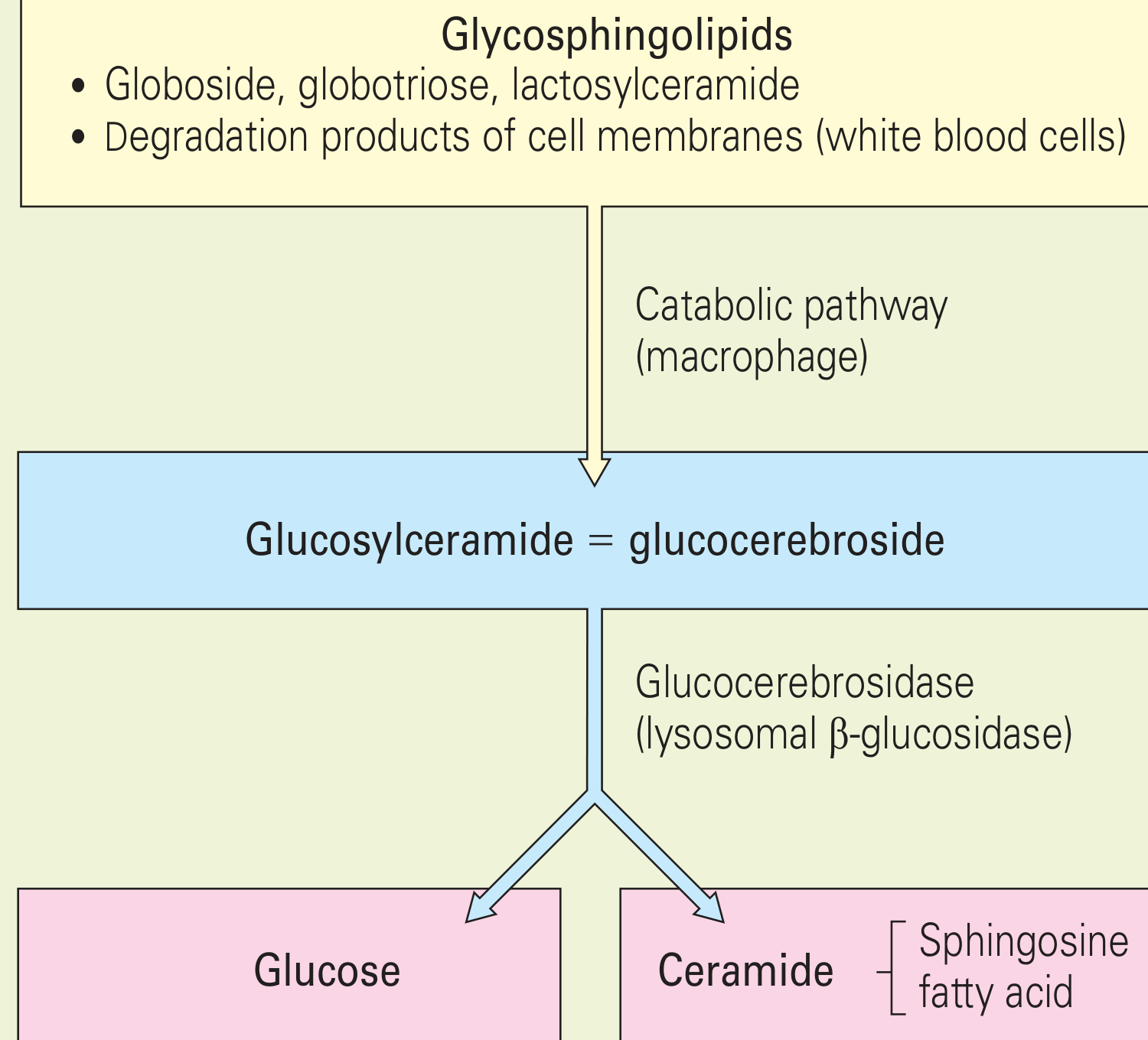
METABOLIC PATHWAY DIAGRAM (from Rheumatology Ch. 213)
1. PEDIATRIC PRESENTATION - AGE-BASED APPROACH
A. NEONATES / PERINATAL LETHAL FORM (OMIM 608013)
- Collodion skin (thickened, parchment-like skin at birth)
- Non-immune hydrops fetalis (massive generalized edema, ascites, pleural effusion)
- Hepatosplenomegaly, thrombocytopenia
- Rarely survives - lethal at or shortly after birth
- GBA mutations: typically null alleles or compound heterozygotes with severe mutations
B. INFANTS (TYPE 2 - Acute Neuronopathic, OMIM 230900)
- 3-6 months: Failure to thrive, feeding difficulties, poor weight gain
- By 6 months: Hepatosplenomegaly becomes obvious
- 3-6 months: Developmental regression begins
- Neurological progression: Hypotonia → spasticity
- Brainstem involvement (cardinal features):
- Trismus (jaw rigidity)
- Strabismus (ocular muscle involvement)
- Bulbar palsy: Dysphagia, drooling, dysphonia
- Opisthotonus (severe extensor rigidity)
- Oculomotor abnormalities - strabismus, then gaze palsies
- Seizures - focal or generalized
- Spasticity and rigidity of limbs
- Recurrent pulmonary infections (due to aspiration from bulbar palsy)
- Death by 2 years (median 9 months), usually from respiratory failure/aspiration pneumonia
- Glucocerebrosidase activity in leukocytes: 0-15% of normal
- Bone marrow biopsy: Gaucher cells
- MRI brain: May show diffuse signal changes, cortical atrophy late
- GBA genotyping: Often L444P/L444P or null alleles
C. YOUNG CHILDREN (TYPE 3 - Subacute/Chronic Neuronopathic, OMIM 231000)
- Visceral disease - Hepatosplenomegaly (often more severe than Type 1)
- Hematologic - Cytopenias (thrombocytopenia > anemia > leukopenia)
- Neurological - Variable; from only SNLGP to:
- Ataxia (cerebellar, prominent in young children ≤5 years)
- Myoclonic epilepsy (progressive myoclonic epilepsy - a common presentation in Japanese/Arab populations)
- Dementia (appears later, >5 years)
- Cognitive impairment - difficulty processing new information, memory problems
- In patients aged ≤5 years: Motor manifestations dominate (ataxia, gaze difficulty, tremors); neurocognitive symptoms appear later
- In older children/adolescents: Cognitive impairment, fatigue, bone pain become more prominent
- Social/functional impacts: Difficulty at school, restricted mobility, declining independence
| Subtype | Age | Dominant feature |
|---|---|---|
| Type 3a | Childhood | Rapidly progressive myoclonic epilepsy + mild visceral disease |
| Type 3b | Early childhood | Massive visceral disease + slowly progressive CNS involvement |
| Type 3c (OMIM 231005) | Childhood | Cardiac valve calcification + corneal opacity + D409H mutation |
D. CHILDREN AND ADOLESCENTS (TYPE 1 - Non-neuronopathic)
- Peak 1: <10-15 years
- Peak 2: ~25 years
- Younger children have greater HSM + cytopenias; bone disease develops progressively
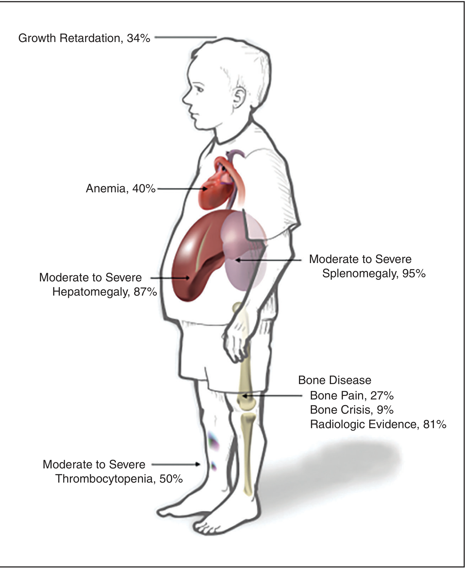
- Growth retardation: 34% of children at diagnosis (see organ involvement diagram above)
- Delayed puberty/pubertal development - important pediatric outcome goal
- Short stature - occurs even without overt malnutrition; multi-factorial (HSM + bone disease + chronic inflammation)
- Erlenmeyer flask deformity develops progressively during the period of rapid skeletal growth and is evident in ~80% of adults - meaning it begins in childhood
- Bone disease may be more aggressive in children due to active bone remodeling
- "H-vertebra" deformity (steplike depressions in vertebral body - same as in sickle cell anemia)
- Gaucheroma - massive focal osteolytic lesion in a metaphysis resembling tumor or aneurysmal bone cyst
2. PEDIATRIC DIAGNOSIS (KEY POINTS FOR PEDIATRICIANS)
When to Suspect Gaucher's in a Child
| Clinical Scenario | Age Group | Relevance |
|---|---|---|
| Unexplained massive splenomegaly | Any age | Most common initial finding (95%) |
| Infant with hepatosplenomegaly + developmental regression | <1 yr | Type 2 - emergency |
| Child with recurrent bone pain + easy bruising | 5-15 yrs | Type 1 |
| Child with anemia + thrombocytopenia + HSM | Any | Think Gaucher before leukemia |
| Neonatal hydrops + collodion skin | Neonatal | Perinatal lethal form |
| Horizontal gaze palsy in a child | 3-8 yrs | Type 3 - earliest neurological sign |
| Family history in Ashkenazi Jewish family | Any | Screen early |
| Child found to have Erlenmeyer flask on X-ray | Any | Strongly suggestive |
Diagnostic Steps in Children
-
FIRST: Enzyme activity - Glucocerebrosidase in peripheral blood leukocytes
- Normal activity: 12.0-17.5 nmol/mg·h (lab-specific reference ranges vary)
- Gaucher: 0-15% of normal (0-3.5 nmol/mg·h)
- Can also use dried blood spot (DBS) - important for newborn screening programs
-
CONFIRM: GBA1 gene sequencing
- Establishes genotype for prognosis and family counseling
- In Ashkenazi Jewish children: 4-mutation panel first (covers 85%)
- In non-Ashkenazi: Full gene sequencing
-
BIOMARKERS for severity and monitoring:
- Chitotriosidase (most widely used) - caution: 6% of Caucasian population are chitotriosidase-deficient (genetic CHIT1 variant - check for this)
- Lyso-Gb1 (glucosylsphingosine) - preferred now as it is NOT affected by CHIT1 polymorphism; correlates well with disease burden; excellent for pediatric monitoring
- ACE, ferritin, tartrate-resistant acid phosphatase
-
IMAGING in children:
- Skeletal survey X-ray: Erlenmeyer flask deformity, vertebral changes
- MRI of femora (coronal T1/T2) + MRI spine - Bone Marrow Burden (BMB) score
- Abdominal MRI - spleen and liver volumes (expressed as multiples of normal for body weight)
- DEXA scan (DXA) - bone mineral density; Z-score used in children (not T-score)
- Organ volumes in children expressed as multiples of normal (MN):
- Hepatomegaly: Moderate >1.25-2.5 MN, Severe >2.5 MN
- Splenomegaly: Moderate >5-15 MN, Severe >15 MN
-
Neurological assessment (mandatory for Types 2 & 3):
- Ophthalmological assessment (horizontal saccades/pursuit - SNLGP)
- EEG (myoclonic epilepsy in Type 3a)
- Formal neuropsychological testing (cognitive function)
- Brain MRI
-
Bone marrow biopsy:
- Not required for diagnosis but may be done when presenting like hematologic malignancy
- Shows pathognomonic Gaucher cells ("crinkled tissue paper" cytoplasm)
- Important: Often the pediatric hematologist performs this first before the diagnosis is suspected
3. PEDIATRIC DIFFERENTIAL DIAGNOSES
| Condition misdiagnosed as | Reason |
|---|---|
| Leukemia/lymphoma | Cytopenias + HSM + bone pain → bone marrow biopsy reveals Gaucher cells |
| ITP | Isolated thrombocytopenia |
| Septic arthritis/osteomyelitis | Bone crisis mimics infection (fever + local pain + tenderness) |
| Portal hypertension | Massive splenomegaly |
| Juvenile idiopathic arthritis | Bone pain and joint involvement |
| Sickle cell anemia | H-vertebra + AVN + bone crises |
- Hemoglobinopathies (thalassemia, sickle cell)
- Osteopetrosis
- Paget disease
- Fibrous dysplasia
- Heavy metal poisoning
- Multiple enchondromatosis
- Gaucher disease
- Niemann-Pick disease
- Thalassemia major
- Portal hypertension
- Visceral leishmaniasis (kala-azar - can closely mimic Gaucher in endemic areas)
- Storage diseases (MPS, GM1 gangliosidosis)
4. PEDIATRIC ERT - SPECIAL CONSIDERATIONS
Indications for Starting ERT in Children (2022 Weinreb et al., PMID 35367141)
- Hemoglobin <11 g/dL (age and sex-adjusted) OR anemia significantly affecting quality of life
- Platelet count <60-120 × 10³/µL (moderate to severe thrombocytopenia)
- Significant hepatomegaly (>1.25 MN) or splenomegaly (>5 MN)
- Bone disease (pain, low BMD, avascular necrosis, bone crisis)
- Growth retardation (below 5th percentile for height)
- Radiologic bone disease on imaging
- Progressive symptoms
ERT Approved Agents for Pediatric Use
| Drug | Age approval | Notes |
|---|---|---|
| Imiglucerase (Cerezyme) | Any age including infants | Most experience; IV every 2 weeks |
| Velaglucerase alfa (VPRIV) | Any age (0-18 yrs per 2026 systematic review, PMID 41645321) | Human cell-line derived; well tolerated; home therapy possible |
| Taliglucerase alfa (Elelyso) | ≥2 years | Plant-derived |
Dosing
- Standard starting dose: 60 IU/kg IV infusion over 2 hours, every 2 weeks
- Children may require dose adjustment based on weight and response
- In severe pediatric cases, higher doses (up to 120 IU/kg) have been used
- Gradual dose reduction to 30 IU/kg q2w possible after stabilization in older patients
Pediatric ERT Therapeutic Targets (Within 1-2 Years of Starting ERT)
| Parameter | Target in Children |
|---|---|
| Hemoglobin | >11.0 g/dL |
| Liver volume | Reduce to within 1.0-1.5x normal; maintain |
| Spleen volume | Reduce to below 2-8x normal |
| Bone mineral density | Improve; attain normal or ideal peak skeletal mass |
| Cortical and trabecular BMD | Increase by year 2 |
| Growth | Achieve normal height per population standards and parental height within 2-3 years |
| Puberty | Achieve normal onset of puberty |
| Bone pain | Lessen or eliminate within 1-2 years |
| Bone crises | Prevent |
| Osteonecrosis | Prevent progression |
| Quality of life | Normalization of schooling, activities |
ERT Response Timeline in Children
- 12-20 weeks: Hematologic variables + organomegaly start improving
- 3 months: Bone pain starts decreasing
- 1-2 years: Imaging shows bone improvement + BMD increases
- 2-3 years: Peak skeletal mass improvement measurable
Velaglucerase Alfa in Pediatrics (Systematic Review 2026, PMID 41645321 - de Las Heras et al.)
- Reviewed 23 studies encompassing 159 pediatric patients (0-18 years)
- Well tolerated in all pediatric age groups including infants
- Improvements in: hematological, visceral, skeletal, biomarker and quality-of-life outcomes in treatment-naive GD1 children
- Home therapy with velaglucerase enhances quality of life for patients AND caregivers
- For GD3 (non-neurological manifestations): Favorable safety, some efficacy - but evidence is limited and exploratory
5. BONE DISEASE IN CHILDREN - EXTRA DETAIL
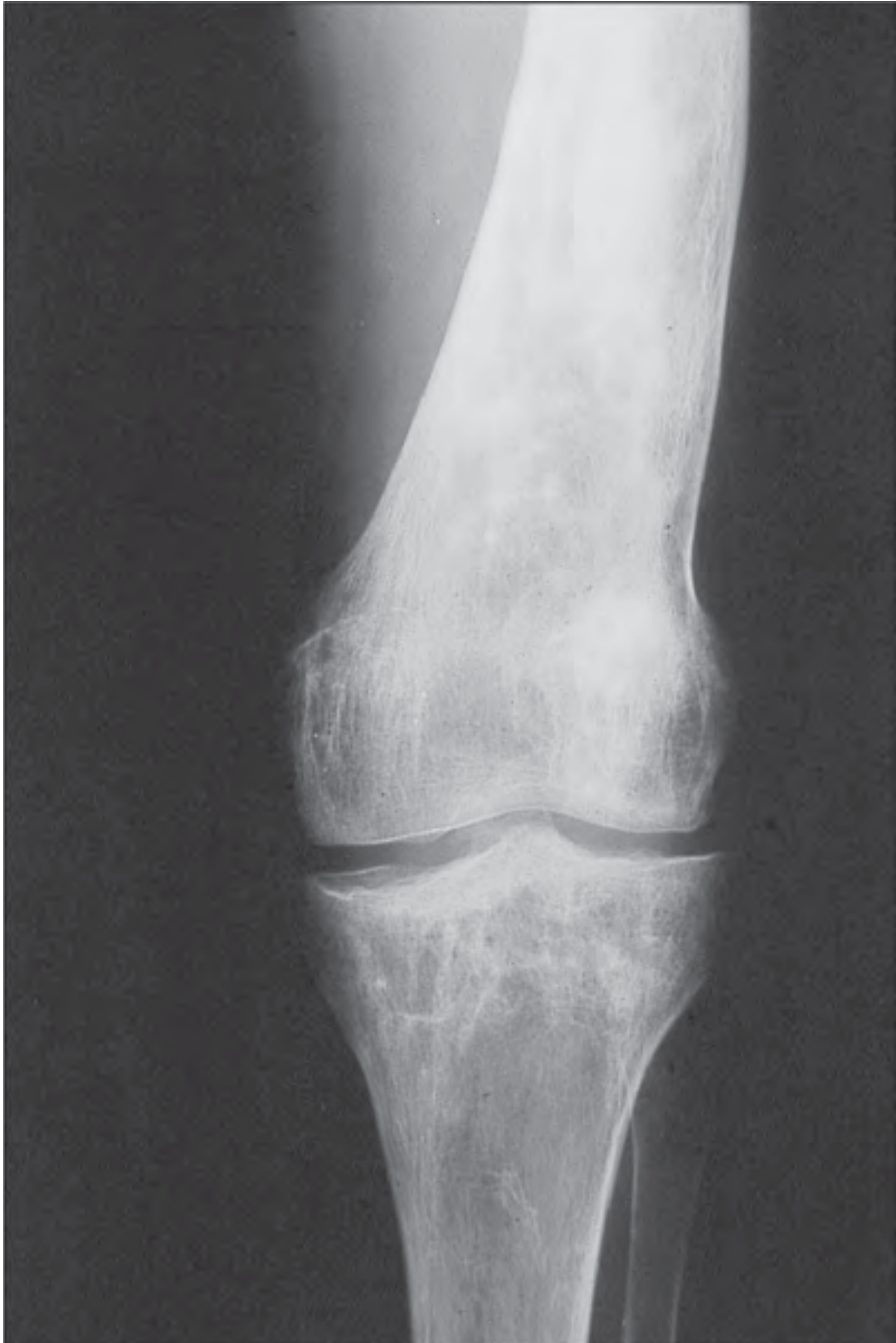
- Develops progressively during childhood's rapid skeletal growth phase
- Evident in ~80% of adults (meaning it starts in childhood)
- Distal femur and proximal tibia most affected
- Very suggestive but not pathognomonic - also seen in thalassemia, osteopetrosis
- Acute episode of excruciating localized bone pain + fever + erythema + leukocytosis
- Represents acute bone infarction (ischemia in Gaucher cell-infiltrated bone)
- Can mimic osteomyelitis or septic arthritis (major diagnostic trap in pediatrics)
- Bone scan (Tc99m): Absent uptake = ischemic crisis (vs increased uptake = osteomyelitis)
- SPECT-CT helps differentiate ischemic crisis from infectious osteomyelitis
- Osteomyelitis occurs in <5% of Gaucher patients but is a real risk
- Occur in 9% of children at diagnosis (see organ involvement chart)
- ERT significantly reduces frequency of bone crises
- Present in 42% of cases before treatment
- Z-score (not T-score) used for children on DXA
- Low bone density in lumbar spine is a strong risk factor for fractures
- Most frequent fractures: vertebral spine (36% of all fractures), then lower extremities
- Early ERT critical to attain normal peak bone mass
6. NEUROLOGICAL FINDINGS - PEDIATRIC FOCUS
Type 2 (Infantile) - CNS Sequence (Adams & Victor's Neurology)
- Abducens palsy (lateral gaze)
- Dysphagia, trismus, rigidity of limbs, dementia
- Opisthotonus
- Spastic quadriplegia
- Death by 2 years
Type 3 (Juvenile) - CNS Manifestations (Adams & Victor's Neurology)
- Features of infantile type: Abducens palsies, dysphagia, trismus, rigidity, dementia
- Features of adult type: Horizontal gaze palsy, diffuse myoclonus, generalized seizures, chronic course
- Diagnosis: Splenomegaly + Gaucher cells + glucocerebroside storage + deficient glucocerebrosidase in leukocytes or fibroblasts
- Motor signs (ataxia, gaze difficulty, tremors) appear in first 3 years and persist beyond 5 years
- Neurocognitive signs appear later in life (school age and beyond)
- For children ≤5 years: Motor > Cognitive
- School impacts: Inability to perform at school, restricted mobility, declining independence
- 58 distinct patient experience concepts identified in GD3 (ataxia, cognitive impairment, fatigue, bone pain most salient)
7. PATHOPHYSIOLOGY - ADDITIONAL CELLULAR MECHANISMS (Rheumatology/Robbins)
- Inflammasome activation: Lysosomal storage + impaired autophagy → inflammasome (multiprotein complex activating caspase-1) → elevated IL-1β and IL-6 → systemic inflammation
- Proinflammatory cytokine release: Macrophages primed to release cytokines in response to stimuli
- Direct hematopoietic impairment: Glucocerebrosidase deficiency directly impairs hematopoietic development
- Erythrophagocytosis by Gaucher cells: Contributes to anemia
- Osteoblast dysfunction: Osteoblast differentiation impaired → reduced bone formation → bone matrix protein and mineral deposition defective
- Uncoupled bone remodeling: Decreased bone formation + increased resorption → osteopenia (decreased osteocalcin in ~50%)
- Wnt/β-catenin pathway downregulation in neuronal progenitors in GD (explains some neurological features)
- Parkinson's link via GD: Impaired macrophage autophagy → accumulation of misfolded proteins (α-synuclein) → Parkinson's risk
8. NEWBORN SCREENING (NBS) FOR GAUCHER DISEASE
- Several countries have piloted or implemented NBS for Gaucher disease using DBS enzyme assay
- Enzyme assay on dried blood spot (DBS) is the method used
- NBS identifies affected and at-risk infants early → early treatment initiation
- Challenges (per PMID 35367141, 2022):
- Many NBS-detected children with GBA mutations remain asymptomatic for years (especially N370S/N370S homozygotes)
- Ethical dilemmas: Pre-symptomatic diagnosis of variable-penetrance disease
- Unclear timing of treatment initiation in pre-symptomatic children
- Cost-benefit issues in heterogeneous populations
- Current consensus: NBS most justified where prevalence is high (Ashkenazi Jewish populations) and where treatment is available
9. DIAGNOSTIC CHALLENGE - GAUCHER vs. HEMATOLOGIC MALIGNANCY IN CHILDREN
- Pallor (anemia)
- Easy bruising/petechiae (thrombocytopenia)
- Bone pain
- Hepatosplenomegaly
- Elevated LDH (from cell turnover)
- Bone marrow with abnormal cells (Gaucher cells may be mistaken for leukemic cells/pseudo-Gaucher cells)
| Feature | Gaucher | Leukemia |
|---|---|---|
| Spleen >> Liver enlargement | Yes (characteristic) | Variable |
| WBC count | Low/normal (leukopenia) | HIGH (leukocytosis) |
| Blast cells on peripheral smear | Absent | Present (in ALL/AML) |
| Bone marrow Gaucher cells | "Crinkled tissue paper" cytoplasm | Blasts with prominent nucleoli |
| Glucocerebrosidase activity | 0-15% normal | Normal |
| Chitotriosidase | Very high | Normal/mildly elevated |
| Lyso-Gb1 | Markedly elevated | Normal |
10. PERIOPERATIVE AND ANESTHETIC CONSIDERATIONS IN CHILDREN
- Thrombocytopenia - platelet count must be checked before any procedure; platelet transfusion may be needed
- Coagulation abnormalities - check coagulation profile (factor V, factor XI deficiency reported)
- Anemia - optimize hemoglobin before elective surgery
- Hepatomegaly - may affect drug metabolism and intraabdominal access
- Massive splenomegaly - rupture risk; avoid trauma to abdomen
- Splenectomy now very rarely needed with ERT availability; if done, risk of post-splenectomy pulmonary hypertension (especially in girls)
11. GENETIC COUNSELING POINTS FOR PEDIATRIC PRACTICE
- Recurrence risk for siblings: 25% (autosomal recessive)
- Carrier testing: Parents are obligate carriers; siblings can be tested (enzyme + molecular)
- Prenatal diagnosis: CVS or amniocentesis - enzyme assay + GBA molecular testing
- Preimplantation genetic testing (PGT): Available at IVF centers
- Asymptomtic N370S/N370S: Up to 40% never develop symptoms - counseling challenge
- GBA carrier status confers ~20-30x increased risk of Parkinson's disease - relevant for parents who are carriers
- Recurrence in siblings of Type 2 (acute neuronopathic) is 25% - counsel parents early regarding poor prognosis; palliative care planning
UPDATED PEDIATRIC APPROACH FLOWCHART
PEDIATRIC CLINICAL SUSPICION
           |
           ▼
┌──────────────────────────────────────────────────────────────┐
│  WHEN TO SUSPECT IN A CHILD:                                 │
│  • Infant (<1 yr): HSM + developmental regression → TYPE 2  │
│  • Child 1-8 yrs: Horizontal gaze palsy → TYPE 3 (early)    │
│  • Any age: Unexplained massive splenomegaly (spleen>>liver) │
│  • Child with bone pain + cytopenias + HSM                   │
│  • "Leukemia workup" with Gaucher cells in marrow            │
│  • Growth retardation + thrombocytopenia                     │
│  • Erlenmeyer flask on X-ray                                 │
│  • Neonatal hydrops + collodion skin                         │
│  • Ashkenazi Jewish family history                           │
└──────────────────────────────────────────────────────────────┘
           |
           ▼
STEP 1: INITIAL WORKUP
┌──────────────────────────────────────────────────────────────┐
│  • CBC + differential (anemia, thrombocytopenia, leukopenia) │
│  • LFTs, coagulation (factors V, XI)                         │
│  • Serum immunoelectrophoresis (monoclonal bands)            │
│  • Skeletal X-ray: Erlenmeyer flask, vertebral changes       │
│  • Abdominal ultrasound (organ size)                         │
│  • Biomarkers: Chitotriosidase + Lyso-Gb1 (preferred pair)  │
│    (ACE, ferritin, tartrate-resistant acid phosphatase)      │
└──────────────────────────────────────────────────────────────┘
           |
           ▼
STEP 2: CONFIRM DIAGNOSIS (MANDATORY)
┌──────────────────────────────────────────────────────────────┐
│  GLUCOCEREBROSIDASE ENZYME ACTIVITY                          │
│  in peripheral blood leukocytes (OR dried blood spot)        │
│                                                              │
│  0-15% of normal = CONFIRMED GAUCHER                         │
│                                                              │
│  THEN: GBA1 gene sequencing                                  │
│  (Ashkenazi: 4-mutation panel first;                        │
│   Non-Ashkenazi: full sequencing)                           │
└──────────────────────────────────────────────────────────────┘
           |
           ▼
STEP 3: CLASSIFY TYPE IN CHILD
┌──────────────────────────────────────────────────────────────┐
│  NEUROLOGICAL ASSESSMENT (mandatory in all children)         │
│                                                              │
│  Age <1 yr + HSM + neurodegeneration?                       │
│          → TYPE 2 (Acute Neuronopathic)                      │
│            Brainstem signs: trismus, strabismus,            │
│            opisthotonus, dysphagia, seizures                 │
│                                                              │
│  Age 3-8 yrs + horizontal gaze palsy + HSM?                 │
│          → TYPE 3 (Subacute Neuronopathic)                   │
│            Subtype: 3a (myoclonic) / 3b (visceral) /        │
│            3c (cardiac + D409H mutation)                     │
│                                                              │
│  No CNS involvement at any age?                              │
│          → TYPE 1 (Non-neuronopathic)                        │
│                                                              │
│  Hydrops fetalis + collodion skin at birth?                  │
│          → PERINATAL LETHAL form                             │
└──────────────────────────────────────────────────────────────┘
           |
           ▼
STEP 4: STAGING/SEVERITY
┌──────────────────────────────────────────────────────────────┐
│  Organ volumes: Abdominal MRI (liver + spleen MN)            │
│  Bone disease: MRI femora + spine (T1/T2) + BMB score        │
│  Bone density: DXA (Z-score in children)                    │
│  Neurological: EEG, brain MRI, saccade testing              │
│  Ophthalmology: horizontal gaze, corneal opacity (Type 3c)   │
│  Pulmonary: Echo (PAH screen), CXR                          │
│  Growth parameters: Height, weight, bone age                 │
│  Pubertal status: Tanner staging                             │
└──────────────────────────────────────────────────────────────┘
           |
           ▼
STEP 5: TREATMENT IN CHILDREN
           |
     ┌─────┴──────────────────┐
     ▼                        ▼
TYPE 1 (symptomatic)         TYPE 2 (Acute Neuronopathic)
TYPE 3 (all children)              │
     │                       PALLIATIVE / SUPPORTIVE CARE
     ▼                       (ERT may be trialed but does
 ┌──────────┐                NOT prevent neurodegeneration)
 │  ERT     │                Seizure management, nutrition,
 │ (IV q2w) │                pulmonary care, family support
 │ 60 IU/kg │
 └──────────┘
     │
     ├── Imiglucerase (Cerezyme) - any age
     ├── Velaglucerase alfa (VPRIV) - 0-18 yrs
     │    [home therapy option for stable children]
     └── Taliglucerase alfa (Elelyso) - ≥2 yrs
     │
     NOTE: SRT (Eliglustat/Miglustat) = NOT for children
     │
     │
     ├── ADJUVANT in children:
     │   • Vitamin D + Calcium supplementation (bone health)
     │   • Physiotherapy (bone pain, mobility)
     │   • Nutritional support (growth)
     │   • Analgesics for bone pain/crises
     │   • Anticonvulsants (Type 3a myoclonic epilepsy)
     │   • Avoid splenectomy if at all possible (increases PAH risk)
     │
     └── HSCT: Considered in Type 3 with life-threatening visceral
             disease unresponsive to ERT; limited neurological benefit

           |
           ▼
STEP 6: MONITORING IN CHILDREN
┌──────────────────────────────────────────────────────────────┐
│  Every 6 months:                                             │
│  • CBC, LFTs, chitotriosidase, lyso-Gb1                     │
│  • Growth parameters (height, weight, Tanner stage)          │
│  • Quality of life / school performance                      │
│                                                              │
│  Every 1-2 years:                                            │
│  • Abdominal MRI (organ volumes)                             │
│  • Neurological evaluation (Types 2 & 3)                    │
│  • EEG (Type 3a)                                             │
│                                                              │
│  Every 2 years:                                              │
│  • DXA scan (Z-score) - bone mineral density                 │
│  • Skeletal MRI (BMB score)                                  │
│  • Ophthalmological assessment                               │
│  • Transition planning in adolescents (to adult services)    │
│                                                              │
│  PEDIATRIC-SPECIFIC TARGETS:                                 │
│  ✓ Normal linear growth & pubertal development               │
│  ✓ Hb >11 g/dL, Plat >120 × 10³/µL                         │
│  ✓ Elimination of bone pain & crises                        │
│  ✓ Normal peak bone mass attained by end of growth           │
│  ✓ Normal school attendance and function                     │
└──────────────────────────────────────────────────────────────┘
PEDIATRIC-SPECIFIC EXAM PEARLS
- Nelson Essentials (9th Ed, 2023): Gaucher listed under "Defects in metabolism of lipids" - classified as sphingolipidosis with glucocerebrosidase deficiency
- Most common lysosomal storage disease overall - and most common sphingolipidosis to present in pediatric practice
- Gaucher vs Niemann-Pick in infants: Both have HSM; Niemann-Pick has cherry-red spot + foamy cells; Gaucher has crinkled-tissue-paper cells, no cherry-red spot
- ERT = macromolecule = cannot cross BBB - this single fact determines why Types 2/3 CNS disease is not treatable with ERT
- SRT is NOT for children - eliglustat/miglustat only for adults
- Bone crisis vs osteomyelitis: Bone scan shows ABSENT uptake in crisis, INCREASED in osteomyelitis
- Horizontal (lateral) gaze palsy in a child = must rule out Type 3 Gaucher (especially in non-Ashkenazi populations)
- D409H mutation = Type IIIC = valve calcification + corneal opacity (cardiovascular form)
- N370S = always Type 1 (never neuronopathic) - at least one N370S allele = protective against CNS involvement
- Lyso-Gb1 (glucosylsphingosine) is the preferred biomarker now - not affected by CHIT1 polymorphism unlike chitotriosidase
- Rheumatology 2-Volume Set (2022), Chapter 213 - "Gaucher Disease" - comprehensive pediatric clinical data
- Adams & Victor's Neurology 12th Ed - Type 2/3 neurological presentation
- Thompson & Thompson Genetics 9th Ed, p. 493-495
- Robbins & Kumar Basic Pathology - lysosomal storage disease pathogenesis
- Weinreb NJ et al. Diagnosis and management of Gaucher in pediatric patients. Mol Genet Metab 2022 (PMID 35367141)
- de Las Heras J et al. Velaglucerase alfa in paediatric patients. Orphanet J Rare Dis 2026 (PMID 41645321) - Systematic Review
- Schiffmann R et al. Gaucher type 3 from infancy through adulthood. Orphanet J Rare Dis 2025 (PMID 40211441)
- Nelson Essentials of Pediatrics 9th Ed (2023) - Marcdante KJ et al., Chapter: Defects in metabolism of lipids
- GeneReviews: Pastores GM, Hughes DA. Gaucher Disease [Updated 2018]. NCBI Bookshelf
So you doesn’t have direct Nelson references
Don’t redo again Just I wanted to know about it
- Harriet Lane Handbook 23rd Ed - but this is a dosing/quick reference, not a textbook
- Red Book 2021 - only for infectious diseases
- Thompson & Thompson Genetics & Genomics in Medicine 9th Ed - has pediatric genetics content
- Rheumatology 2-Vol Set (Ch. 213) - which contains an excellent, detailed Gaucher chapter with pediatric-specific tables and data
- Adams & Victor's Neurology 12th Ed - for Type 2/3 neurological features
- Robbins Pathology - for pathogenesis
- GeneReviews (Pastores & Hughes) - via web extract - this IS the reference Nelson itself cites
- PubMed pediatric literature - PMID 35367141, 41645321, 40211441
Do u have swaimans Peds neurology
- Adams and Victor's Principles of Neurology, 12th Ed
- Bradley and Daroff's Neurology in Clinical Practice
- Eric Kandel's Principles of Neural Science, 6th Ed
- Harriet Lane Handbook 23rd Ed
- Red Book 2021 (infectious diseases only)
So orris is basically for adult medicine not for Pediatrics
- Internal Medicine (Harrison's 22E, Goldman-Cecil)
- Surgery (Campbell, Bailey & Love, Schwartz)
- Neurology (Adams & Victor, Bradley & Daroff)
- Cardiology, Nephrology, Pulmonology, Gastroenterology
- Genetics (Thompson & Thompson, Emery's)
- Radiology, Pathology (Robbins), Biochemistry
- Rheumatology, Dermatology, Orthopedics
- Nelson's Textbook of Pediatrics
- Swaiman's Pediatric Neurology
- Zitelli's Atlas of Pediatric Diagnosis
- Forfar & Arneil's Textbook of Pediatrics
- Rudolph's Pediatrics
- Fenichel's Clinical Pediatric Neurology
- Volpe's Neurology of the Newborn
- Only Harriet Lane (dosing reference) and Red Book (infectious disease) present
- Genetic/metabolic disease mechanisms (Emery's, Thompson & Thompson)
- Pathology and biochemistry of pediatric diseases
- PubMed literature search for current pediatric evidence
- GeneReviews and OMIM data via web search
- Adult references that contain pediatric tables/sections
It’s of 100 mb still can I upload
- The sandbox can handle files up to 20 MB for the
pubmed_fulltexttool specifically - For user-uploaded attachments, the limit depends on the platform/interface you're using to access Orris - not something I control
-
Try uploading - if it's under the platform's limit, it will work. If it fails, you'll get an error message immediately.
-
If 100 MB is too large, you can:
- Upload specific chapters only (e.g., just the metabolic/storage disease chapter from Nelson) - which would be much smaller, maybe 2-5 MB
- Split the PDF into chapters using any free PDF splitter (ilovepdf.com, etc.)
- Upload just the relevant chapter for the topic you're studying
-
Most practical approach: Extract just the chapter you need (e.g., Chapter on Lysosomal Storage Diseases / Sphingolipidoses from Nelson) and upload that single chapter PDF - it would be well under any size limit.
If o get chance to suggest app developers how can I go about
Suggestions for Orris - Pediatric Medicine Gap
1. CRITICAL MISSING TEXTBOOKS
- Nelson's Textbook of Pediatrics (21st Ed) - the bible of pediatrics
- Swaiman's Pediatric Neurology (6th Ed)
- Volpe's Neurology of the Newborn (6th Ed)
- Forfar & Arneil / Lissauer & Carroll Illustrated Textbook of Paediatrics
- Fenichel's Clinical Pediatric Neurology
- Zitelli & Davis Atlas of Pediatric Physical Diagnosis
- Avery's Diseases of the Newborn (neonatology)
- Fanaroff & Martin's Neonatal-Perinatal Medicine
- Park's Pediatric Cardiology (already have Park's PSM - but not cardiology)
- Pediatric Infectious Diseases (Feigin & Cherry)
- Pediatric Endocrinology (Sperling)
- Pediatric Hematology-Oncology (Nathan & Oski)
2. UPLOAD SIZE LIMIT
- Increase attachment limit beyond current threshold
- Allow chapter-by-chapter PDF uploads by users for personal reference
3. SPECIALTY TAGGING
- Currently the library is tagged by specialty but pediatrics is barely represented
- Suggest a dedicated "Pediatrics" specialty filter with adequate coverage
4. HOW TO SUBMIT THIS FEEDBACK
- Look for a "Feedback" or "Suggest a feature" button in the app
- Contact through support email or in-app chat
- If there's a community forum or Discord, post it there - developer teams respond well to specific, structured requests like the list above