
Desending tracts
- Corticospinal tract (lateral and anterior) - voluntary movement
- Rubrospinal tract - motor coordination
- Vestibulospinal tract - balance and posture
- Reticulospinal tract (pontine and medullary) - posture and autonomic functions
- Tectospinal tract - reflex head turning to visual/auditory stimuli
- Corticobulbar tract - voluntary control of cranial nerve nuclei
descending tracts spinal cord motor pathways diagram
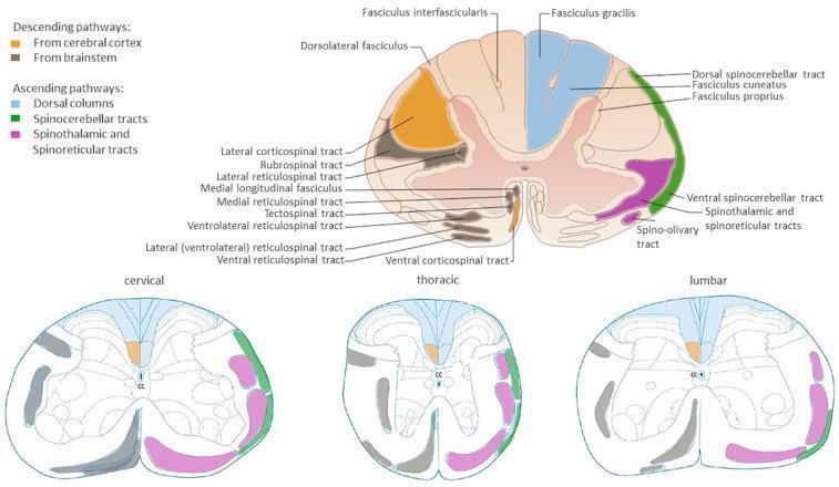
Anatomical diagrams illustrating the organization of major white matter tracts in the human spinal cord across cervical, thoracic, and lumbar cross-sections. A primary large-scale diagram provides detailed labeling of pathways, while three comparative smaller diagrams show regional variations. Descending (motor) pathways are color-coded in orange (corticospinal tracts from the cerebral cortex) and grey/black (brainstem-derived tracts including rubrospinal, reticulospinal, and tectospinal tracts). Ascending (sensory) pathways are highlighted in blue for dorsal columns (fasciculus gracilis and cuneatus), green for dorsal and ventral spinocerebellar tracts, and pink for the spinothalamic and spinoreticular tracts. The diagrams illustrate key neuroanatomical landmarks such as the central canal (CC), dorsal horns, and ventral horns. Educational focus is placed on the somatotopic arrangement and the relative change in white matter volume versus grey matter across different spinal levels, highlighting the clinical relevance for localized spinal cord injury and neurological deficit mapping.
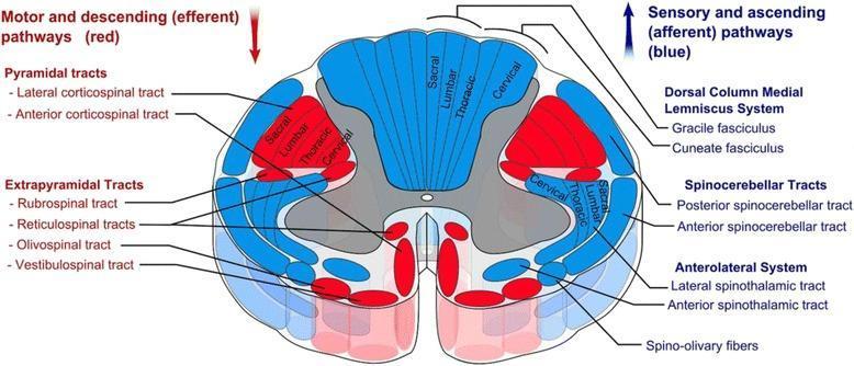
This medical anatomical diagram illustrates a transverse section of the human spinal cord, specifically detailing the organization of white matter tracts. The visual uses a color-coded schema to differentiate between motor and sensory pathways: descending (efferent) motor tracts are highlighted in red, while ascending (afferent) sensory tracts are shown in blue. Key anatomical structures identified include the Pyramidal tracts (lateral and anterior corticospinal tracts), Extrapyramidal tracts (rubrospinal, reticulospinal, olivospinal, and vestibulospinal), the Dorsal Column-Medial Lemniscus system (gracile and cuneate fasciculi), Spinocerebellar tracts, and the Anterolateral system (lateral and anterior spinothalamic tracts). A significant educational feature of the diagram is the depiction of somatotopic organization, showing the concentric arrangement of sacral, lumbar, thoracic, and cervical fibers within the lateral corticospinal and dorsal column tracts. The image provides a comprehensive overview of spinal cord neuroanatomy, useful for understanding clinical localization of spinal cord lesions and the functional topography of the central nervous system.
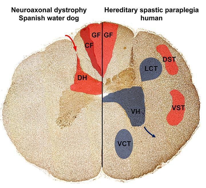
This Comparison Diagram presents a cross-sectional illustration of the spinal cord, contrasting the localization of axonal spheroids and neuronal loss in Neuroaxonal Dystrophy (NAD) and Hereditary Spastic Paraplegia (HSP). The diagram is split into two halves: the left represents a Spanish Water Dog with NAD, and the right represents human HSP. Ascending sensory pathways are highlighted in red, including the Gracile Fasciculus (GF), Cuneate Fasciculus (CF), and Dorsal Horn (DH) on the NAD side, and GF, Dorsal Spinocerebellar Tract (DST), and Ventral Spinocerebellar Tract (VST) on the HSP side. Descending motor pathways, affected only in the HSP model, are highlighted in blue, depicting the Lateral Corticospinal Tract (LCT), Ventral Corticospinal Tract (VCT), and Ventral Horn (VH). The visual demonstrates that in NAD, pathology is restricted to sensory, ascending tracts in the dorsal funiculus and dorsal horn. In contrast, human HSP shows involvement of both sensory pathways and descending motor pathways (pyramidal tracts and ventral horns). This diagram illustrates the pathophysiology of upper motor neuron and sensory system degeneration relevant to neurology and neurohistology.
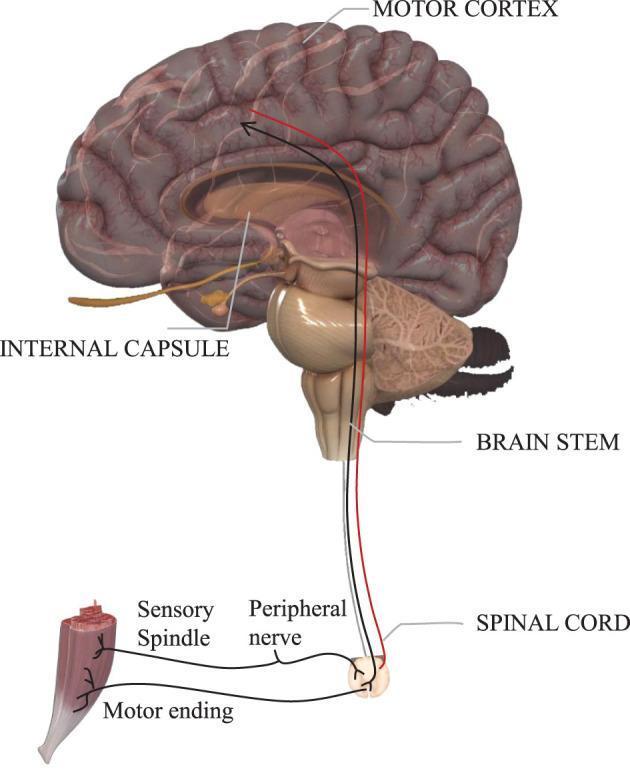
An anatomical diagram illustrating the descending motor pathways from the brain to the skeletal muscle. The visual highlights the relationship between the central and peripheral nervous systems. Key structures labeled include the motor cortex (the origin of voluntary movement), the internal capsule, the brainstem, and the spinal cord. Two distinct neural tracts are color-coded: a red pathway representing the pyramidal tract for voluntary muscle control, and a black pathway representing involuntary or extrapyramidal motor systems. These pathways descend through the brainstem into the spinal cord, where they synapse with a peripheral nerve. The diagram further details the innervation of a muscle fiber, showing the motor ending (neuromuscular junction) and a sensory spindle, illustrating the feedback loop between muscle tension and the spinal cord. This infographic is designed to teach the neuroanatomical distinction between voluntary and involuntary motor control and the physical sequence of nerve conduction.
Descending Tracts of the Spinal Cord
Classification at a Glance
| System | Tracts | Function |
|---|---|---|
| Lateral | Lateral corticospinal, Rubrospinal | Voluntary, fine, distal limb movements |
| Medial | Anterior corticospinal, Vestibulospinal (medial + lateral), Reticulospinal (pontine + medullary), Tectospinal | Posture, balance, axial/trunk control, head-eye reflexes |
I. LATERAL MOTOR SYSTEM
1. Lateral Corticospinal Tract (LCST)
- Origin: Primary motor cortex (area 4), premotor cortex (area 6), and somatosensory parietal cortex. About 2/3 of fibres arise from frontal motor areas.
- Course: Axons converge in the corona radiata → posterior limb of the internal capsule → crus cerebri (midbrain) → anterior pons (small bundles) → forms the medullary pyramid at the base of the medulla.
- Decussation: At the pyramidal decussation (cervicomedullary junction) - ~85-90% of fibres cross to the contralateral side.
- Location in cord: Lateral funiculus (lateral white column).
- Termination: Entire cord (predominantly cervical and lumbosacral enlargements) - synapses on lower motor neurons (LMNs) in the lateral anterior horn.
- Function: Voluntary movement of contralateral upper and lower limbs, especially fine distal movements (fingers, hands).
2. Rubrospinal Tract
- Origin: Red nucleus, magnocellular division, in the midbrain tegmentum.
- Decussation: Ventral tegmental decussation in the midbrain (immediately after leaving the red nucleus).
- Location in cord: Lateral column, just anterior to the LCST.
- Termination: Cervical cord only - synapses on interneurons in the anterior horn.
- Function: Facilitates flexor muscles and inhibits extensors of the upper limb. Assists fine motor control of the arm. Note: In humans its role is uncertain - it may be functionally minor compared to primates.
II. MEDIAL (VENTROMEDIAL) MOTOR SYSTEM
3. Anterior (Ventral) Corticospinal Tract
- Origin: Primary motor cortex and supplementary motor area (SMA).
- Course: Fibres that did NOT decussate at the pyramidal decussation remain ipsilateral.
- Location in cord: Medial aspect of the anterior (ventral) column.
- Decussation: Crosses at the level of termination (segmental, in the anterior commissure).
- Termination: Cervical and upper thoracic cord - synapses on LMNs in the medial anterior horn.
- Function: Bilateral control of axial (neck, trunk) and girdle muscles. Coordinates posture.
4. Vestibulospinal Tracts (VST)
| Medial VST | Lateral VST | |
|---|---|---|
| Origin | Medial and inferior vestibular nuclei | Lateral vestibular nucleus (Deiters' nucleus) |
| Laterality | Bilateral | Ipsilateral |
| Termination | Cervical + upper thoracic cord | Entire cord (to lumbar) |
| Function | Head and neck positioning; stabilizes the eyes as the body moves | Maintains upright posture; facilitates extensor (antigravity) motor neurons of the legs |
5. Reticulospinal Tracts
| Pontine (Medial) Reticulospinal | Medullary (Lateral) Reticulospinal | |
|---|---|---|
| Origin | Pontine reticular formation | Medullary reticular formation |
| Projection | Ipsilateral (mostly) | Bilateral |
| Function | Facilitates antigravity extensor stretch reflexes; helps maintain standing posture against gravity | Inhibits antigravity extensors; modulates voluntary and reflex activity |
| Clinical relevance | After cortical stroke, the medullary reticulospinal loses its inhibitory tone → pontine reticulospinal dominates → spasticity + extensor posturing (decerebrate rigidity pattern) |
6. Tectospinal Tract
- Origin: Superior colliculus (optic tectum) of the midbrain, which receives visual, auditory, and somatosensory inputs.
- Decussation: Dorsal tegmental decussation immediately after leaving the superior colliculus.
- Location in cord: Near anterior median fissure, alongside the medial longitudinal fasciculus (MLF).
- Termination: Cervical cord only - synapses on interneurons in the anterior horn.
- Function: Reflex postural movements of head and neck in response to visual/auditory stimuli (orienting response - e.g., turning the head toward a sudden sound or moving object). Functional role in humans is uncertain.
Key Concept: UMN vs LMN
| Feature | UMN Lesion | LMN Lesion |
|---|---|---|
| Tone | Increased (spasticity) | Decreased (flaccidity) |
| Reflexes | Hyperreflexia | Hyporeflexia/areflexia |
| Babinski | Present | Absent |
| Wasting | Absent (disuse only) | Present (denervation atrophy) |
| Fasciculations | Absent | Present |
Diagram of Spinal Cord Tract Positions
Summary Table
| Tract | Origin | Decussation | Location in Cord | Termination | Function |
|---|---|---|---|---|---|
| Lateral corticospinal | Motor cortex | Pyramidal decussation (cervicomedullary junction) | Lateral funiculus | Entire cord (cervical + lumbosacral) | Contralateral limb voluntary movement |
| Anterior corticospinal | Motor cortex + SMA | Segmental (anterior commissure) | Anterior funiculus | Cervical + upper thoracic | Bilateral axial/girdle muscles |
| Rubrospinal | Red nucleus (midbrain) | Ventral tegmental decussation | Lateral funiculus | Cervical cord | Arm flexion; uncertain in humans |
| Lateral VST | Lateral vestibular nucleus | None (ipsilateral) | Anterior funiculus | Entire cord | Postural balance, leg extensors |
| Medial VST | Medial/inferior vestibular nuclei | Bilateral | Anterior funiculus | Cervical + upper thoracic | Head/neck positioning |
| Pontine reticulospinal | Pontine reticular formation | Ipsilateral | Anterior funiculus | Entire cord | Facilitates extensors, posture |
| Medullary reticulospinal | Medullary reticular formation | Bilateral | Lateral funiculus | Entire cord | Inhibits extensors; modulates posture |
| Tectospinal | Superior colliculus | Dorsal tegmental decussation | Anterior funiculus | Cervical cord | Reflex head-eye orientation |