
Total thyroidectomy in detail and radio iodine ablation in detail according to standard ENT textbook in simplified manner
total thyroidectomy surgical technique anatomy recurrent laryngeal nerve
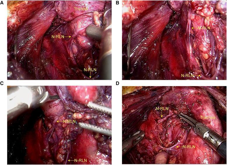
A series of four intraoperative endoscopic photographs (A-D) documenting a right-sided non-recurrent laryngeal nerve (NRLN) during an endoscopic thyroidectomy via a total areolar approach. Panel A shows the nerve (labeled N-RLN) in a position where it could be mistaken for the superior thyroid artery. Panel B illustrates the vertical and horizontal dissection of the cord-like, grayish-white nerve structure adjacent to the carotid sheath. Panel C highlights the nerve being manipulated with surgical forceps as it courses horizontally toward the larynx, posterior to the thyroid gland. Panel D provides a broader view of the surgical field, demonstrating the relationship of the NRLN to the trachea and its horizontal entry into the larynx below the inferior horn of the thyroid cartilage. The images show typical surgical anatomy of the central neck compartment, including muscle fibers, vascular structures, and the dissected thyroid bed. This content is intended for surgical residents and endocrine surgeons to illustrate the identification of an anatomical variant associated with an aberrant right subclavian artery (arteria lusoria).
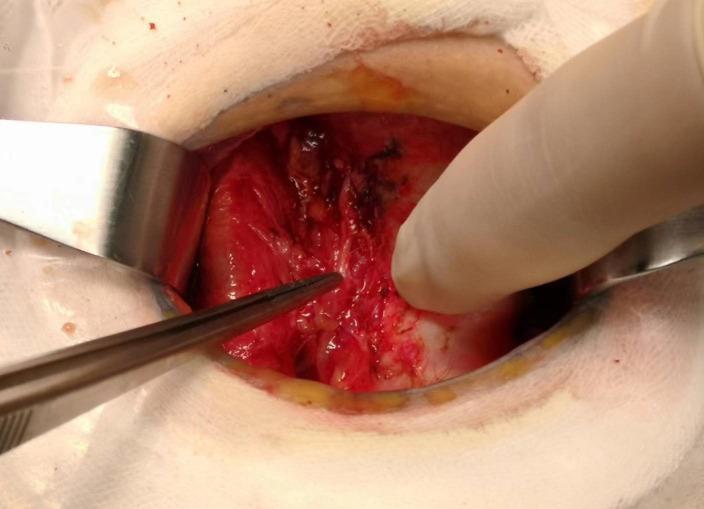
This clinical photograph captures a surgical field during an endocrine procedure, specifically a total thyroidectomy or central neck dissection. The image shows the anatomical exposure of the right tracheoesophageal groove. A metal retractor is used to displace the strap muscles and lateral tissues, providing visualization of the deeper cervical structures. Fine surgical forceps are used to point toward a thin, white, cord-like structure which is identified as the right recurrent laryngeal nerve (RLN). A gloved finger provides counter-traction and palpation of the surrounding tissue, likely assessing the proximity of the nerve to the trachea or thyroid bed. The tissue exhibits a reddish, raw appearance characteristic of fresh dissection, with some surgical sutures and darkening of tissue indicating localized hemostasis. The educational focus is the identification and preservation of the recurrent laryngeal nerve to prevent postoperative vocal cord paralysis. This image demonstrates the critical surgical anatomy required for surgeons and medical students to understand nerve mapping and safety during thyroid and parathyroid surgeries.
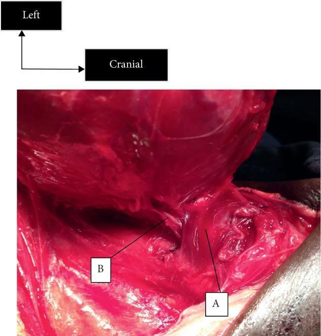
This clinical intraoperative photograph displays the surgical anatomy of the cervical region during a thyroidectomy, specifically highlighting the relationship between the cricopharyngeal muscle and the recurrent laryngeal nerve (RLN). Label A identifies the cricopharyngeal muscle, which appears as a distinct band of reddish, striated muscle tissue. Label B identifies the left recurrent laryngeal nerve, characterized as a thin, white, cord-like structure. The image demonstrates the critical anatomical landmark where the RLN (B) courses superiorly to enter the larynx, passing deep to or near the lower border of the cricopharyngeal muscle (A). An orientation guide in the upper left corner indicates the left and cranial directions. This visual serves as an educational reference for surgeons to identify the nerve's entry point into the larynx, which is essential for preventing iatrogenic injury during thyroid and parathyroid procedures. The surgical field shows the exposure of the deep neck structures after the mobilization of the thyroid gland.
Total Thyroidectomy - In Detail
1. SURGICAL ANATOMY
Position and Structure
- The thyroid gland lies anterior to the cricoid cartilage and inferior to the thyroid cartilage
- Two lobes joined at midline by the isthmus
- In 1 in 4 patients, a pyramidal lobe extends superiorly from the isthmus (thyroglossal duct remnant)
- Layers from superficial to deep: skin → dermis → platysma → sternohyoid/omohyoid → sternothyroid → pretracheal fascia → thyroid
Blood Supply
- 2 arteries per lobe: Superior thyroid artery (from external carotid) + Inferior thyroid artery (from thyrocervical trunk)
- 3 veins per lobe: Superior, middle, inferior thyroid veins (inferior usually a plexus, not a single vessel)
Key Nerves - The Two "Critical Nerves"
- Innervates the cricothyroid muscle (controls high-pitch voice projection)
- Travels lateral to the superior thyroid pedicle, crosses it medially to enter the cricothyroid
- Cernea Classification (how close the nerve is to the superior pole):
- Type 1: Crosses >1 cm above the superior pole (safest)
- Type 2A: Crosses within 1 cm above the superior pole
- Type 2B: Crosses below the superior border of the superior pole (most at risk)
- Innervates all intrinsic laryngeal muscles except cricothyroid
- Left RLN: Loops around the aortic arch, ascends in the tracheoesophageal groove
- Right RLN: Loops around the subclavian artery, ascends more laterally and obliquely
- ⚠️ Non-recurrent laryngeal nerve (0.5% prevalence): Occurs when the right subclavian artery originates directly from the aortic arch (aberrant "arteria lusoria") - the nerve then runs nearly horizontal across the field instead of vertically
- 3 most dangerous zones for RLN injury:
- Posterior to the inferior pole of the thyroid
- At the intersection with the inferior thyroid artery
- Behind the Ligament of Berry
Parathyroid Glands
- Classically 4 glands (2 superior, 2 inferior) per patient
- Superior glands: Lie posterior to the RLN plane, within 2-5 mm of the nerve; 80% symmetrically located
- Inferior glands: Lie anterior to the RLN plane, more variable, 7-10 mm from the nerve
- Ectopic glands in 15% of patients - locations include retroesophageal, intrathyroidal, carotid sheath, thyro-thymic ligament, or mediastinum
- ⚠️ Up to 25% of superior glands are supplied by the superior thyroid artery (not inferior) - ligating the superior artery too far from the gland can devascularize them
2. PATIENT POSITIONING AND EXPOSURE
- Position: Supine, head raised ~15 degrees, neck extended with posterior shoulder roll. Avoid excessive extension (uncomfortable postoperatively); avoid inadequate extension (crowds field).
- Incision: Collar (Kocher) incision along Langer's lines or within an existing neck crease, typically 2-3 cm above the clavicular heads. A preoperative ultrasound by the surgeon helps optimize placement.
- Skin flaps: Carried through platysma; subplatysmal flaps raised inferiorly to the clavicular heads and superiorly to the thyroid cartilage using sharp + blunt dissection.
- Strap muscles: Lifted off the thyroid capsule at midline and separated posteriorly along avascular planes. The middle thyroid vein is then ligated between the thyroid and internal jugular vein (IJV).
- For large/firm glands: The sternothyroid muscle may be divided. Always divide it high (near thyroid cartilage insertion) since ansa cervicalis innervation enters inferiorly.
3. STEP-BY-STEP TECHNIQUE
Step 1 - Isthmus and Pyramidal Lobe
- Begin by isolating the draining veins of the isthmus and pyramidal lobe
- Early isthmus division facilitates mobilization (especially through small <4 cm incisions for lobectomy)
Step 2 - Superior Pole Dissection
- Develop the avascular cricothyroid space (Space of Reeve) between the superior pole and cricothyroid muscle
- Superior thyroid artery is isolated through blunt, closed-tip dissection (excessive spreading causes bleeding)
- Identify the EBSLN (visually or with nerve monitor) before ligating the superior pedicle
- Divide the pedicle as close to the gland as possible to keep the nerve safe
Step 3 - Inferior Pole
- Retract the inferior pole superiorly to expose inferior thyroid vein branches
- Ligate these close to the thyroid gland - straying lateral/deep risks RLN injury and devascularizes the lower parathyroid
Step 4 - Preservation of Parathyroid Glands
- After dividing venous drainage, rotate the thyroid lobe medially
- Expose the tracheoesophageal groove and carotid sheath
- Identify the inferior thyroid artery and RLN posteriorly
- In 80% of cases, both parathyroid glands are supplied by the inferior thyroid artery
- Dissect immediately along the thyroid capsule, dividing the tertiary branches of the inferior thyroid artery individually - this preserves parathyroid blood flow
- If a parathyroid is intrathyroidal, the capsule is entered focally to liberate it (preserves vascular pedicle)
- ⚠️ Truncal ligation of the inferior thyroid artery without first identifying the parathyroids is strongly discouraged
Step 5 - RLN Identification and Protection
- To safely find the nerve: spread perpendicular to the nerve axis through fatty/nodal tissue posterior to the thyroid
- Once found, use a blunt hemostatic clamp (tip upward) to separate the nerve from superficial structures along its course
- The Tubercle of Zuckerkandl (posterior thickened thyroid projection) must be mobilized to expose the nerve coursing within the fissure between it and the remaining gland
- Divide the Ligament of Berry only after full nerve visualization
Step 6 - Intraoperative Neuromonitoring (IONM)
- Receiving electrode on the endotracheal tube senses vocal cord signaling; grounding electrode on skin
- Negative predictive value approaches 100% (loss of signal reliably predicts injury risk)
- Positive predictive value <50% (signal loss does not always mean permanent injury)
- Used to: confirm visual nerve assignment, map branching pattern, ensure signal during retraction at Ligament of Berry, and confirm at the vagus and tracheoesophageal groove at start and end
- Has not been proven to prevent nerve injury, but assists decision-making
4. CLOSURE
- No routine drainage needed after thyroidectomy
- Valsalva maneuver is used to inspect for bleeding sites
- Strap muscles reapproximated in the midline with absorbable suture, leaving a 1 cm gap inferiorly (prevents blood/fluid from compressing the trachea)
- Platysma closed with absorbable suture; skin closed subcuticularly
5. COMPLICATIONS
| Complication | Details |
|---|---|
| RLN injury | Permanent injury <0.5% in experienced hands; transient higher. Causes hoarseness (unilateral) or airway compromise (bilateral) |
| EBSLN injury | Loss of high-pitch voice; subtle; missed unless tested |
| Hypoparathyroidism | Hypocalcemia (transient in up to 30%, permanent ~1-2%); give calcium + Vit D |
| Hemorrhage | Rare but life-threatening if hematoma compresses trachea |
| Hypothyroidism | Expected after total thyroidectomy; requires lifelong T4 replacement |
Radioiodine (RAI / ¹³¹I) Ablation - In Detail
1. WHAT IS IT AND HOW DOES IT WORK?
- ¹³¹I is an unsealed radionuclide of iodine that is taken up by thyroid tissue (both normal and differentiated thyroid cancer) in the same way as dietary stable iodine
- After a total/near-total thyroidectomy, residual thyroid tissue called the thyroid remnant remains - this remnant preferentially takes up ¹³¹I over cancer tissue
- ¹³¹I destruction of this residual tissue is called Radioiodine Remnant Ablation (RRA)
- Destroys microscopic foci of cancer in the remnant
- Allows detection of residual/metastatic disease on whole-body scan (remnant uptake would otherwise mask it)
- Aids interpretation of serum thyroglobulin (Tg) levels during follow-up - Tg becomes a reliable tumor marker only after all normal thyroid tissue is gone
- May reduce local recurrence and improve survival (evidence mixed)
2. INDICATIONS (Risk Stratification)
| Category | Criteria | RAI? |
|---|---|---|
| Low risk | Unifocal/multifocal papillary or follicular thyroid cancer <1 cm, histologically standard, no angioinvasion, no extrathyroidal extension | NOT indicated |
| High risk | Tumors >4 cm, gross extrathyroidal spread, or distant metastases | Indicated (ablation dose + treatment dose) |
| Intermediate risk | Tumors 1-4 cm, intermediate features | Individualized decision at MDT |
- Aggressive histology (tall cell, poorly differentiated, diffuse sclerosing PTC)
- Widely invasive follicular thyroid cancer
- Extracapsular invasion
- Multiple involved lymph nodes or high ratio of involved:uninvolved nodes
- Large individual node size
3. PATIENT PREPARATION - TSH STIMULATION
Method 1 - Thyroid Hormone Withdrawal (THW)
- Patient stops thyroxine (T4) 4-6 weeks before RAI
- Patient becomes hypothyroid → TSH rises naturally
- Drawback: Significant hypothyroid symptoms (fatigue, depression, cognitive impairment, reduced quality of life)
Method 2 - Recombinant Human TSH (rhTSH / Thyrogen™)
- Patient stays on thyroxine throughout
- rhTSH (0.9 mg) injected intramuscularly (deep into buttock) on Days 1 and 2
- ¹³¹I administered on Day 3
- Preferred whenever possible to avoid hypothyroid morbidity
- Hypopituitarism (cannot raise own TSH)
- Functional metastases suppressing TSH
- Severe ischaemic heart disease
- History of psychiatric illness triggered by hypothyroidism
- Advanced disease or frailty
4. LOW-IODINE DIET AND PREPARATION
- Before ¹³¹I administration, the total body iodine pool must be reduced so that ¹³¹I uptake is maximized
- Low daily iodine intake increases the effective radiation dose delivered by ¹³¹I
- Duration: Usually 1-2 weeks (no universal consensus on duration)
- Eliminate: iodinated IV contrast media, amiodarone, and other iodine-rich sources before treatment
5. DOSES OF ¹³¹I
| Purpose | Activity |
|---|---|
| RRA (remnant ablation) for pT1-2 N0 R0 (low/intermediate risk) | 1.1 GBq |
| RRA for pT3 and/or N1 disease | Individualized by MDT |
| Treatment (high risk / metastatic disease) | 3.7-5.5 GBq |
| rhTSH pre-stimulation licensed range for ablation | 1.1-3.7 GBq |
6. PRE-ABLATION SCAN
- Not routinely indicated but may be used to assess remnant size
- If a large remnant is seen: consider further surgery before RAI, or prescribe prophylactic corticosteroids
- Preferred isotopes for pre-ablation scan: ⁹⁹ᵐTc pertechnate or ¹²³I (NOT ¹³¹I)
- Reason: Using ¹³¹I for the diagnostic scan causes "stunning" - a reduction in uptake of the subsequent therapy dose
7. POST-TREATMENT WHOLE-BODY SCAN
- Performed 2-10 days after RAI administration
- More sensitive than diagnostic scans (higher activity used)
- SPECT-CT added to planar gamma camera imaging allows precise anatomical localization of iodine uptake
- Physiological ¹³¹I uptake is seen in: salivary glands, genitourinary tract, GI tract, and sinuses (these are normal findings)
8. SPECIAL CONSIDERATION - HURTHLE CELL TUMORS
- Hurthle cell (oncocytic) lesions are generally poorly responsive to radioiodine
- Distant metastases from Hurthle cell tumors are often resistant to RAI therapy
9. THYROID HORMONE REPLACEMENT AFTER RAI
- If patient was on THW: Start thyroid hormones 1-3 days after RAI administration
- If patient received rhTSH: Can stay on thyroxine throughout the entire procedure
10. SIDE EFFECTS AND TOXICITY OF ¹³¹I
| Side Effect | Notes |
|---|---|
| Neck discomfort/swelling | Due to inflammatory response in remnant - most likely with large remnants; treat with analgesics or prophylactic corticosteroids |
| Altered taste, nausea | Common; vomiting uncommon; prophylactic antiemetics not routinely needed |
| Sialadenitis | ¹³¹I excreted by salivary tissue causes inflammation; encourage high fluid intake; long-term: parotid swelling, xerostomia, parotid duct massage helps |
| Lacrimal gland dysfunction | Less common |
| Radiation cystitis | Reduce risk by high fluid intake during isolation period |
| Gastritis | From GI tract excretion |
| Bone marrow suppression | Peaks 4-6 weeks post-treatment; more likely with extensive skeletal metastases or prior EBRT/chemo |
| Gonadal exposure | Via blood and excretion in urine/feces |
Reproductive Considerations
- Females: Amenorrhea/oligomenorrhea for 4-10 months; defer pregnancy for 6 months (BTA) / 6-12 months (ATA) post-treatment; breastfeeding must be stopped at least 8 weeks before RAI (prevents excessive breast tissue uptake)
- Males: Defer fathering a child for 4 months; offer sperm banking as infertility is possible
11. ISOLATION REQUIREMENTS
- Patients must remain in an isolation ward following RAI
- Must avoid contact with adults and children for 14-25 days after treatment
- This is one of the important inconveniences to counsel patients about beforehand
SUMMARY COMPARISON
| Feature | Total Thyroidectomy | Radioiodine Ablation |
|---|---|---|
| Purpose | Remove thyroid gland entirely | Destroy residual thyroid tissue post-surgery |
| When needed | Thyroid cancer, large goiter, Graves', etc. | After total thyroidectomy for differentiated thyroid cancer |
| Key risk | RLN injury, hypoparathyroidism | Sialadenitis, gonadal exposure, bone marrow suppression |
| Prerequisite | - | Total thyroidectomy must be done first |
| Lifelong consequence | Requires T4 replacement | Tg becomes reliable tumor marker |