
Development of pancreas and annular pancreas
Development of the Pancreas and Annular Pancreas
1. Overview
2. The Two Pancreatic Buds
Dorsal Pancreatic Bud
- Appears first, slightly cranial to the ventral bud
- Develops within the dorsal mesentery (dorsal mesogastrium)
- Forms the tail, body, and superior portion of the head of the pancreas
- Contains the dorsal duct, which becomes the distal portion of the main pancreatic duct (of Wirsung) and the entire accessory duct (of Santorini)
- Molecular induction: FGF2 and Activin (a TGF-β family member), produced by the notochord and dorsal aortic endothelium, repress SHH expression in the gut endoderm destined to become the dorsal bud, thereby allowing pancreatic specification
Ventral Pancreatic Bud
- Arises as an outgrowth of the hepatic diverticulum, near the entry of the bile duct into the duodenum
- Has a bilobed (bifid) origin - the left ventral bud normally regresses; only the right portion completes rotation
- Grows within the ventral mesentery
- Forms the uncinate process and inferior part of the head
- Contains the ventral duct, which becomes the proximal portion of the main pancreatic duct (of Wirsung)
- Molecular induction: the ventral bud is induced by visceral mesoderm
Both processes lead to upregulation of PDX-1 (Pancreatic and Duodenal Homeobox 1 gene), the master regulator of pancreatic development.
3. Rotation and Fusion (5th-8th weeks)
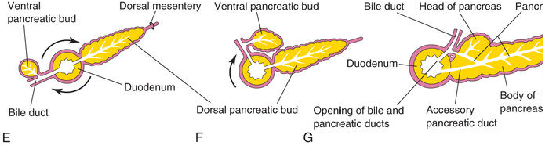
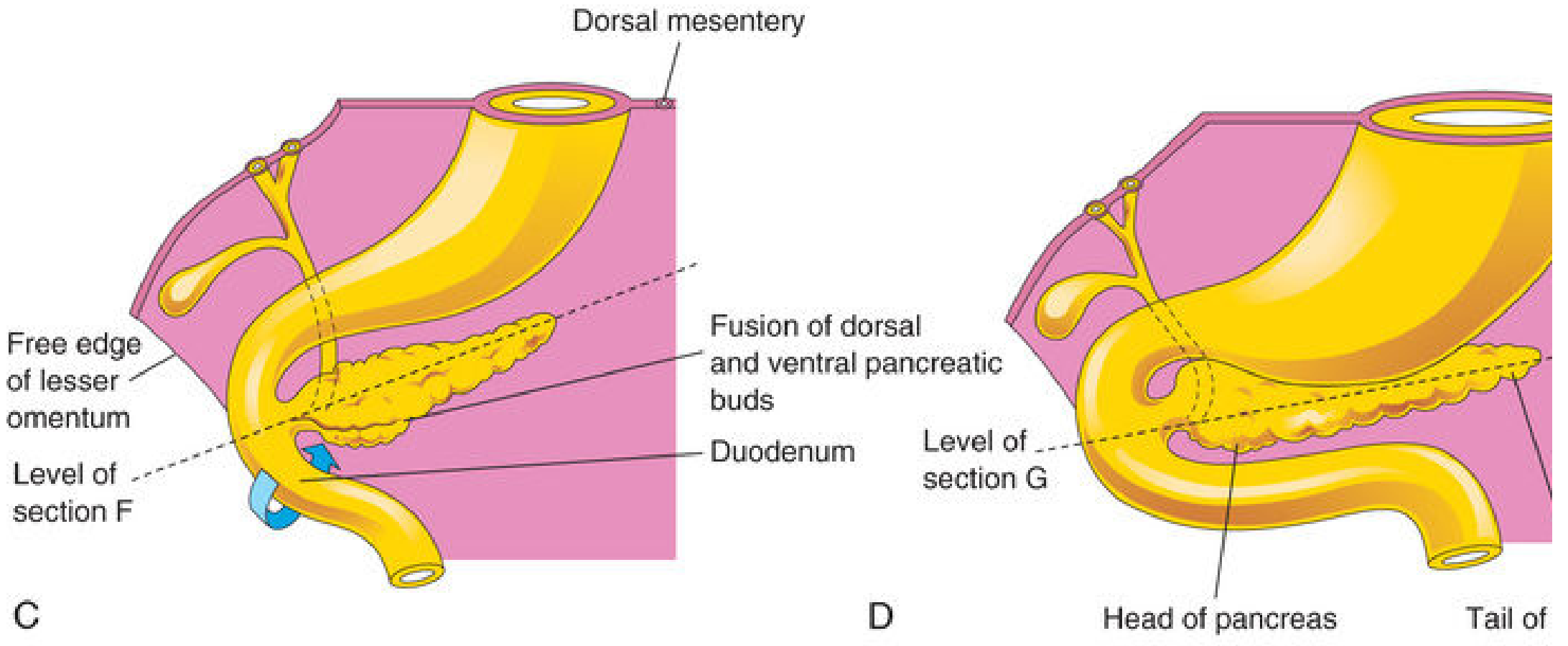
4. Duct System After Fusion
| Duct | Formed From | Opens Into |
|---|---|---|
| Main pancreatic duct (of Wirsung) | Entire ventral duct + distal part of dorsal duct | Major duodenal papilla (with bile duct) |
| Accessory pancreatic duct (of Santorini) | Proximal part of dorsal duct | Minor duodenal papilla (~2 cm cranial to main duct) |
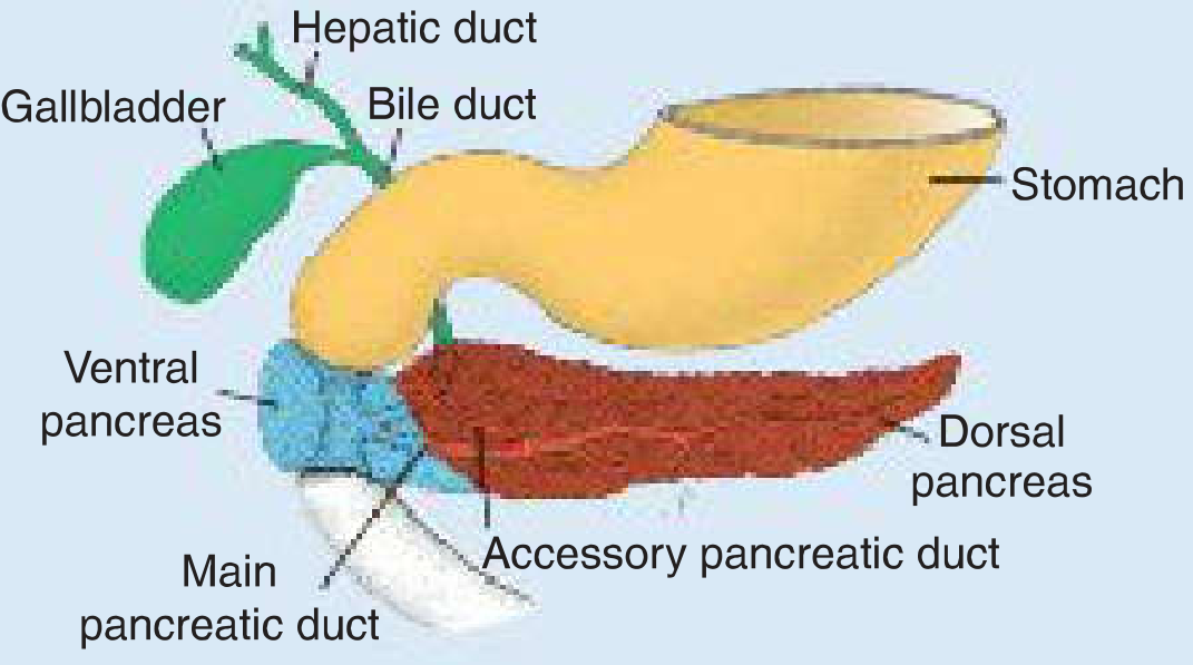
5. Cellular Differentiation
- Pancreatic islets of Langerhans develop from parenchymal pancreatic tissue in the 3rd month; insulin secretion begins at ~5th month
- Islet cell specification is controlled by paired homeobox genes:
- PAX4 + PAX6 co-expression → β cells (insulin), δ cells (somatostatin), γ cells (pancreatic polypeptide)
- PAX6 alone → α cells (glucagon)
- Visceral mesoderm surrounding the pancreatic buds forms the pancreatic connective tissue and stroma
Islet Cell Proportions (mature pancreas)
| Cell Type | Proportion | Hormone |
|---|---|---|
| Beta (β) | 50-80% | Insulin, amylin |
| PP / F cells | 10-35% | Pancreatic polypeptide |
| Alpha (α) | 5-20% | Glucagon |
| Delta (δ) | ~5% | Somatostatin |
| Epsilon (ε) | Rare | Ghrelin |
6. Final Position
7. Annular Pancreas
Definition
Embryologic Mechanism
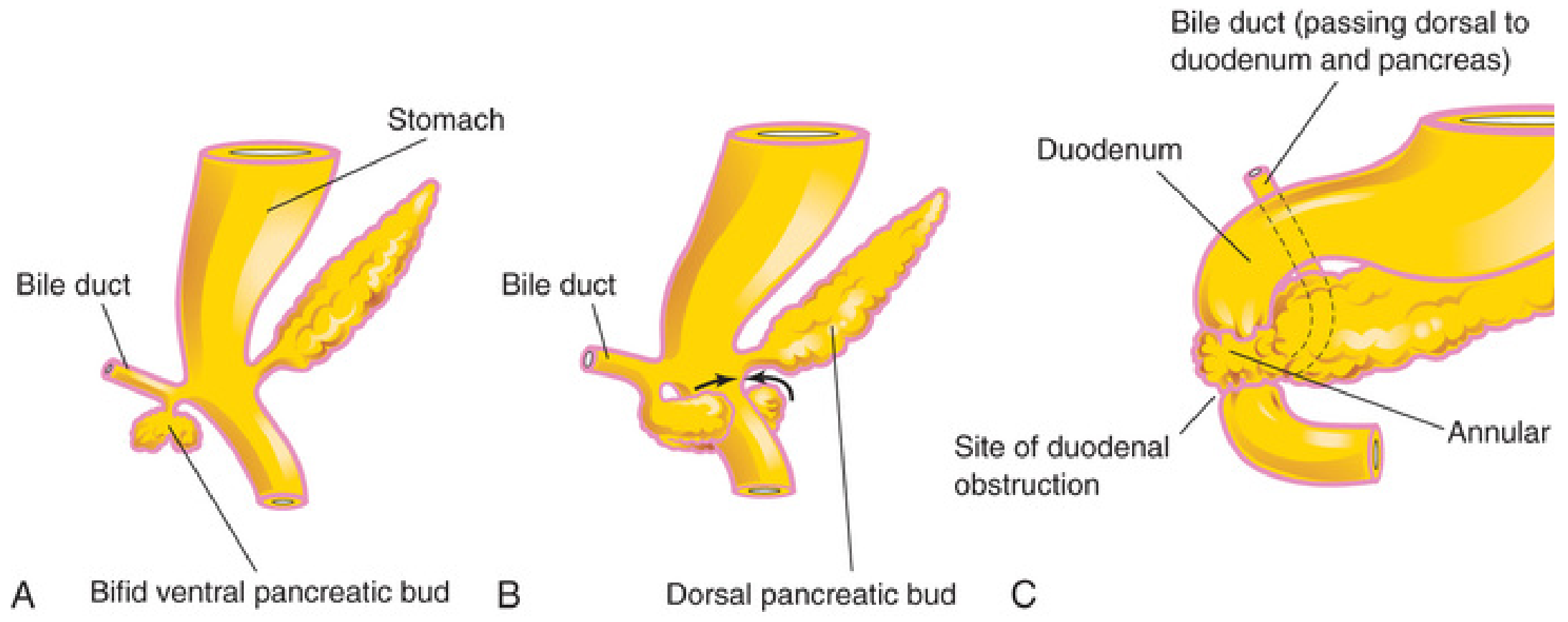
- The ventral bud splits (becomes bifid) - the right portion migrates normally (posteriorly), but the left portion migrates in the opposite direction (anteriorly). The two segments then encircle the duodenum and fuse with the dorsal bud, forming a ring.
- Failure of the left ventral bud to regress - persistence of the left ventral lobe contributes to annular tissue.
Associations and Genetics
- Trisomy 21 (Down syndrome) - most common association
- Cardiac defects
- Intestinal malrotation
- Duodenal atresia
- Genitourinary anomalies
- Tracheoesophageal fistula
- Females affected more frequently than males
- Familial cases reported (siblings, identical twins); microduplication on chromosome 6q24.2 (utrophin gene) identified in familial cases
- Shh and Ihh gene defects show 42-85% incidence of annular pancreas in mouse models
Clinical Presentation
| Age Group | Symptoms |
|---|---|
| Neonates / Infants | Non-bilious vomiting, feeding intolerance, failure to thrive; complete or partial duodenal obstruction |
| Adults (4th-7th decades) | Abdominal pain, pancreatitis, biliary obstruction, nausea, vomiting, bloating |
- Prenatal: duodenal obstruction prevents the fetus from swallowing amniotic fluid → polyhydramnios; may be detected on prenatal ultrasound
- Inflammation (pancreatitis) in the annular tissue can worsen or precipitate obstruction
Investigations
- Children: abdominal X-ray (classic "double bubble" sign), ultrasound, upper GI series
- Adults: CT scan, MRCP, or ERCP; diagnosis sometimes made at laparotomy
- Classic radiological sign: mid-duodenal stricture with proximal dilatation on barium contrast upper GI series
Treatment
- Duodenoduodenostomy - surgical bypass of the obstructed segment; treatment of choice in children and many adults
- The annular tissue must never be divided (contains functional pancreatic tissue and the pancreatic duct may run through the annulus)
- Complex pancreatic surgery may be required in adults
- Long-term risks: increased risk of acute/recurrent pancreatitis and pancreaticobiliary neoplasia (including duodenal carcinoma); ongoing surveillance recommended
8. Other Developmental Anomalies of the Pancreas
| Anomaly | Mechanism | Clinical Significance |
|---|---|---|
| Pancreas divisum | Failure of dorsal and ventral duct systems to fuse (up to 10% of population) | Accessory duct becomes main conduit; possible recurrent pancreatitis |
| Ectopic pancreatic tissue | Separate nests of pancreatic tissue | Found in gastric mucosa, proximal duodenum, jejunum, pyloric antrum, Meckel's diverticulum; usually asymptomatic |
| Pancreatic agenesis | Failure of bud formation; PDX-1 mutations | Rare; associated with neonatal diabetes |
- The Developing Human: Clinically Oriented Embryology (Moore et al.), Ch. 11 - pancreatic buds and annular pancreas
- Langman's Medical Embryology, Ch. 15 - molecular regulation, PAX4/PAX6 signaling, annular pancreas mechanism
- Sleisenger and Fordtran's Gastrointestinal and Liver Disease, Ch. 55 - embryonic/fetal development, islet cell proportions, annular pancreas clinical details
- Gray's Anatomy for Students, Ch. 4 - bifid ventral bud mechanism, polyhydramnios
development of interatrial septum
Development of the Interatrial Septum
1. Background: The Primordial Atrium
2. Step 1 - Formation of Septum Primum and Foramen Primum (Ostium Primum)
- As it descends, it only partially divides the atrium
- The gap between its free crescentic lower edge and the endocardial cushions is called the foramen primum (ostium primum)
- This foramen acts as a right-to-left shunt, allowing oxygenated blood from the placenta (entering the right atrium) to pass into the left atrium
- The septum primum's lower edge carries a mesenchymal cap that will eventually fuse with the endocardial cushions
Molecular basis: A distinct population of extracardiac progenitor cells from the second heart field migrates through the dorsal mesocardium to complete the lateral septum. Sonic Hedgehog (Shh) signaling plays a critical role in this migration.
3. Step 2 - Formation of Foramen Secundum (Ostium Secundum) by Apoptosis
- This ensures that right-to-left blood flow is maintained even as the foramen primum is closing below
- The foramen primum then completely closes as the mesenchymal cap of the septum primum fuses with the fused AV endocardial cushions, forming a primordial AV septum
4. Step 3 - Appearance of Septum Secundum
- It grows downward from the roof of the right atrium, descending with an upper and lower limb
- It never forms a complete partition - it always leaves a gap
- Its anterior (lower) limb extends to the septum in the AV canal
- The left venous valve and septum spurium fuse with the right side of the septum secundum, contributing to its formation
- As it grows, its free concave edge overlaps the foramen secundum from the right side
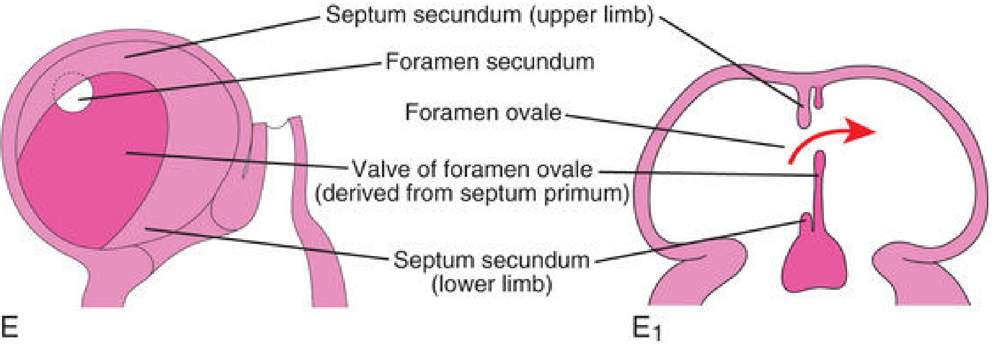
5. Step 4 - Foramen Ovale and Its Valve
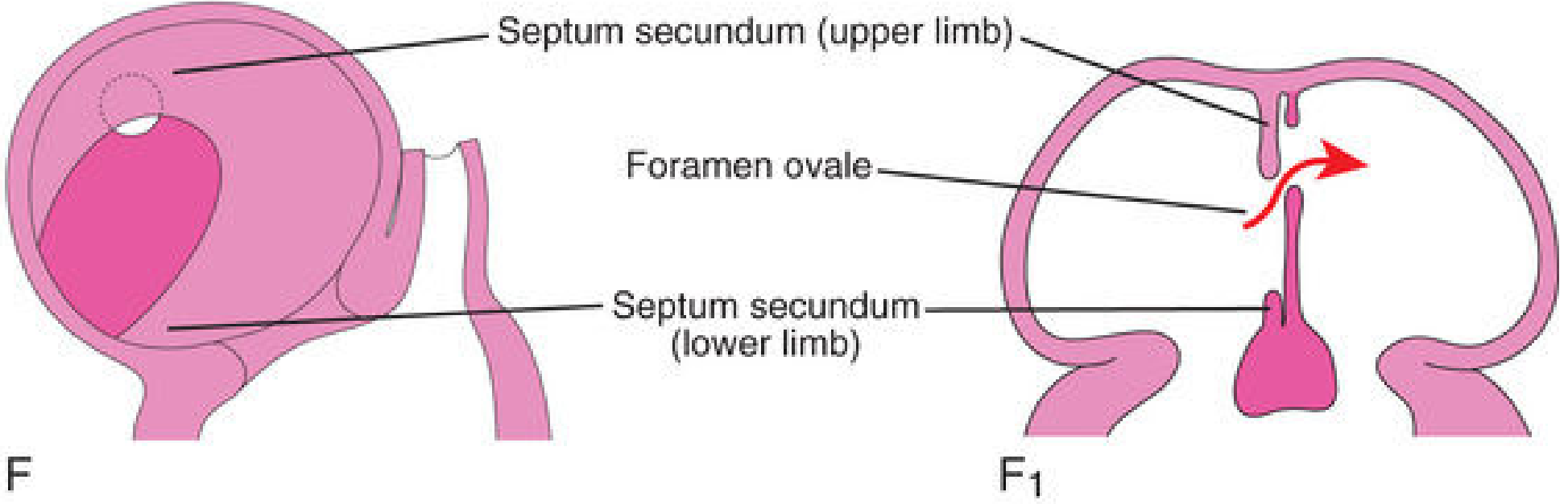
- This opening aligns with (is continuous with) the foramen secundum in the septum primum
- Together they form an obliquely elongated cleft-like passage through which blood flows from right to left
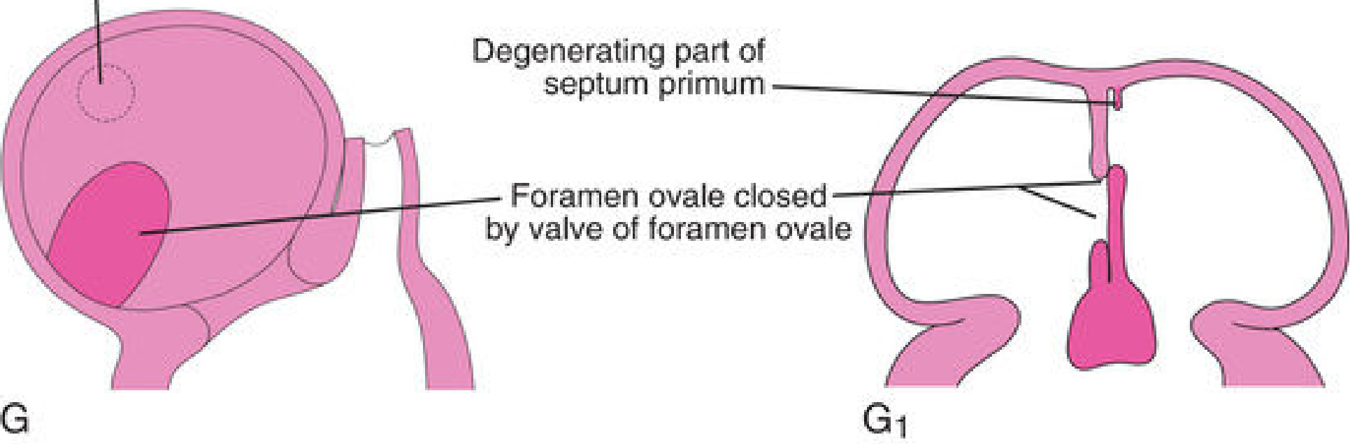
- The remaining inferior portion of the septum primum (whose upper part has degenerated) becomes the valve of the foramen ovale
- This valve mechanism only opens when right atrial pressure > left atrial pressure
6. Fetal Blood Flow Through the Foramen Ovale
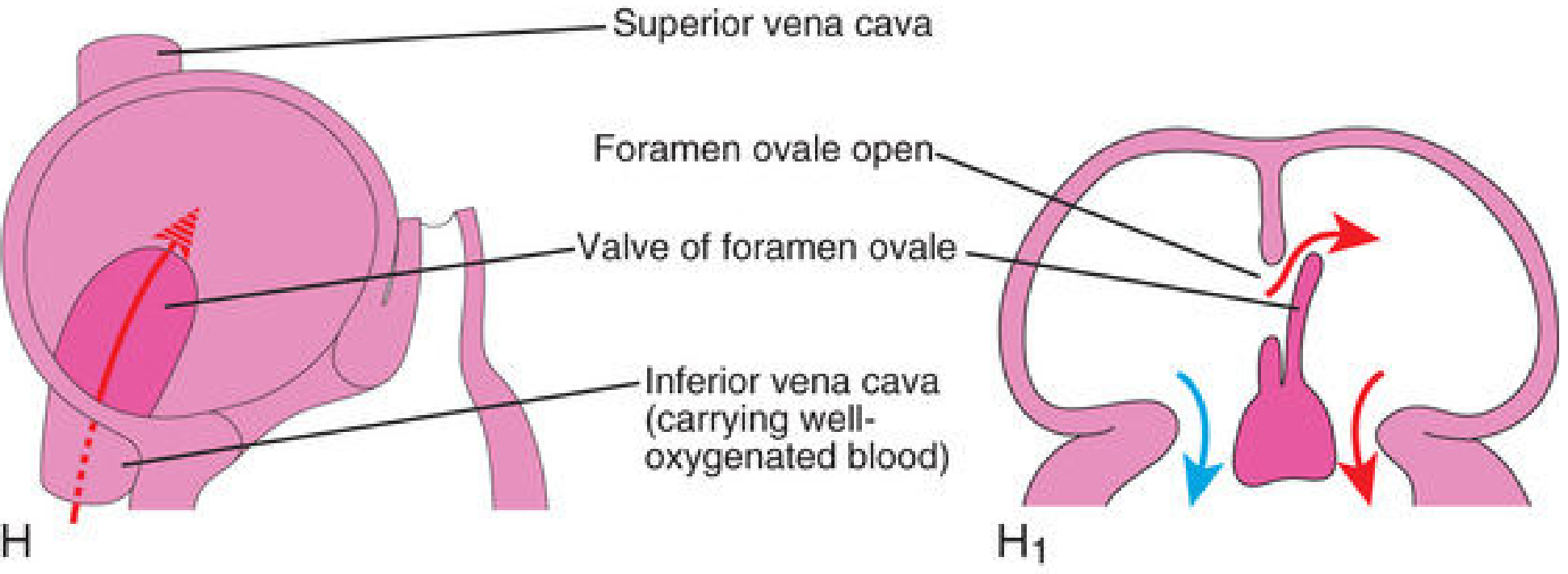
- Lungs are non-functional; pulmonary vascular resistance is high
- Right atrial pressure > left atrial pressure
- The valve of the foramen ovale is pushed open, allowing right-to-left shunting
- Well-oxygenated blood from the inferior vena cava (carrying placental blood) is directed by the crista dividens (Eustachian valve of the IVC) straight through the foramen ovale into the left atrium
- This bypasses the non-functional pulmonary circuit
7. Closure at Birth - Formation of Fossa Ovalis
- Lungs expand with the first breath
- Pulmonary vascular resistance drops
- Left atrial pressure rises above right atrial pressure
- The valve of the foramen ovale is pressed against the septum secundum from the left side, functionally closing the foramen ovale
- Within months, the valve permanently fuses with the septum secundum by fibrous adhesion, completing the interatrial septum
- The floor = remnant of the septum primum (the old valve of the foramen ovale)
- The raised rim = the limbus fossa ovalis = lower free edge of the septum secundum
8. Summary Table: Sequential Events
| Stage | Week | Event | Structure Formed |
|---|---|---|---|
| 1 | ~4th week | Septum primum grows down from atrial roof | Foramen primum (ostium primum) below its edge |
| 2 | ~5th week | Apoptosis perforates upper septum primum | Foramen secundum (ostium secundum) |
| 3 | ~5th week | Septum primum fuses with endocardial cushions | Foramen primum obliterated |
| 4 | ~6th week | Septum secundum grows down to right of septum primum | Foramen ovale (opening in septum secundum) |
| 5 | ~7th week | Upper septum primum degenerates | Valve of foramen ovale (lower septum primum remnant) |
| 6 | Fetal life | Foramen ovale open; right-to-left shunting | Foramen ovale + its valve |
| 7 | At birth | Left atrial pressure rises | Functional then permanent closure of foramen ovale |
| 8 | Postnatal | Fusion of valve to septum secundum | Fossa ovalis |
9. Atrial Septal Defects (ASDs) - Developmental Basis
Types and Mechanisms
| Type | Frequency | Location | Mechanism |
|---|---|---|---|
| Ostium secundum ASD | ~90% | Oval fossa region | Excessive resorption of septum primum OR inadequate formation of septum secundum |
| Ostium primum ASD (endocardial cushion defect) | ~5% | Adjacent to AV valves | Failure of septum primum to fuse with endocardial cushions; often with AV valve and VSD abnormalities |
| Sinus venosus defect | ~5% | Near SVC or IVC entrance | Defect in incorporation of sinus venosus; often with anomalous pulmonary venous return |
| Common atrium | Rare | Entire septum absent | Complete failure of atrial septation |
Patent Foramen Ovale (PFO)
- Present in ~25-30% of adults
- Results from failure of the valve of the foramen ovale to permanently fuse with the septum secundum after birth
- Not a true ASD - the channel is still valved; no fixed opening
- Can allow paradoxical embolism (venous clot crossing to arterial circulation)
- Associated with cryptogenic stroke in young adults
10. Adult Derivatives Summary
| Embryonic Structure | Adult Derivative |
|---|---|
| Septum primum (lower remnant) | Floor of fossa ovalis (valve of foramen ovale) |
| Septum secundum (free edge) | Limbus fossa ovalis (annulus ovalis) |
| Foramen ovale | Fossa ovalis (closed) |
- The Developing Human: Clinically Oriented Embryology (Moore et al.) - Ch. 13, Fig. 13.13 series
- Langman's Medical Embryology - Ch. 13, septum formation and Figs. 13.13/13.16
- Robbins, Cotran & Kumar Pathologic Basis of Disease - Ch. 12, ASD classification
- Color Atlas of Human Anatomy Vol. 2 (Internal Organs) - septum primum/secundum sequence
3. Development of CNS and Clinical Aspects [ANENCEPHALY , SPINA BIFIDA , CRANIORACHISCHICIS] 4. Midgut Rotation 5. Development of Kidneys and its clinical aspects 6. Descent of testis and undescended testis 7. Pharyngeal Arches derivatives 8. Development of Face and Facial Clefts
3. Development of CNS and Clinical Aspects
Neural Tube Formation (Neurulation)
- Neural plate forms (day 18) from the dorsal ectoderm, induced by notochord signals (Chordin, Noggin, Follistatin - BMP antagonists)
- Neural folds rise on either side of the neural groove
- Fusion of the neural folds begins at the cervical level (~day 22) and proceeds cranially and caudally (like a zipper)
- The open cranial end = anterior (rostral) neuropore - closes on day 25
- The open caudal end = posterior (caudal) neuropore - closes on day 27
- Neural crest cells migrate away from the dorsal margins of the neural folds before fusion
Three Brain Vesicles (Week 4)
| Primary Vesicle | Secondary Vesicle | Adult Structure |
|---|---|---|
| Prosencephalon | Telencephalon | Cerebral hemispheres, basal ganglia, olfactory bulbs |
| Diencephalon | Thalamus, hypothalamus, retina | |
| Mesencephalon | Mesencephalon | Midbrain (superior/inferior colliculi, cerebral peduncles) |
| Rhombencephalon | Metencephalon | Pons, cerebellum |
| Myelencephalon | Medulla oblongata |
Spinal Cord Development
- Mantle layer (gray matter) - neuroblasts forming alar plate (sensory) and basal plate (motor)
- Marginal layer (white matter) - axons and myelination
- Roof plate and floor plate - organizer regions
Clinical Aspects: Neural Tube Defects (NTDs)
A. Anencephaly
- Mechanism: Failure of the anterior (rostral) neuropore to close (day 25)
- Result: Absence of the forebrain, calvaria, and scalp; the brain tissue that does form is exposed and degenerates into a vascular, hemorrhagic mass (area cerebrovasculosa)
- Incidence: ~1/1,000 births; females > males (3:1)
- Associations: Polyhydramnios (fetus cannot swallow - absent swallowing reflex due to absent brain centers)
- Prenatal diagnosis: Elevated AFP in maternal serum and amniotic fluid; absent cranial vault on ultrasound
- Outcome: Always fatal - most are stillborn or die within hours/days of birth
- Anencephaly is also called meroanencephaly (partial) or sometimes holoanencephaly
B. Spina Bifida
- Mechanism: Failure of the posterior (caudal) neuropore to close (day 27), resulting in incomplete closure of the vertebral arch
- Most common site: Lumbosacral region (L5-S1)
- Risk factors: Folic acid deficiency, valproate exposure, maternal diabetes
| Type | Features | Severity |
|---|---|---|
| Spina Bifida Occulta | Vertebral arch defect only; skin intact; tuft of hair, lipoma, or dimple may overlie it; spinal cord and meninges not herniated | Least severe; often asymptomatic |
| Meningocele | Meninges herniate through defect as a CSF-filled sac; spinal cord stays in canal; skin usually covers sac | Moderate; surgery possible |
| Myelomeningocele | Both spinal cord/nerve roots AND meninges herniate; cord exposed or covered by thin membrane | Most severe common form; neurological deficits below level |
| Myeloschisis (Rachischisis) | Spinal cord completely open/flat; no covering at all | Severe; complete neurological loss |
- Lower limb paralysis and sensory loss below the lesion
- Bladder and bowel dysfunction (neurogenic bladder)
- Associated with Arnold-Chiari malformation type II (downward displacement of cerebellar vermis and medulla through foramen magnum) in >90% of cases
- Hydrocephalus (due to obstruction of CSF flow)
- Treatment: surgical closure within 24-48 hours of birth; VP shunt for hydrocephalus
C. Craniorachischisis (Craniorachischisis Totalis)
- The most severe of all NTDs
- Mechanism: Failure of neural tube closure affecting both the cranial AND spinal regions - essentially the entire neural tube fails to close
- Results in anencephaly combined with open spina bifida (rachischisis) - a continuous open defect from the head to the lumbar or sacral spine
- The entire neuraxis is exposed; the brain is absent and the spinal cord lies open
- Always lethal - incompatible with extrauterine life
- Associated with extreme cases of folic acid deficiency; some cases linked to VANGL1/VANGL2 mutations (planar cell polarity pathway)
- Diagnosis: prenatal ultrasound + elevated AFP
4. Midgut Rotation
The Midgut Loop
- Cranial limb: Forms small intestinal loops (future jejunum and ileum)
- Caudal limb: Develops the cecal diverticulum (primordium of cecum and appendix)
- The apex is attached to the yolk stalk (vitello-intestinal duct / omphalomesenteric duct)
- The entire loop is supplied by the superior mesenteric artery (SMA), which forms the axis of rotation
Rotation (270° Total, Counterclockwise)
Stage 1 - In the umbilical cord (6th-10th weeks): 90° rotation
- While herniated, the loop rotates 90° counterclockwise (viewed from front)
- The cranial limb (small intestine) swings to the right
- The caudal limb (large intestine) swings to the left
- The cranial limb elongates to form jejunal and ileal coils
Stage 2 - On return to abdomen (10th week): 180° more rotation
- During the 10th week, as the abdominal cavity enlarges (liver and kidneys relatively decrease), the intestines return to the abdomen
- The small intestine returns first, passing posterior to the SMA, occupying the central abdomen
- The large intestine returns next, undergoing a further 180° counterclockwise rotation
- Total rotation = 270° counterclockwise
Final Positions After Return
- Cecum initially lies in the right upper quadrant, then descends to the right iliac fossa
- Ascending colon and descending colon fuse with the posterior abdominal wall → retroperitoneal
- Duodenum, head of pancreas become retroperitoneal
- The sigmoid colon retains its mesentery (remains intraperitoneal)
Clinical Aspects of Midgut Rotation
| Condition | Mechanism | Features |
|---|---|---|
| Non-rotation | Gut returns but doesn't rotate; small intestine on right, colon on left | Often asymptomatic; risk of volvulus |
| Malrotation | Incomplete rotation (only 90-180° instead of 270°) | Duodenum fails to cross the midline; Ladd's bands compress duodenum; bilious vomiting in neonates |
| Midgut volvulus | Narrow SMA mesenteric base (due to malrotation) allows twisting | Surgical emergency; intestinal ischemia/necrosis |
| Reversed rotation | Loop rotates clockwise instead of counterclockwise | Colon lies posterior to SMA; obstruction |
| Omphalocele | Gut fails to return to abdomen; covered by peritoneal sac | Associated with chromosomal anomalies |
| Gastroschisis | Abdominal wall defect (usually right of umbilicus); no peritoneal covering | Not covered; bowel exposed to amniotic fluid |
| Meckel's diverticulum | Persistence of vitello-intestinal duct | "Rule of 2s": 2% incidence, 2 inches long, 2 feet from ileocecal valve, 2 types of ectopic tissue (gastric/pancreatic) |
5. Development of Kidneys and Clinical Aspects
Three Sequential Kidneys
| Kidney | Week | Functional? | Fate |
|---|---|---|---|
| Pronephros | Week 3 | Non-functional (vestigial) | Completely degenerates by week 5; pronephric duct becomes mesonephric duct |
| Mesonephros | Weeks 4-8 | Temporarily functional excretory organ | Partially regresses; mesonephric (Wolffian) duct persists in males |
| Metanephros | Week 5 onwards | Permanent kidney | Persists; urine production begins ~week 9-10 |
Development of the Metanephros (Permanent Kidney)
1. Ureteric Bud (Metanephric Diverticulum)
- Outgrowth from the mesonephric (Wolffian) duct near its entry into the cloaca
- Key signaling: GDNF (from metanephric mesenchyme) activates the RET receptor on the mesonephric duct, driving ureteric bud outgrowth
- Undergoes dichotomous branching ~12-15 generations, forming:
- Ureter
- Renal pelvis
- Major calyces (from 1st 5 generations dilate)
- Minor calyces
- Collecting ducts
2. Metanephric Mesenchyme (Metanephric Blastema)
- Condensing mesenchyme surrounds the ureteric bud
- Undergoes mesenchymal-to-epithelial conversion (renal tubulogenesis)
- Induces branching of the ureteric bud
- Key genes: PAX2, WT1, GDNF, LIM1
- Forms: glomeruli, Bowman's capsule, proximal tubule, loop of Henle, distal tubule
Ascent of the Kidney
- The metanephros initially develops in the pelvis near the sacrum
- It gradually "ascends" to its lumbar position (L1-L3) by weeks 6-9
- This "ascent" is actually due to differential growth of the lumbar and sacral regions of the embryo; the kidney itself does not actively move
- As it ascends, it is vascularized by a succession of transient aortic sprouts at progressively higher levels; the final pair (upper lumbar) become the definitive renal arteries
- The kidneys also rotate 90° medially during ascent (hilum faces medially in the adult)
Clinical Aspects of Kidney Development
| Anomaly | Mechanism | Features |
|---|---|---|
| Renal agenesis (unilateral) | Failure of ureteric bud to form or to induce metanephric mesenchyme; associated with GDNF/RET mutations | Contralateral kidney undergoes compensatory hypertrophy; usually asymptomatic |
| Bilateral renal agenesis (Potter sequence) | Both kidneys fail to develop | No urine output → severe oligohydramnios → Potter facies (flattened nose, recessed chin, low-set ears), pulmonary hypoplasia, limb deformities; lethal |
| Horseshoe kidney | Inferior poles fuse across midline during ascent; ascent arrested at inferior mesenteric artery level (L4-5) | Most common renal fusion anomaly (1/400); associated with Turner syndrome; increased risk of pelviureteric junction obstruction |
| Ectopic kidney | Incomplete ascent | May be pelvic, iliac, or crossed; risk of obstruction and infection |
| Duplex kidney (duplicated ureter) | Premature bifurcation of ureteric bud | Upper pole ureter inserts ectopically (below normal orifice in males, may drain ectopically in females); lower pole ureter prone to vesicoureteric reflux ("Weigert-Meyer rule") |
| Polycystic kidney disease | Failure of connecting tubules to properly unite with collecting ducts OR cystic dilation of tubules | Autosomal recessive (ARPKD - PKHD1 gene, neonates) and autosomal dominant (ADPKD - PKD1/PKD2 genes, adults) |
| Multicystic dysplastic kidney | Abnormal ureteric bud - metanephric mesenchyme signaling | Noncommunicating cysts; usually involutes; contralateral kidney compensates |
| Renal tubular acidosis, renal hypoplasia | Various gene mutations (PAX2, HNF1B, UMOD) | Part of "congenital anomalies of kidney and urinary tract (CAKUT)" spectrum |
6. Descent of Testis and Undescended Testis
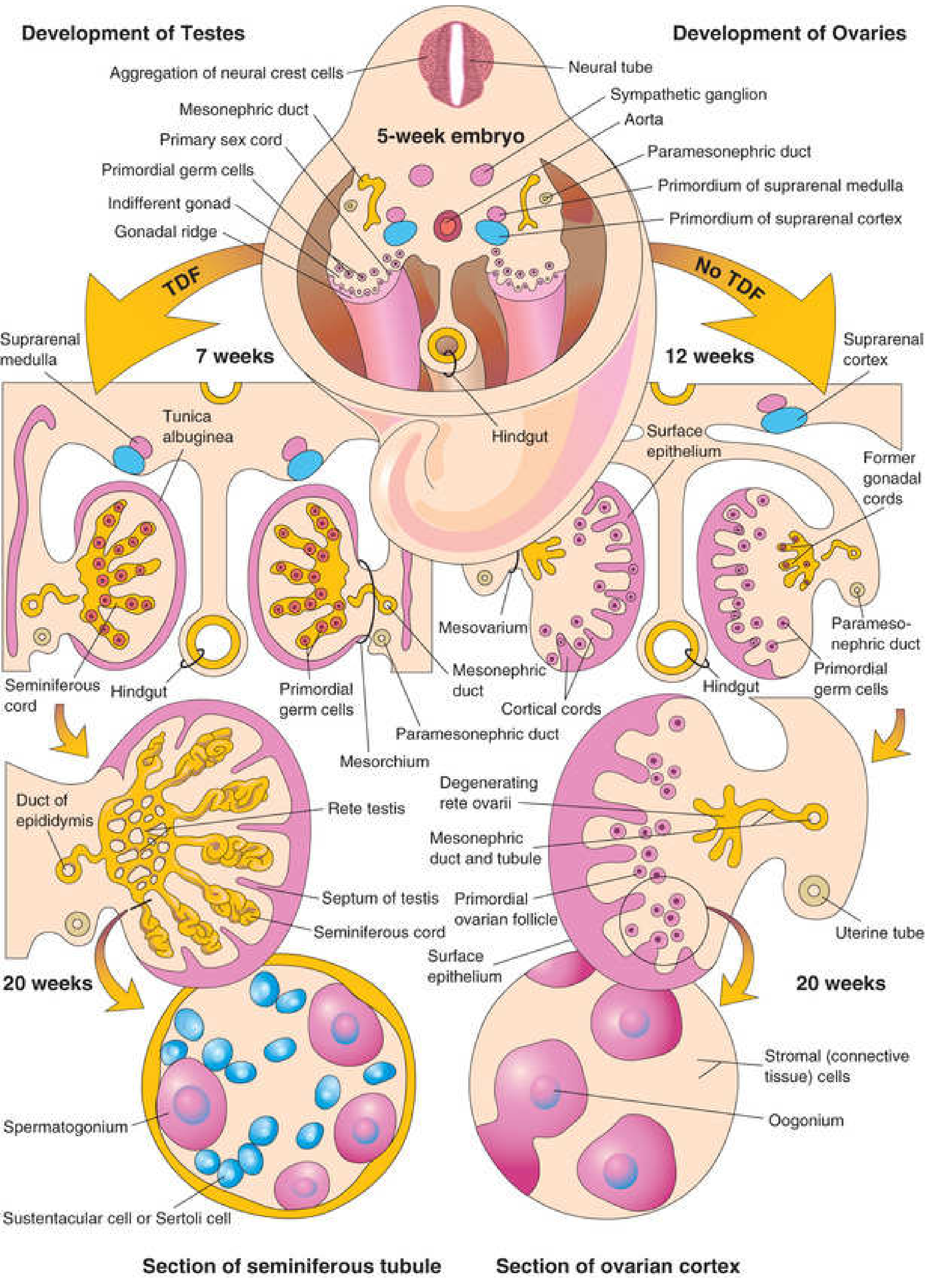
Development of the Testis
- The indifferent gonad develops on the gonadal (genital) ridge (medial part of the urogenital ridge) from intermediate mesoderm, beginning at week 5
- Primordial germ cells migrate from the yolk sac wall along the dorsal mesentery to reach the gonadal ridge by week 6
- In a 46,XY embryo, the SRY gene on the Y chromosome produces Testis-Determining Factor (TDF):
- Induces gonadal cords → seminiferous cords → extend into medulla → form rete testis
- A dense fibrous capsule, the tunica albuginea, develops - the hallmark of testicular development
- Sertoli cells (from surface epithelium) form sustentacular cells in seminiferous tubules; secrete AMH (Anti-Müllerian Hormone) → causes regression of paramesonephric (Müllerian) ducts
- Leydig cells (from mesenchyme between tubules) begin secreting testosterone by week 8 → virilizes mesonephric (Wolffian) ducts → form epididymis, ductus deferens, seminal vesicles
Descent of the Testis
Phase 1 - Transabdominal Descent (Weeks 8-15)
- By end of 2nd month, the testis and mesonephros share the urogenital fold
- The gubernaculum (lower gonadal ligament) forms after mesonephros regression
- Passes below the genital ducts
- Pierces the abdominal wall through the inguinal canal
- Ends in the labioscrotal swelling (scrotum primordium)
- Traction from gubernaculum + rapid body growth (faster than gonad growth) causes the testis and epididymis to slide down the posterior trunk wall external to the peritoneum
- By start of 3rd month, testis reaches the entrance of the inguinal canal
- Hormones involved: Insulin-like factor 3 (INSL3) from Leydig cells + its receptor RXFP2 on gubernaculum drive this phase; also involves AMH
Phase 2 - Transinguinal (Inguinoscrotal) Descent (Weeks 26-32, completed near birth)
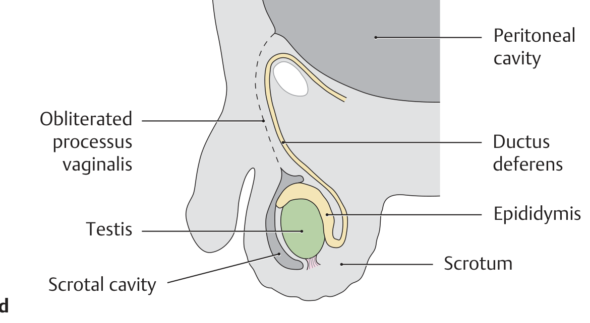
- The processus vaginalis forms as a funnel-shaped evagination of peritoneum anterior to the gubernaculum, extending into the scrotal swelling
- The testis passes through the inguinal canal into the scrotum
- The processus vaginalis carries the layers of the abdominal wall → forms coverings of the spermatic cord and testis
- After descent, the processus vaginalis obliterates except for a small space that becomes the tunica vaginalis testis (visceral and parietal layers)
- Hormones: testosterone (and its DHT metabolite) and genitofemoral nerve (CGRP) are required for this phase
Undescended Testis (Cryptorchidism)
- Definition: Testis that fails to complete its normal descent into the scrotum
- Incidence: 3-4% of full-term male infants; 30% of premature males; most descend spontaneously by 3 months (incidence 1% by 1 year)
- Unilateral in 90% of cases; right side more common; bilateral in 10%
Types
| Type | Description |
|---|---|
| True undescended | Testis in the normal path of descent (abdominal, inguinal canal, or high scrotal) |
| Ectopic testis | Testis deviated from normal path (perineal, femoral, superficial inguinal pouch) |
| Retractile testis | Testis in scrotum but pulled up by cremasteric reflex; not truly undescended |
Complications of Cryptorchidism
- Infertility - failure of spermatogenesis (requires temperature ~2°C below core body temperature); bilateral cryptorchidism = near-certain infertility if untreated
- Malignancy - 4-10x increased risk of testicular germ cell tumours (mainly seminoma); even the contralateral descended testis has increased risk in bilateral cases
- Torsion - undescended testis more prone to torsion
- Indirect inguinal hernia - persistent processus vaginalis; associated in ~90% of cases
- Psychological impact
Treatment
- Orchidopexy (surgical fixation in scrotum): recommended between 6-18 months of age (ideally by 12-18 months)
- Hormonal therapy: hCG or GnRH analogues (limited role; variable success)
- Early treatment reduces (but does not eliminate) risk of malignancy and preserves fertility potential
7. Pharyngeal Arch Derivatives
Overview
- A mesenchymal core (neural crest cells + mesodermal mesenchyme) → skeletal/connective tissue
- A muscle component → specific muscles
- An artery (aortic arch artery)
- A nerve (cranial nerve)
Derivatives by Arch
Arch 1 (Mandibular Arch)
- Meckel's cartilage (proximal part): contributes to mandibular ramus; leaves behind:
- Sphenomandibular ligament
- Anterior malleolar ligament
- Malleus (except manubrium)
- Incus (except long process)
- Maxillary process (dorsal portion of arch 1):
- Premaxilla, maxilla, zygoma, part of temporal bone
- Muscles of mastication: masseter, temporalis, medial and lateral pterygoids
- Mylohyoid, anterior belly of digastric
- Tensor tympani, tensor veli palatini
Arch 2 (Hyoid Arch / Reichert's Cartilage)
- Proximally: Styloid process, manubrium of malleus, long process of incus, stapes suprastructure (footplate mostly from otic capsule)
- Middle: Stylohyoid ligament (remnant after central part withers)
- Distally: Superior body and lesser cornu of hyoid bone
- All muscles of facial expression
- Stapedius, stylohyoid, posterior belly of digastric
Arch 3
- Inferior body and greater cornu of hyoid bone
- Stylopharyngeus (only muscle from arch 3)
Arch 4 and Arch 6 (Arches 4 and 6 fuse in humans)
- Arch 4: Superior laryngeal nerve branch of vagus
- Arch 6: Recurrent laryngeal nerve branch of vagus
- Laryngeal cartilages: thyroid, cricoid, arytenoid, corniculate, cuneiform cartilages
- Arch 4 (superior laryngeal n.): cricothyroid, levator veli palatini, pharyngeal constrictors
- Arch 6 (recurrent laryngeal n.): all intrinsic muscles of the larynx (except cricothyroid)
Summary Table
| Arch | Nerve | Skeletal Derivatives | Muscle Derivatives |
|---|---|---|---|
| 1 (Mandibular) | CN V3 | Malleus, incus, mandible, maxilla, zygoma, sphenomandibular lig. | Mastication muscles, mylohyoid, ant. digastric, tensor tympani, tensor veli palatini |
| 2 (Hyoid) | CN VII | Stapes, styloid, stylohyoid lig., lesser cornu & sup. body of hyoid | Facial expression, stapedius, stylohyoid, post. digastric |
| 3 | CN IX | Greater cornu & inf. body of hyoid | Stylopharyngeus |
| 4 | CN X (superior laryngeal) | Thyroid/cricoid cartilage | Cricothyroid, pharyngeal constrictors, levator veli palatini |
| 6 | CN X (recurrent laryngeal) | Arytenoid, corniculate, cuneiform cartilages | All intrinsic laryngeal muscles (except cricothyroid) |
Pharyngeal Pouches and Clefts
| Pouch | Derivatives |
|---|---|
| 1st pouch | Tubotympanic recess → auditory (Eustachian) tube + middle ear cavity; tympanic membrane |
| 2nd pouch | Crypts of palatine tonsil; surface epithelium forms the tonsillar fossa |
| 3rd pouch | Inferior parathyroid glands (dorsal wing) + thymus (ventral wing) |
| 4th pouch | Superior parathyroid glands (dorsal wing) + ultimobranchial body/C-cells of thyroid (ventral wing) |
| Cleft | Derivatives |
|---|---|
| 1st cleft | External auditory meatus |
| 2nd, 3rd, 4th clefts | Normally buried by overgrowth of 2nd arch; if persist → branchial (cervical) cyst/sinus/fistula |
8. Development of Face and Facial Clefts
The Five Facial Processes
- Frontonasal prominence (1 - unpaired) - forms forehead, bridge of nose, medial and lateral nasal processes
- Maxillary processes (2 - one on each side, from 1st pharyngeal arch)
- Mandibular processes (2 - one on each side, from 1st pharyngeal arch)
Formation of the Nose
- Nasal placodes (thickenings of ectoderm) appear on the inferolateral part of the frontonasal prominence at week 4
- Each nasal placode is surrounded by a horseshoe-shaped elevation:
- Medial nasal process (inner limb)
- Lateral nasal process (outer limb)
- The placode invaginates to form the nasal pit
Formation of the Upper Lip and Primary Palate
- The maxillary processes grow medially and approach the medial nasal processes
- The two medial nasal processes fuse with each other → intermaxillary segment (philtrum of lip, premaxilla, primary palate)
- The maxillary process merges with the medial nasal process on each side → forms the lateral parts of the upper lip
- The lower lip and jaw form by merging of the two mandibular processes
Formation of the Secondary Palate (Weeks 6-12)
- Palatine shelves (horizontal extensions of maxillary processes) initially grow vertically downward on each side of the tongue
- At ~7th-8th week, the shelves elevate horizontally (above the tongue) and fuse with each other in the midline
- They also fuse with the nasal septum above and with the primary palate anteriorly
- The primary and secondary palate fuse at the incisive foramen (its remnant)
- Fusion completes by week 12
Clinical Aspects: Facial Clefts
Cleft Lip (Cheiloschisis)
- Mechanism: Failure of maxillary process to fuse with medial nasal process on one or both sides
- Occurs at ~week 6-7
- Unilateral cleft lip: Most common (left side > right); gap in the upper lip lateral to the midline
- Bilateral cleft lip: Both maxillary processes fail to fuse; central portion (philtrum/premaxilla) projects forward
- Can be complete (extending into the nostril) or incomplete
- Incidence: ~1/700-1000 births; males > females; left > right
- Genetics: Multifactorial (IRF6, MTHFR, TGFA, BRCA3 gene loci); associated with folic acid deficiency, retinoic acid, phenytoin, smoking
Cleft Palate (Palatoschisis)
- Mechanism: Failure of palatine shelves to elevate and/or fuse
- Occurs at ~weeks 8-12
- Can be isolated (without cleft lip) - then females > males
- Types:
- Cleft of soft palate only
- Cleft of both hard and soft palate
- Submucous cleft palate (muscle fails to fuse but mucosa intact)
- Consequences: feeding difficulties (cannot generate suction), speech problems (hypernasal voice), recurrent otitis media (Eustachian tube dysfunction), dental abnormalities
Cleft Lip with Cleft Palate
- Most common combined form; the cleft lip creates a communication that allows the tongue to fill the space, preventing palatal shelf elevation → secondary cleft palate
- More severe; left unilateral, right unilateral, or bilateral
Oblique Facial Cleft
- Rare; results from failure of maxillary process to merge with lateral nasal process
- Cleft extends from upper lip to the eye (nasolacrimal groove fails to close)
Median Cleft of Upper Lip (True Median Cleft)
- Very rare; failure of the two medial nasal processes to merge with each other
- Results in a central notch in the upper lip; associated with holoprosencephaly
Macrostomia / Microstomia
- Macrostomia: failure of maxillary and mandibular processes to fully merge (lateral corner of mouth extends too far)
- Microstomia: excessive merging
Summary: Key Fusion Events and Corresponding Clefts
| Fusion Failure | Resulting Defect |
|---|---|
| Maxillary process + medial nasal process | Cleft lip |
| Palatine shelves fail to elevate/fuse with each other | Cleft palate |
| Maxillary process + lateral nasal process | Oblique facial cleft |
| Two medial nasal processes | Median cleft lip |
| Mandibular processes | Mandibular (median) cleft (rare) |
- The Developing Human: Clinically Oriented Embryology (Moore et al.) - Ch. 11 (midgut), Ch. 12 (kidneys, testis), Ch. 13 (neural tube defects), Ch. 9 (pharyngeal arches), Ch. 10 (face)
- Langman's Medical Embryology - Ch. 13 (neural tube), Ch. 15 (midgut rotation)
- Campbell-Walsh-Wein Urology - Ch. 20 (kidney development, molecular mechanisms)
- Cummings Otolaryngology Head and Neck Surgery - Ch. 202 (pharyngeal arch derivatives)
- Thieme General Anatomy and Musculoskeletal System - testis descent diagrams
type and classification of chromosomes
Types and Classification of Chromosomes
1. Chromosome Structure - Key Landmarks
- p arm (short arm) - from French petit = small
- q arm (long arm) - the longer arm below the centromere
- Telomeres - specialized repeat sequences (TTAGGG)n at both ends; stabilize chromosomes; shorten with each cell division (cellular aging mechanism)
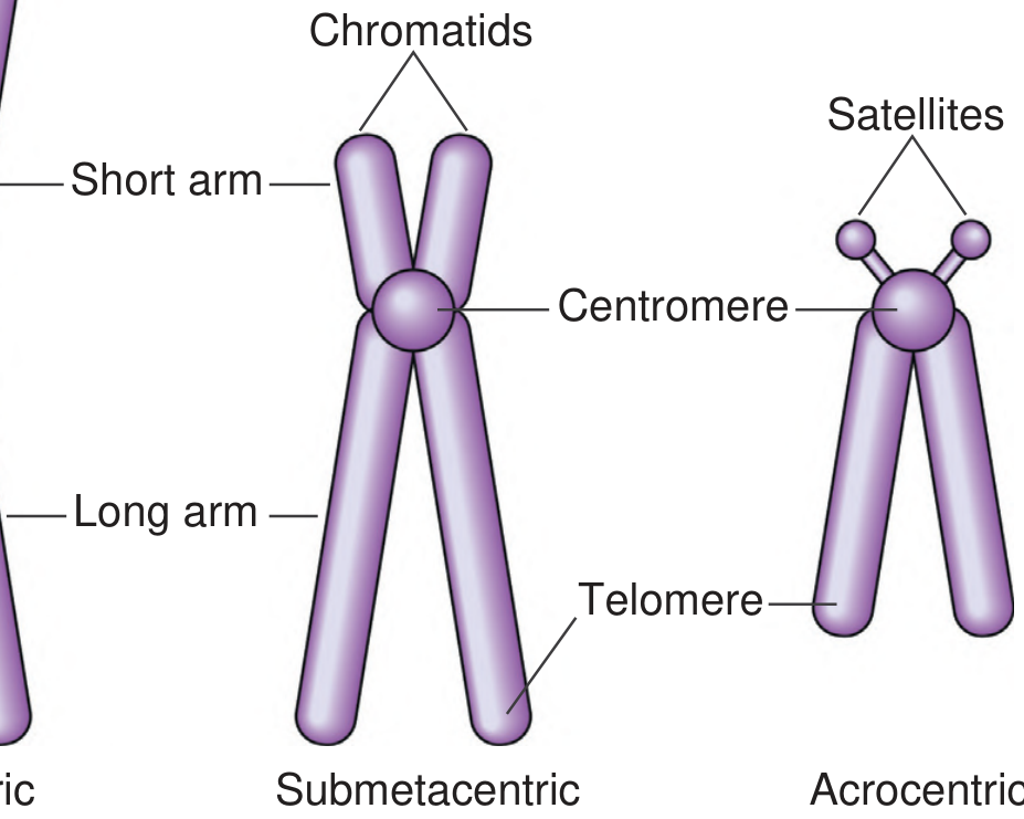
- Satellites - small rounded bodies attached by a thin stalk to the short arm of acrocentric chromosomes; contain multiple copies of rRNA genes that form the nucleolus
2. Classification Based on Centromere Position
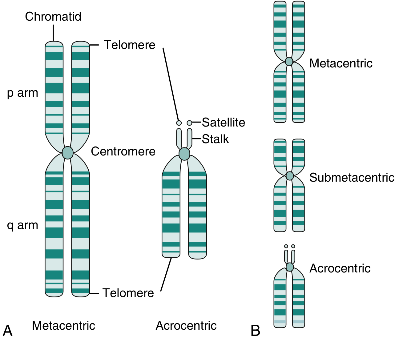
Type 1 - Metacentric
- Centromere is located at or near the middle of the chromosome
- p arm and q arm are of roughly equal length
- Chromosome appears X-shaped at metaphase
- Arm ratio (p:q) ≈ 1:1
- Human examples: Chromosomes 1, 3, 16, 19, 20
Type 2 - Submetacentric
- Centromere is positioned between the middle and the end (off-center)
- q arm is longer than the p arm
- Chromosome appears slightly J-shaped or L-shaped
- Arm ratio (p:q) = intermediate (p arm noticeably shorter but present)
- Human examples: Chromosomes 2, 4, 5, 6, 7, 8, 9, 10, 11, 12, 17, 18, X
Type 3 - Acrocentric
- Centromere is located near one end (tip) of the chromosome
- The p arm is very short or vestigial (no significant p arm)
- These short p arms contain stalks and satellites (rRNA-encoding regions; sites of nucleolus organizer regions - NORs)
- Chromosome appears V-shaped or almost rod-like
- Prone to Robertsonian translocations (fusion at centromeres between two acrocentrics)
- Human examples: Chromosomes 13, 14, 15, 21, 22 (the 5 acrocentric pairs)
Note: The Y chromosome is also considered acrocentric/subacrocentric.
Type 4 - Telocentric
- Centromere is at the very tip (terminus) of the chromosome
- Only one arm exists; no p arm at all
- Not found in normal human karyotype (no human chromosome is truly telocentric)
- Common in other species (e.g., rodents)
- Can arise in humans as an abnormality (e.g., isochromosome)
3. Denver Classification (1960)
- Overall length (longest to shortest)
- Position of the centromere
- Presence or absence of satellites
| Group | Chromosomes | Type | Characteristics |
|---|---|---|---|
| A | 1, 2, 3 | Metacentric / submetacentric | Largest; 1 and 3 metacentric; 2 submetacentric |
| B | 4, 5 | Submetacentric | Large; short p arms |
| C | 6-12, X | Submetacentric | Medium-sized; X chromosome resembles group C |
| D | 13, 14, 15 | Acrocentric | Medium; satellites on short arms; NOR-bearing |
| E | 16, 17, 18 | Metacentric/submetacentric | Shorter; 16 is metacentric; 17 and 18 submetacentric |
| F | 19, 20 | Metacentric | Short; both metacentric |
| G | 21, 22, Y | Acrocentric | Smallest; satellites on 21 and 22; Y has no satellites |
Note: Chromosome 21 is actually slightly shorter than chromosome 22 (misnaming was recognized after banding techniques were developed), but the numbering was retained by convention.
4. Classification by Function/Content
| Type | Description |
|---|---|
| Autosomes | Chromosomes 1-22 (22 pairs); identical in both sexes |
| Sex chromosomes (allosomes) | X and Y; determine sex (XX = female, XY = male) |
| Euchromatic chromosomes | Lightly staining; contain actively expressed genes |
| Heterochromatic regions | Darkly staining; repetitive, inactive DNA |
5. Classification by Chromatin Type
| Type | Staining | DNA Content | Function |
|---|---|---|---|
| Euchromatin | Light (G-band pale) | GC-rich; gene-rich | Actively transcribed genes |
| Constitutive heterochromatin | Dark (always condensed) | Repetitive sequences; centromeres, telomeres | Structural; no genes |
| Facultative heterochromatin | Dark (conditionally condensed) | Inactive X chromosome (Barr body) | Silenced genes; can be reactivated |
6. Chromosome Banding - Modern Classification Aid
| Banding Technique | Stain | Appearance | Basis |
|---|---|---|---|
| G-banding (most common) | Giemsa | Dark (AT-rich) and light (GC-rich) bands | Standard karyotype |
| Q-banding | Quinacrine (fluorescent) | Similar pattern to G-bands; Y chromosome fluoresces brightly | Used in sex determination |
| R-banding | Reverse Giemsa | Reverse of G-bands; light bands are dark | Highlights regions poorly seen by G |
| C-banding | Giemsa (alkaline) | Centromeres and constitutive heterochromatin | Identify centromere polymorphisms |
| NOR-banding | Silver stain | Stains satellites of acrocentric chromosomes (D and G groups) | Identifies nucleolar organizer regions |
7. Chromosome Nomenclature (ISCN)
[chromosome number][arm][region][band].[subband]
14q32.1 = chromosome 14, long arm (q), region 3, band 2, subband 1 (where IGH immunoglobulin gene resides)| Symbol | Meaning |
|---|---|
| p | Short arm |
| q | Long arm |
| del | Deletion |
| dup | Duplication |
| inv | Inversion |
| t | Translocation |
| r | Ring chromosome |
| i | Isochromosome |
| + / - | Gain/loss of chromosome |
| pter / qter | Terminal end of p or q arm |
8. Summary Diagram of Types
| Type | Centromere Position | p:q Ratio | Shape | Human Examples |
|---|---|---|---|---|
| Metacentric | Middle | ~1:1 | X-shaped | 1, 3, 16, 19, 20 |
| Submetacentric | Off-center | p < q | J/L-shaped | 2, 4-12, 17, 18, X |
| Acrocentric | Near tip | p very small + satellites | V-shaped | 13, 14, 15, 21, 22, Y |
| Telocentric | At tip | No p arm | Rod-shaped | Not in humans (normal) |
- Emery's Elements of Medical Genetics and Genomics - Ch. 3 (chromosome morphology, Denver classification, banding)
- Henry's Clinical Diagnosis and Management by Laboratory Methods - Ch. 71 (chromosome structure, types)
- Quick Compendium of Clinical Pathology, 5th ed. - Ch. 7.2 (classification by arm length, banding)
- Basic Medical Biochemistry: A Clinical Approach, 6th ed. - Ch. 18 (chromosome nomenclature, ISCN table)
8. Numerical chromosomal anomalies. 9. Structural chromosomal anomalies.
8. Numerical Chromosomal Anomalies
Definition
- Aneuploidy - chromosome number is NOT an exact multiple of the haploid set (n = 23)
- Polyploidy - chromosome number IS an exact multiple of the haploid set, but greater than diploid
Causes of Numerical Anomalies
1. Nondisjunction (Principal Cause)
- Meiosis I nondisjunction: Both homologs go to the same cell → both daughter cells abnormal → one gamete gets 2 copies, one gets 0 copies of that chromosome
- Meiosis II nondisjunction: Sister chromatids fail to separate → one gamete gets 2 copies, one is normal, one gets 0 copies
- Mitotic nondisjunction: Occurs after fertilization in somatic cell divisions → produces mosaicism
2. Anaphase Lag
3. Dispermy
A. ANEUPLOIDY
Types of Aneuploidy
| Term | Formula | Chromosome Number | Description |
|---|---|---|---|
| Nullisomy | 2n - 2 | 44 | Loss of a complete homologous pair; always lethal |
| Monosomy | 2n - 1 | 45 | Loss of one chromosome from a pair |
| Trisomy | 2n + 1 | 47 | Gain of one extra chromosome |
| Double trisomy | 2n + 2 | 48 | Two pairs trisomic |
| Tetrasomy | 2n + 2 | 48 | Four copies of one chromosome |
Aneuploidy occurs in 3-4% of clinically recognized pregnancies - the most common and clinically significant numeric chromosomal abnormality.
AUTOSOMAL TRISOMIES (viable)
Trisomy 21 - Down Syndrome
- Karyotype: 47,XX,+21 or 47,XY,+21
- Incidence: ~1 in 700-800 live births (most common autosomal trisomy)
- Cause:
- 95% - nondisjunction (usually Meiosis I; 90-95% maternal origin)
- 3-4% - Robertsonian translocation (usually 14;21); not age-related; inheritable
- 1-2% - mosaicism (milder phenotype)
- Maternal age effect: Strong positive correlation; risk rises sharply after age 35 (see table below)
| Maternal Age | Incidence of Trisomy 21 |
|---|---|
| 20-24 years | 1 in 1400 |
| 30-34 years | 1 in 700 |
| 35 years | 1 in 350 |
| 37 years | 1 in 225 |
| 39 years | 1 in 140 |
| 41 years | 1 in 85 |
| 43 years | 1 in 50 |
| 45+ years | 1 in 30 |
- Hypotonia - first sign in newborn
- Brachycephaly with flat occiput
- Flat nasal bridge
- Upward slanting palpebral fissures
- Epicanthic folds
- Protruding tongue (macroglossia relative to small oral cavity)
- Single transverse palmar (simian) crease
- 5th finger clinodactyly (incurved)
- Short, broad hands
- Sandal-gap deformity (wide space between 1st and 2nd toes)
- Congenital heart disease in ~50% (most common: AV septal defect, VSD)
- Duodenal atresia ("double bubble" sign - more common than other disorders)
- Cognitive/intellectual disability - moderate (IQ typically 25-75)
- Short stature
- Increased risk of leukemia (ALL, AML) - 10-20x higher risk
- Alzheimer disease - virtually all develop neuropathological changes by age 40 (APP gene on chromosome 21)
- Atlanto-axial instability
- Average life expectancy >50 years with modern care
Trisomy 18 - Edwards Syndrome
- Karyotype: 47,XX,+18 or 47,XY,+18
- Incidence: 1 in 8,000 live births
- Prognosis: Very poor; 1-year survival ~6-12%; most die within first weeks/months
- Features:
- Severe intellectual disability and growth retardation
- Prominent occiput
- Low-set, malformed ears
- Micrognathia (small jaw)
- Short sternum
- Clenched fists with overlapping fingers (index over 3rd, 5th over 4th)
- Hypoplastic nails
- Rocker-bottom feet
- Ventricular septal defect (most common cardiac defect)
Trisomy 13 - Patau Syndrome
- Karyotype: 47,XX,+13 or 47,XY,+13
- Incidence: 1 in 12,000 live births
- Prognosis: Worse than trisomy 18; most die within first weeks
- Features:
- Severe intellectual disability
- Severe CNS malformations (holoprosencephaly - failure of forebrain to divide)
- Sloping forehead
- Microphthalmia (small eyes) or anophthalmia
- Bilateral cleft lip ± cleft palate
- Scalp defects (aplasia cutis)
- Polydactyly (post-axial - extra digits on little finger/toe side)
- Malformed, low-set ears
- Posterior prominence of heels (rocker-bottom feet)
- Cardiac defects (VSD, ASD, PDA)
SEX CHROMOSOME TRISOMIES
Klinefelter Syndrome (47,XXY)
- Most common sex chromosome aneuploidy in males; 1 in 600-1000 male births
- Not usually detected until puberty (no characteristic findings in infants)
- Sex chromatin (Barr body) positive - one inactive X present in nuclei
- Features:
- Tall stature with long limbs
- Small, firm testes (hyalinization of seminiferous tubules)
- Azoospermia / infertility (primary)
- Gynecomastia (breast development)
- Hypogonadism - low testosterone
- Reduced body hair and beard
- Mild intellectual disability (IQ slightly below normal) or learning difficulties
- Increased risk of breast cancer, autoimmune disease
- Variants: 48,XXXY; 49,XXXXY (greater severity with each extra X)
Triple X Syndrome (47,XXX) - Trisomy X
- Incidence: 1 in 1000 females
- Usually asymptomatic and fertile; often undiagnosed
- Two Barr bodies in cell nuclei
- Mild intellectual disability, tall stature, menstrual irregularities possible
- Variants: 48,XXXX and 49,XXXXX (tetrasomy/pentasomy X) - serious cognitive deficiency and physical abnormalities
47,XYY Syndrome (Jacob's Syndrome)
- Incidence: 1 in 1000 males
- Usually tall stature, normal fertility
- May have mild learning difficulties or behavioral issues
- Often undiagnosed; discovered incidentally
- Early association with aggression has been disproven
MONOSOMIES
Turner Syndrome (45,X) - Monosomy X
- Only viable monosomy in humans; all autosomal monosomies are lethal
- Incidence: 1 in 8,000 female live births
- Most common cytogenetic abnormality in spontaneous abortions (~18% of chromosomally abnormal abortions); only ~1% of 45,X conceptuses survive to birth
- Karyotype variants: 45,X (most common); 45,X/46,XX mosaic (milder); 46,X,i(Xq) isochromosome; ring X
- Error origin: ~75% paternal (missing paternal X or Y in sperm)
- Barr body negative (no inactive X)
- Features:
- Short stature (most consistent feature)
- Webbing of neck (pterygium colli) - due to cystic hygroma in fetus
- Low posterior hairline
- Broad ("shield") chest with widely spaced nipples
- Cubitus valgus (increased carrying angle)
- Lymphedema of hands and feet at birth
- Primary amenorrhea and streak gonads (ovarian dysgenesis)
- Infertility in most (90%); occasional spontaneous pregnancies in mosaics
- Coarctation of the aorta (15-20%) - most common cardiac defect
- Bicuspid aortic valve
- Normal intelligence (may have specific deficits in spatial perception)
- Short 4th metacarpal, high arched palate
- Increased risk of aortic dissection (dilated aorta)
B. POLYPLOIDY
Triploidy (69 chromosomes)
- Three complete haploid sets (3n)
- Most common type of polyploidy in humans
- Cause: Most frequently dispermy (fertilization by 2 sperms); or failure of one meiotic division → diploid gamete
- Clinical features: Severe intrauterine growth restriction with head-body disproportion; most abort in 1st trimester
- Account for ~20% of chromosomally abnormal spontaneous abortions
- Very rarely born alive; not compatible with survival
Tetraploidy (92 chromosomes)
- Four complete haploid sets (4n)
- Cause: Failure of the first cleavage division of the zygote; subsequent divisions produce cells with 92 chromosomes
- Outcome: Abort very early; often recover only an empty chorionic sac ("blighted ovum")
C. MOSAICISM
- Results in a milder phenotype than non-mosaic trisomy/monosomy
- Example: 45,X/46,XX mosaic Turner syndrome - less severe features than pure 45,X
- Example: Mosaic Down syndrome (46,XX/47,XX+21) - milder intellectual disability
- The earlier the mitotic error, the greater the proportion of abnormal cells
9. Structural Chromosomal Anomalies
Definition and Causes
- Ionizing radiation (X-rays, gamma rays)
- Viral infections
- Mutagenic drugs and chemicals (chemotherapy agents, alkylating agents)
- Recombination errors at segmental duplications
Classification of Structural Anomalies
BALANCED vs. UNBALANCED
| Type | Total DNA content | Phenotype |
|---|---|---|
| Balanced | Normal amount, rearranged | Usually phenotypically normal carrier; but risk of unbalanced offspring |
| Unbalanced | Net gain or loss of chromosomal material | Usually abnormal phenotype (birth defects, intellectual disability) |
1. Deletion
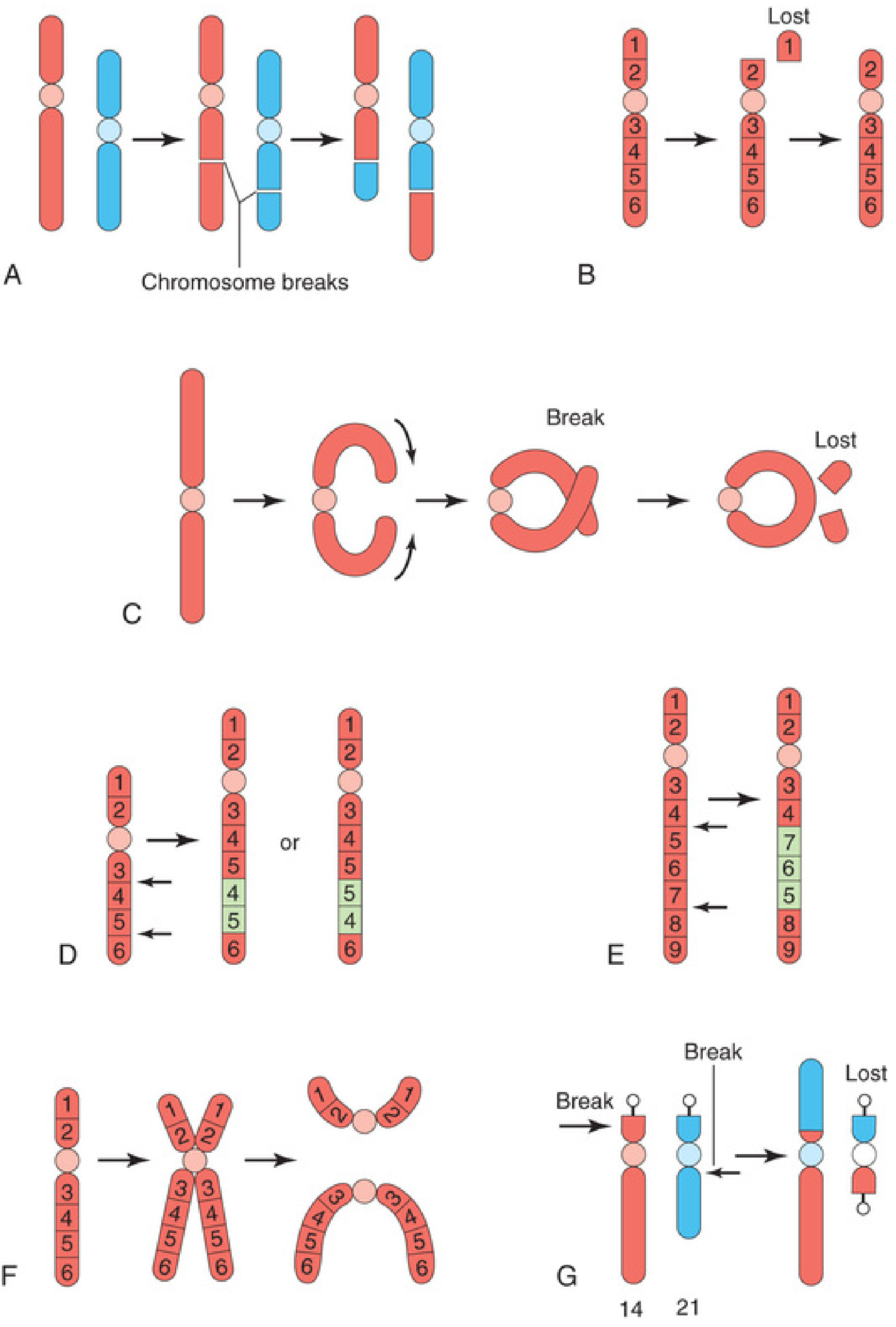
- Terminal deletion: Single break near end of chromosome; terminal segment is lost (Fig. 20.13B)
- Interstitial deletion: Two breaks; middle segment is lost and the two ends rejoin
- Cause: Terminal deletion of short arm of chromosome 5 (5p-)
- Features:
- Characteristic cat-like cry in infancy (mewing sound - due to laryngeal hypoplasia)
- Microcephaly
- Severe intellectual disability
- Hypertelorism (widely spaced eyes)
- Congenital heart disease
- Low birth weight; low set ears
2. Duplication
- More common than deletions and less harmful (no net loss of genetic material)
- The repeated segment may be in:
- Direct orientation (tandem duplication)
- Inverted orientation (inverted duplication)
- Usually causes cognitive impairment or birth defects due to gene dosage imbalance
- May involve part of a gene, a whole gene, or a series of genes
3. Inversion
| Type | Definition | Consequence if Inherited |
|---|---|---|
| Paracentric inversion | Both breaks within one arm (centromere NOT included); segment is inverted within that arm (Fig. 20.13E) | Recombinant chromosomes (dicentric or acentric) - usually inviable offspring; carrier usually phenotypically normal |
| Pericentric inversion | Breaks in both arms (centromere IS included); segment including centromere is inverted | Carriers at risk of having offspring with duplications and deletions due to unequal crossing-over at meiosis; may change arm ratio |
- Inversion carriers are usually phenotypically normal (balanced rearrangement)
- Risk is to offspring who may inherit unbalanced recombinant chromosomes
4. Translocation
A. Reciprocal Translocation
- Segments are exchanged between two non-homologous chromosomes (Fig. 20.13A)
- Balanced carriers are phenotypically normal
- At meiosis, the 4-chromosome translocation quadrivalent segregates → risk of unbalanced gametes
- Most common structural chromosomal abnormality: 1 in 1000 in general population
- Reciprocal translocation between chromosomes 9 and 22
- BCR-ABL fusion oncogene → constitutively active tyrosine kinase
- Present in >95% of Chronic Myeloid Leukemia (CML); also in ALL
B. Robertsonian Translocation
- Fusion of two acrocentric chromosomes at their centromeres (Fig. 20.13G)
- Both short arms (satellite-containing) are lost (usually the small rDNA-containing short arms)
- Results in one large derivative chromosome instead of two
- Net chromosome count = 45 (but functionally balanced - no critical genes lost from small arms)
- Most common: involves chromosomes 13, 14, 15, 21, 22
- Most clinically significant: rob(14;21) - carrier has 45 chromosomes but is phenotypically normal
| Carrier Karyotype | Offspring Risk |
|---|---|
| rob(14;21) carrier | 1/3 Down syndrome (47 chromosomes, extra 21 material), 1/3 carrier like parent, 1/3 normal |
| Accounts for 3-4% of Down syndrome cases | Risk independent of maternal age (unlike nondisjunction trisomy) |
Babies born to balanced Robertsonian translocation carriers must be investigated regardless of maternal age.
5. Isochromosome
- Results in one arm being duplicated and the other arm being lost
- Most common: i(Xq) - isochromosome of the long arm of X
- In Turner syndrome: 46,X,i(Xq) - monosomy for Xp (short arm), trisomy for Xq (long arm)
- Accounts for ~15% of Turner syndrome cases
- Other examples: i(12p) in germ cell tumors; i(17q) in medulloblastoma and leukemia
6. Ring Chromosome
- Net loss of genetic material from both ends (unbalanced)
- Rings are unstable - can be lost during cell division → mosaicism
- Found associated with all chromosomes; described in Turner syndrome (45,X with ring X), trisomy 18, and various deletion syndromes
- Rings may also form from supernumerary chromosomes (extra marker chromosomes)
7. Microdeletions and Microduplications (Contiguous Gene Syndromes)
| Syndrome | Chromosome | Type | Parental Origin | Clinical Features |
|---|---|---|---|---|
| Prader-Willi | del 15q11-13 | Microdeletion | Paternal | Hypotonia (neonatal), hyperphagia/obesity, hypogonadism, short stature, small hands/feet, mild intellectual disability |
| Angelman | del 15q11-13 | Microdeletion | Maternal | Severe intellectual disability, seizures, ataxia, inappropriate laughter ("happy puppet"), microcephaly |
| DiGeorge / Velocardiofacial (22q11.2 deletion) | del 22q11.2 | Microdeletion | Either | Cardiac outflow defects, hypocalcemia (absent parathyroids), T-cell immunodeficiency (thymic aplasia), palatal anomalies, characteristic facial features |
| Williams | del 7q11.23 | Microdeletion | Either | Supravalvular aortic stenosis, hypercalcemia, "cocktail party" personality, intellectual disability, elfin facies |
| Miller-Dieker | del 17p13.3 | Microdeletion | Either | Lissencephaly (smooth brain), severe intellectual disability, seizures |
| Cri du Chat | del 5p | Deletion (may be micro or macro) | Either | Cat-like cry, microcephaly, intellectual disability (discussed above) |
Imprinting-Related Disorders (Prader-Willi / Angelman)
- PWS = paternal deletion (or maternal uniparental disomy) - paternal alleles are normally active in this region
- AS = maternal deletion (or paternal uniparental disomy) - maternal alleles (UBE3A gene) are normally active
Summary: Structural Anomalies at a Glance
| Anomaly | Balanced? | Mechanism | Key Clinical Relevance |
|---|---|---|---|
| Deletion | No | Single or double break + loss | Cri du chat (5p-), 22q11 deletion, 1p36 deletion |
| Duplication | Usually No | Segment repeated | Charcot-Marie-Tooth (PMP22 dup 17p12) |
| Inversion | Yes (carrier) | Two breaks + inversion | Carrier at risk; offspring may be unbalanced |
| Reciprocal translocation | Yes (carrier) | Exchange between 2 chromosomes | Offspring unbalanced; BCR-ABL in CML |
| Robertsonian translocation | Yes (functional) | Fusion of 2 acrocentrics | Familial Down syndrome (14;21) |
| Isochromosome | No | Transverse centromere division | i(Xq) in Turner syndrome; i(12p) in testicular tumors |
| Ring chromosome | No | Loss of both telomeres + join | Turner syndrome variant |
| Microdeletion | No | Submicroscopic; contiguous gene loss | PWS, Angelman, DiGeorge, Williams |
- The Developing Human: Clinically Oriented Embryology (Moore et al.) - Ch. 20 (numerical and structural anomalies, trisomy tables, mosaicism, polyploidy)
- Thompson & Thompson Genetics and Genomics in Medicine, 9th ed. - Ch. 6 (autosomal trisomies, Down syndrome, Klinefelter, mechanism tables)
- Robbins, Cotran & Kumar Pathologic Basis of Disease - Ch. 5 (structural abnormalities, nondisjunction, anaphase lag)
- Henry's Clinical Diagnosis and Management by Laboratory Methods - Ch. 71 (aneuploidy, chromosome classification)
- Basic Medical Biochemistry: A Clinical Approach, 6th ed. - Ch. 18 (nondisjunction, gene dosage)
answer the following questions of physiology in detail
. Describe the Sino-aortic reflex & its importance 18. Renin-Angiotensin system in regulation of blood pressure 19. Explain Arterial pulse – Aug 1999 (OS) 20. Explain the causes of each wave in Jugular venous pulse – Oct 2008 21. Coronary circulation 22. Explain Cushing’s reflex 23. Explain Cerebral circulation 24. What microcirculation & explain the pressures in it 25. Explain Triple response (Reaction) 26. Describe the manifestations of congestive heart failure 27. Functions of lymphatic circulation. 28. Explain the factors affecting venous return. 29. Explain baroreceptor reflex mechanism. 30. Explain Chemoreceptor reflex. 31. Explain Bainbridge reflex 32. Explain Sympathetic vasodilator system. 33. Explain Autonomic regulation of cardiovascular function. 34. Explain Autoregulation of blood flow. 35. Explain Blood- brain barrier 36. Explain Measurement of coronary blood flow.
Cardiovascular Physiology - Detailed Answers
17. Sino-Aortic Reflex and Its Importance
Receptor Locations
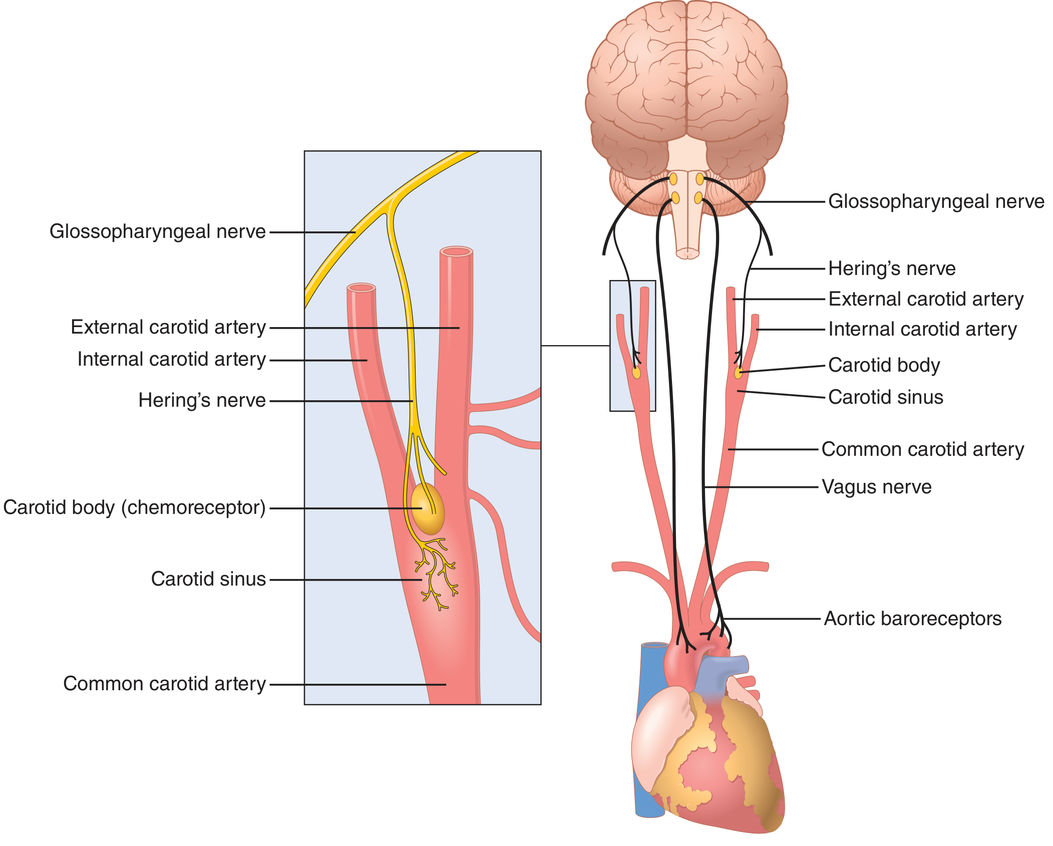
- Carotid sinus - a small dilation of the internal carotid artery just above the bifurcation of the common carotid; afferents travel via Hering's nerve (branch of CN IX - Glossopharyngeal nerve)
- Aortic arch - in the wall of the aortic arch; afferents travel via the aortic depressor nerve (branch of CN X - Vagus nerve)
- Both sets of afferents terminate at the Nucleus Tractus Solitarius (NTS) in the medullary brainstem
Neural Pathway
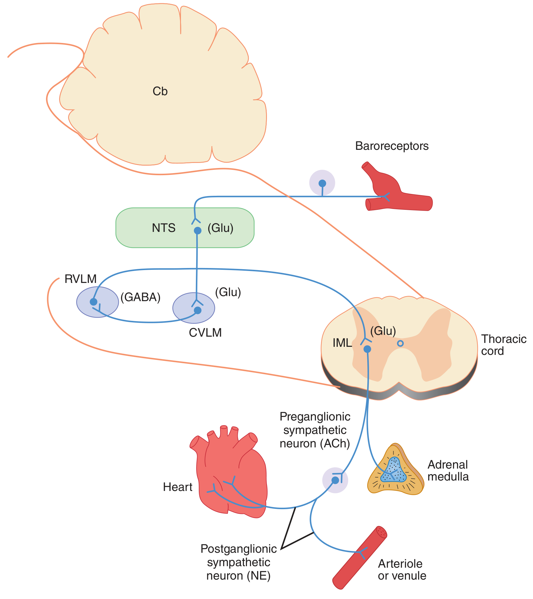
- Baroreceptors stretch → increased firing rate
- Afferents (CN IX, X) → NTS (release glutamate)
- NTS → Caudal Ventrolateral Medulla (CVLM) (excitatory, glutamate)
- CVLM neurons release GABA → inhibit Rostral Ventrolateral Medulla (RVLM) (sympathetic vasomotor center)
- NTS also excites vagal motor nuclei (nucleus ambiguus + dorsal motor nucleus)
- ↓ Sympathetic outflow → vasodilation, venodilation
- ↑ Vagal tone → bradycardia, ↓ cardiac output
- ↓ Blood pressure (restored toward normal)
- Opposite occurs: ↓ baroreceptor firing → ↓ GABA on RVLM → ↑ sympathetic outflow → tachycardia, vasoconstriction, ↑ BP
Properties of Baroreceptors
- Not stimulated below 50-60 mmHg; maximally active at ~180 mmHg
- Aortic receptors operate ~30 mmHg higher than carotid receptors
- More sensitive to pulsatile (changing) pressure than to sustained constant pressure
- Respond in fractions of a second - fastest BP regulation mechanism
Importance of the Sino-Aortic Reflex
- Minute-to-minute BP regulation - most important short-term buffer of arterial pressure
- Postural hypotension prevention - when standing up, gravity pools blood downward; baroreceptors immediately activate to maintain BP
- Operating range: Most effective near normal MAP of 100 mmHg - a small change causes a large corrective response
- Resetting: With sustained hypertension, baroreceptors reset upward (adapt) - hence they are not effective for long-term BP regulation (that role belongs to the kidneys)
- Clinical use: Carotid sinus massage activates baroreceptors → useful for terminating SVT (vagal effect)
18. Renin-Angiotensin-Aldosterone System (RAAS) in Regulation of Blood Pressure
Sequence of Events
↓ Renal perfusion pressure / ↓ Na+ at macula densa / Sympathetic activation
↓
Juxtaglomerular (JG) cells of kidney → secrete RENIN
↓
RENIN cleaves Angiotensinogen (liver) → Angiotensin I (10 amino acids)
↓
ACE (Angiotensin Converting Enzyme, lung mainly) cleaves Ang I → ANGIOTENSIN II (8 amino acids)
↓ ↓ ↓ ↓
Vasoconstriction Aldosterone ADH release Thirst center
of arterioles (adrenal cortex) (posterior (hypothalamus)
↑TPR → ↑MAP ↑Na+ reabsorption pituitary) ↑water intake
↑K+ excretion ↑water retention
↑H+ excretion ↑blood volume
Three Stimuli for Renin Release
- ↓ Renal perfusion pressure - sensed by stretch receptors in JG cells (baroreceptor mechanism)
- ↓ NaCl delivery to macula densa - sensed by the macula densa (tubuloglomerular feedback)
- ↑ Sympathetic activity - via β1 receptors on JG cells (direct neural stimulation)
Actions of Angiotensin II
- Vasoconstriction of arterioles (AT1 receptors) → ↑ TPR → ↑ MAP
- Aldosterone release from adrenal cortex zona glomerulosa → ↑ Na+ reabsorption in distal tubule/collecting duct → ↑ ECF volume → ↑ blood volume → ↑ BP
- ADH (vasopressin) release from posterior pituitary → ↑ water reabsorption
- Thirst stimulation → ↑ water intake
- Direct renal effects - ↑ Na+/H+ antiporter in proximal tubule → ↑ Na+ reabsorption
- Cardiac and vascular hypertrophy (long-term structural effects)
Clinical Significance
- RAAS blockade is a cornerstone of hypertension and heart failure treatment:
- ACE inhibitors (e.g., captopril, enalapril) - block Ang I → Ang II conversion
- ARBs (e.g., losartan) - block AT1 receptors
- Aldosterone antagonists (e.g., spironolactone)
- Renin inhibitors (aliskiren)
- Renovascular hypertension: Renal artery stenosis → kidney "perceives" low pressure → excess renin → excess Ang II → hypertension
- Primary hyperaldosteronism (Conn's syndrome): Autonomous aldosterone secretion → Na+ retention → hypertension
19. Arterial Pulse
Normal Arterial Pulse Waveform
Peak (systolic pressure ~120 mmHg)
/
Anacrotic /
notch / ← Anacrotic limb (upstroke)
/ ← Dicrotic notch (aortic valve closure)
/ \___/
/ ← Dicrotic wave
---- ← Diastolic pressure (~80 mmHg)
- Anacrotic limb (percussion wave) - rapid upstroke; represents rapid ejection of blood into aorta during ventricular systole
- Systolic peak - maximum pressure (~120 mmHg); represents peak LV ejection
- Anacrotic notch - small notch on the upstroke (seen in aortic stenosis); represents initial ejection before valve fully opens
- Dicrotic notch - sharp notch on the descending limb; caused by closure of the aortic valve at end of systole; separates systole from diastole
- Dicrotic wave - small secondary wave after the dicrotic notch due to rebound of blood from the closed aortic valve (elastic recoil)
- Diastolic decay - gradual fall to diastolic pressure (~80 mmHg) as blood runs off through peripheral vessels
Normal Pulse Characteristics
- Rate: 60-100 beats/min
- Rhythm: Regular
- Volume/Character: Normal (not bounding or weak)
- Pulse pressure = Systolic - Diastolic = ~40 mmHg
- Transmission velocity: ~4-5 m/s (faster than blood flow)
Clinical Variants of Arterial Pulse
| Type | Features | Causes |
|---|---|---|
| Pulsus paradoxus | BP drops >10 mmHg during inspiration | Cardiac tamponade, constrictive pericarditis |
| Pulsus alternans | Alternating strong and weak beats | Severe LV failure |
| Pulsus bisferiens | Two systolic peaks | Aortic regurgitation + stenosis, HOCM |
| Water-hammer pulse (Corrigan's) | Bounding; rapid rise and fall | Aortic regurgitation |
| Pulsus parvus et tardus | Small amplitude, slow upstroke | Aortic stenosis |
| Collapsing pulse | Rapidly rising and falling | PDA, thyrotoxicosis, fever |
Sites of Palpation
20. Jugular Venous Pulse (JVP) - Waves and Their Causes
Waves of JVP
| Wave/Descent | Timing | Cause |
|---|---|---|
| 'a' wave | Just before carotid pulse; pre-systolic | Atrial systole (right atrial contraction); also due to cessation of venous inflow causing pressure rise |
| x descent | After 'a' wave; during systole | Atrial relaxation + downward displacement of AV valves during ventricular systole |
| 'c' wave | Interrupts x descent; at onset of systole | Transmitted manifestation of closure and bulging of tricuspid valve into right atrium during isovolumetric ventricular contraction |
| x' descent | After 'c' wave | Continuation of atrial relaxation (downward pulling of AV ring during ventricular ejection) |
| 'v' wave | End of systole; mirrors systole | Passive filling of right atrium against closed tricuspid valve during ventricular systole (venous return with closed AV valve) |
| y descent | After 'v' wave; in diastole | Opening of tricuspid valve → blood rushes from RA to RV; atrial pressure falls |
Mnemonic
- 'a' = Atrial systole
- 'c' = Closure (tricuspid) / Carotid (coincides with carotid pulse)
- 'v' = Venous filling
Abnormalities of JVP Waves
| Abnormality | Wave Change | Cause |
|---|---|---|
| Absent 'a' wave | No 'a' wave | Atrial fibrillation (no organized atrial contraction) |
| Giant 'a' wave | Large 'a' wave | Tricuspid stenosis, pulmonary hypertension, RV hypertrophy (increased resistance to RA emptying) |
| Cannon 'a' wave | Very large, irregular 'a' wave | Complete heart block, VT (atrium contracts against closed tricuspid valve) |
| Absent x descent | No x | Atrial fibrillation |
| Giant 'v' wave | Large 'v' wave | Tricuspid regurgitation (blood regurgitates during systole), VSD |
| Absent y descent | Plateau JVP | Cardiac tamponade, constrictive pericarditis (Kussmaul's sign) |
| Elevated JVP | Raised baseline | Right heart failure, tricuspid stenosis, cardiac tamponade |
Normal JVP Measurement
- Normal JVP = 3-8 cm H2O
- Measured at 45° with patient inclined; vertical height of pulsation above sternal angle
- Internal jugular vein used (not external, due to valves)
21. Coronary Circulation
Anatomy
- Left coronary artery (LCA): Arises from left aortic sinus; divides into:
- LAD (Left Anterior Descending): Supplies anterior LV wall, anterior IV septum, apex
- LCx (Left Circumflex): Supplies lateral and posterior LV wall
- Right coronary artery (RCA): Arises from right aortic sinus; supplies:
- Right ventricle, SA node (60%), AV node (80%), posterior IV septum (in right dominant)
- Dominance: Determined by which artery supplies the posterior descending artery (PDA); right dominant in 85%
Normal Coronary Blood Flow
- Total flow: ~250 mL/min at rest (5% of cardiac output)
- Can increase 4-5x during maximal exercise (coronary reserve)
- LV perfusion: Occurs mainly in diastole (extravascular compression during systole opposes flow)
- RV perfusion: Occurs in both systole and diastole (lower wall stress)
- Coronary perfusion pressure = Aortic diastolic pressure - LVEDP
Regulation of Coronary Blood Flow
- Metabolic (most important): Local hypoxia, ↑CO2, ↑H+, adenosine (main vasodilator), lactate → vasodilation of coronary arterioles; heart extracts ~65-75% of O2 at rest (highest of any organ), so increased demand must come from increased flow
- Autoregulation: Coronary flow remains constant over perfusion pressures of 60-180 mmHg (myogenic + metabolic mechanisms); lost below 40 mmHg
- Neural: Sympathetic (α1 → mild vasoconstriction; β2 → vasodilation); vagal (mild vasodilation)
- Humoral: Endothelin-1 (vasoconstriction), NO/prostacyclin (vasodilation), adenosine
- Physical factors: Heart rate (↑HR → ↑demand; also shortens diastolic perfusion time)
Coronary Vascular Reserve
- Difference between autoregulated (basal) flow and maximally vasodilated flow
- Normally 3-5x basal flow
- Reduced when coronary stenosis is severe → signs of ischemia
Measurement of Coronary Blood Flow
- Fick principle - Coronary flow = MVO2 / (AO2 - CVO2); requires coronary sinus catheterization
- Thermodilution - cold saline injected into coronary; temperature change measured
- Coronary angiography - anatomical assessment; fractional flow reserve (FFR) measures functional significance
- Doppler flow wire - placed in coronary artery; measures velocity and flow reserve
- TIMI frame count - frames needed for dye to travel a standard coronary artery distance
- Myocardial perfusion imaging (MPI) - thallium-201 or Tc-99m sestamibi SPECT; non-invasive
- PET scanning - most accurate for measuring absolute myocardial blood flow (mL/min/g)
- Cardiac MRI with perfusion imaging - detects regional perfusion defects
22. Cushing's Reflex
Mechanism
- Raised ICP (tumor, hemorrhage, edema, hydrocephalus) → compression of cerebral arteries
- → ↓ Cerebral blood flow → cerebral ischemia
- → ↑ CO2, ↓ O2, ↓ pH in brain tissue
- → Medullary chemoreceptors detect these changes (CO2/H+ most important stimulus)
- → Maximal sympathetic discharge → intense peripheral vasoconstriction → marked ↑ MAP
- → The rise in MAP is an attempt to overcome the raised ICP and restore cerebral perfusion
- → ↑ MAP stretches the baroreceptors → reflex bradycardia (vagal response)
Classic Triad (Cushing's Triad)
- Hypertension (widened pulse pressure, markedly ↑ systolic BP)
- Bradycardia (reflex via baroreceptors)
- Irregular respirations (Cheyne-Stokes or ataxic breathing due to brainstem compression)
Clinical Significance
- Indicates impending brainstem herniation (transtentorial or tonsillar)
- A pre-terminal sign - emergency neurosurgical decompression required
- BP may rise to 250-300 mmHg in severe cases
- The key distinguishing feature: bradycardia with hypertension (unlike shock where tachycardia accompanies hypotension)
23. Cerebral Circulation
Normal Values
- Total cerebral blood flow (CBF): 750 mL/min (~15% of resting cardiac output)
- CBF/100g tissue: ~50 mL/100g/min
- Brain weight: ~1400g but receives disproportionately high blood supply
Anatomy
- Circle of Willis: Formed by anterior communicating artery + bilateral anterior, middle, posterior cerebral arteries + posterior communicating arteries; provides collateral flow
- Internal carotid arteries (anterior circulation): Supply 80% of cerebral blood flow
- Vertebral-basilar arteries (posterior circulation): Supply posterior fossa, cerebellum, brainstem
Regulation of Cerebral Blood Flow
-
Metabolic control (most important):
- CO2/H+ - primary regulator; ↑PaCO2 → vasodilation → ↑CBF (most potent vasodilator); ↓PaCO2 (hyperventilation) → vasoconstriction → ↓CBF; used clinically to treat acute ↑ICP
- O2: Hypoxia (PaO2 <50 mmHg) → vasodilation; hyperoxia → vasoconstriction (mild)
- pH: Acidosis → vasodilation; alkalosis → vasoconstriction
- Adenosine, K+, NO - local vasodilators
-
Autoregulation: CBF remains constant between MAP of 60-150 mmHg; below 60 mmHg, CBF falls (ischemia); above 150 mmHg, autoregulation breaks down (hypertensive encephalopathy)
-
Neural control: Relatively minor; sympathetic (α1) and parasympathetic innervation present but override metabolic mechanisms is limited
-
Blood-Brain Barrier effect: Many circulating vasoactive substances (catecholamines, serotonin, angiotensin) cannot affect cerebral vessels because they cannot cross the BBB
Special Features
- No significant O2 storage - neurons die within 4-6 minutes of ischemia
- Active hyperemia: When neurons fire, local CBF increases proportionally
- Venous drainage: Via cerebral veins → dural venous sinuses → internal jugular veins; no valves; therefore ICP is affected by venous pressure
- Monro-Kellie doctrine: Total intracranial volume (brain + blood + CSF) is constant; increase in one component must come at the expense of another
24. Microcirculation and Pressures
Definition
Components
| Vessel | Diameter | Function |
|---|---|---|
| Arterioles (resistance vessels) | 10-100 μm | Primary site of vascular resistance; control blood flow to capillary beds |
| Metarterioles | 10-20 μm | Connect arterioles to capillaries; have precapillary sphincters |
| Precapillary sphincters | - | Rings of smooth muscle at the origin of capillaries; key regulators of capillary perfusion |
| Capillaries (exchange vessels) | 5-10 μm | Site of gas, nutrient, and waste exchange |
| Thoroughfare channels | 10-20 μm | Direct channels from metarterioles to venules (bypass capillaries) |
| Venules (capacitance vessels) | 10-200 μm | Collect blood from capillaries; site of leukocyte rolling and diapedesis |
Pressures in the Microcirculation
| Site | Pressure |
|---|---|
| Aorta | ~100 mmHg (mean) |
| Large arteries | 90-95 mmHg |
| Arterioles | 60-85 mmHg |
| Arteriolar end of capillary | ~35 mmHg |
| Venous end of capillary | ~15 mmHg |
| Venules | 10-15 mmHg |
| Right atrium | ~0 mmHg |
Starling's Law of Capillary Exchange
- Pc = capillary hydrostatic pressure (pushes fluid OUT) = ~35 mmHg (arterial end), ~15 mmHg (venous end)
- Pi = interstitial hydrostatic pressure (pushes fluid IN) = ~-3 mmHg (slightly negative)
- πc = plasma oncotic pressure (pulls fluid IN) = ~28 mmHg (dominant force against filtration)
- πi = interstitial oncotic pressure (pulls fluid OUT) = ~8 mmHg
- Kf = filtration coefficient, σ = reflection coefficient
Vasomotion
25. Triple Response (Lewis's Triple Response)
Three Components (in order of appearance)
| Component | Time of Onset | Appearance | Mechanism |
|---|---|---|---|
| 1. Red Line (Local Redness) | Immediately (5-30 sec) | Narrow red line following the stroke | Direct mechanical stimulation → release of local mediators → capillary vasodilation; NOT mediated by nerves |
| 2. Flare (Red Flare/Erythema) | 30-60 sec | Spreading redness/erythema around the red line (2-5 cm radius) | Axon reflex - impulses travel antidromically (backwards) along sensory C-fibers → release of neuropeptides (Substance P, CGRP) → arteriolar vasodilation; ABOLISHED by local anesthetic |
| 3. Wheal (Edema) | 2-5 min | Pale, raised, pitting swelling over the red line | Release of histamine (and other vasoactive mediators) from mast cells → ↑ capillary permeability → plasma protein and fluid leak into interstitium; also bradykinin, serotonin, prostaglandins |
Mediators Involved
- Histamine (main mediator of wheal)
- Substance P and CGRP (from C-fibers; mediate flare)
- Bradykinin, serotonin, prostaglandins (contribute to all three)
- NO (vasodilator)
Clinical Importance
- Leprosy testing (Histamine test): In leprosy, sensory nerve damage → absent flare (axon reflex lost); red line and wheal still present
- Allergic reactions: Exaggerated wheal and flare
- Skin test for allergies: Same mechanism
- Understanding neurogenic inflammation
- Dermatographism: Exaggerated triple response to light touch - skin writing
26. Manifestations of Congestive Heart Failure (CHF)
Pathophysiology
- ↓ Cardiac Output → compensatory mechanisms activate (RAAS, SNS, ADH) → initially beneficial but eventually maladaptive
- ↑ Filling pressures → backward failure → congestion of pulmonary and systemic venous circulation
- Cardiac remodeling: Eccentric hypertrophy → dilation → further ↓ function
LEFT HEART FAILURE - Manifestations (Pulmonary Congestion)
| Symptom/Sign | Mechanism |
|---|---|
| Dyspnea (exertional initially) | ↑ Pulmonary venous pressure → interstitial edema → ↓ lung compliance → ↑ work of breathing |
| Orthopnea | Recumbency → ↑ venous return → worsens pulmonary congestion; patient needs extra pillows |
| Paroxysmal Nocturnal Dyspnea (PND) | Nocturnal reabsorption of peripheral edema → fluid load to heart; plus ↓ adrenergic support during sleep |
| Pulmonary edema (acute severe) | Capillary pressure >25 mmHg → massive fluid transudation into alveoli → frothy pink sputum |
| Cough / cardiac cough | Pulmonary venous congestion irritates airways |
| S3 gallop | ↑ Filling pressure → rapid ventricular filling → ventricular wall vibration |
| Pulsus alternans | Alternating strong/weak beats due to poor LV function |
| Cardiomegaly on CXR | Ventricular dilation and hypertrophy |
| Basal crepitations | Interstitial pulmonary edema |
| Pleural effusion | ↑ Pulmonary capillary pressure |
RIGHT HEART FAILURE - Manifestations (Systemic Venous Congestion)
| Symptom/Sign | Mechanism |
|---|---|
| Peripheral pitting edema | ↑ Systemic venous pressure + Na+/water retention → fluid accumulation in gravity-dependent areas |
| Raised JVP | ↑ Right atrial filling pressure |
| Hepatomegaly (congestive) | ↑ Hepatic venous pressure → liver enlargement; tender liver |
| Ascites | ↑ Portal venous pressure + hypoalbuminemia |
| Anorexia, nausea | Gut edema and hepatic congestion |
| Right-sided S3 | ↑ RV filling pressure |
| Tricuspid regurgitation | Dilated RV → tricuspid annular dilation |
General/Combined Manifestations
| Feature | Mechanism |
|---|---|
| Fatigue and weakness | ↓ Cardiac output → ↓ O2 delivery to muscles |
| Cardiac cachexia | ↑ TNF-α, poor nutrition, ↑ metabolic rate |
| Tachycardia | Compensatory sympathetic activation |
| Oliguria | ↓ Renal perfusion → RAAS activation |
| Nocturia | Reabsorption of edema fluid at night → ↑ urine output |
| Dilutional hyponatremia | Excess ADH, excess water retention |
| Cheyne-Stokes respiration | Oscillating hypercapnia/hypocapnia; disturbed sleep |
NYHA Functional Classification
- Class I: No symptoms with ordinary activity
- Class II: Slight limitation; symptoms on moderate exertion
- Class III: Marked limitation; symptoms on minimal exertion
- Class IV: Symptoms at rest; unable to carry on any activity
27. Functions of Lymphatic Circulation
1. Return of Excess Interstitial Fluid (Most Important)
- ~2-4 L of fluid filters out of capillaries daily in excess of reabsorption
- Lymphatics collect this excess fluid → return it to venous circulation (at thoracic duct → left subclavian vein; right lymphatic duct → right subclavian vein)
- Prevents edema: Lymphatic obstruction (e.g., filariasis) → massive lymphedema
2. Return of Proteins
- ~100g of plasma protein leaks from capillaries daily
- Cannot be reabsorbed by capillaries (too large) → lymphatics are the only route back to blood
- Without this return, plasma oncotic pressure would fall → severe edema
3. Absorption of Fats from Gut
- Dietary lipids (as chylomicrons) are absorbed by lacteals (specialized lymphatics in intestinal villi)
- Transported via chyle in thoracic duct → into blood (bypasses portal circulation)
- Lymph in the thoracic duct becomes milky (chyle) after a fatty meal
4. Immune Defense
- Lymph nodes along lymphatic vessels: filter lymph, remove bacteria, foreign particles, cancer cells
- Lymphocytes and macrophages in nodes mount immune responses
- Sentinel lymph nodes in cancer staging
5. Transport of Hormones and Other Substances
- Some large molecules, hormones, and cytokines too large for capillaries are transported via lymphatics
Mechanism of Lymph Flow
- Intrinsic pumping: Rhythmic contraction of lymphangion (smooth muscle in lymphatic walls)
- Extrinsic pumping: Skeletal muscle contraction (muscle pump), arterial pulsations, respiratory movements (thoracic pressure changes), intestinal peristalsis
- One-way valves prevent backflow
- Total thoracic duct lymph flow: ~1-3 L/day
28. Factors Affecting Venous Return
Factors That Increase Venous Return
| Factor | Mechanism |
|---|---|
| 1. Skeletal muscle pump | Contraction of limb muscles compresses veins → propels blood toward heart; one-way venous valves prevent backflow |
| 2. Respiratory pump | Inspiration → ↓ intrathoracic pressure → ↑ thoracic venous caliber → ↑ suction effect on venous blood; diaphragm descent compresses abdominal veins |
| 3. Venoconstriction (sympathetic) | Sympathetic stimulation → venoconstriction → ↓ venous compliance → mobilizes blood from venous reservoirs (~70% of blood volume is in veins) → ↑ VR |
| 4. Low right atrial pressure | Anything that ↓ RAP (e.g., ↑ cardiac output, ↓ cardiac tamponade) → ↑ pressure gradient for VR |
| 5. Arterial vasodilation | ↑ Capillary flow → more blood reaches venous side |
| 6. Exercise | Combination of muscle pump + ↑ sympathetic tone + ↑ respiration |
| 7. Hypervolemia | ↑ Blood volume → ↑ MSFP → ↑ VR |
| 8. Elevation of lower limbs | Gravity aids venous return from legs |
| 9. Trendelenburg position | Head-down tilting → gravity shifts blood toward thorax |
Factors That Decrease Venous Return
| Factor | Mechanism |
|---|---|
| Hypovolemia (hemorrhage, dehydration) | ↓ Blood volume → ↓ MSFP → ↓ VR |
| ↑ RAP (cardiac tamponade, tension pneumothorax) | ↓ Venous pressure gradient |
| Venodilation (nitroglycerin, SNP) | Pools blood in venous capacitance vessels |
| Venous obstruction (DVT, compression) | Blocks venous return |
| Gravity (prolonged standing) | Blood pools in leg veins |
| Positive pressure ventilation (IPPV) | ↑ Intrathoracic pressure → compresses great veins → ↓ VR → ↓ CO (important clinical concern) |
| Valsalva maneuver | ↑ Intrathoracic pressure → compresses veins → ↓ VR |
| Raised intra-abdominal pressure | Compresses abdominal veins; reduces effective VR |
29. Baroreceptor Reflex Mechanism
Rapid Summary
- NTS → CVLM (Glu) → RVLM (GABA inhibition) → ↓ sympathetic outflow
- NTS → vagal nuclei → ↑ vagal outflow to heart
- ↓ HR (vagal bradycardia)
- ↓ Contractility (↓ sympathetic)
- Vasodilation (↓ sympathetic vasoconstriction)
- ↓ BP (restored toward normal)
30. Chemoreceptor Reflex
Peripheral Chemoreceptors
- Carotid bodies - at bifurcation of common carotid; innervated by CN IX (most important)
- Aortic bodies - near aortic arch; innervated by CN X
- ↓ PaO2 (<60 mmHg) - primary stimulus for peripheral chemoreceptors
- ↑ PaCO2
- ↓ pH
- ↓ Blood flow (stagnant hypoxia)
- Direct effect: Bradycardia (vagal activation) + vasoconstriction (sympathetic)
- These effects are usually masked by secondary effects of hyperventilation (↑ ventilation activates lung inflation receptors → tachycardia)
- Net effect with hypoxia + normal ventilation: ↑ HR, ↑ BP, ↑ ventilation
Central Chemoreceptors
- This is the mechanism of the Cushing reflex (see Q22)
Clinical Application
- Sleep apnea: Peripheral chemoreceptors detect hypoxia → arousal + cardiovascular response
- Carotid endarterectomy: May damage carotid body → impaired hypoxic ventilatory response
- Chronic COPD: Peripheral chemoreceptors (O2 drive) become the main driver of ventilation; giving 100% O2 can dangerously suppress respiration
31. Bainbridge Reflex
Definition
Mechanism
- Distension of right atrium and great veins (superior and inferior vena cava) → stretch receptors (mechanoreceptors) in atrial wall are activated
- Afferents: Vagal afferents (CN X) → medullary cardiovascular center
- Response: Inhibition of vagal efferents to the SA node → tachycardia (↑ HR)
- Also: slight increase in ventricular contractility and renin release (via renal nerves)
Physiological Significance
- When large volumes of blood suddenly return to the right heart (e.g., during inspiration, exercise, transfusion) → atria distend → Bainbridge reflex → ↑ HR to pump the extra volume
- This is the positive chronotropic response to volume loading
- Explains why HR temporarily increases during inspiration (increased venous return) - contributing to sinus arrhythmia (opposite direction to baroreceptor reflex during respiratory cycle)
Bainbridge vs. Baroreceptor Reflex
| Feature | Bainbridge | Baroreceptor |
|---|---|---|
| Receptor | Low-pressure atrial stretch receptors | High-pressure arterial baroreceptors |
| Stimulus | ↑ Venous return / ↑ atrial pressure | ↑ Arterial BP |
| Response | ↑ HR (tachycardia) | ↓ HR (bradycardia) |
32. Sympathetic Vasodilator System
Definition
Key Features
- The vasomotor sympathetic nervous system is predominantly vasoconstrictor (via NE + α1 receptors)
- However, there is a distinct subset of sympathetic fibers that are vasodilatory
Types of Sympathetic Vasodilation
- Postganglionic sympathetic fibers that release Acetylcholine (ACh) instead of NE
- ACh → muscarinic receptors on vascular smooth muscle → vasodilation
- Present in skeletal muscle arterioles
- Activated by the cortex (higher centers) in anticipation of exercise ("alerting response")
- NOT critical for routine regulation; may participate in vasodilator response at onset of exercise
- Epinephrine from adrenal medulla activates β2 receptors on skeletal muscle arterioles → vasodilation
- Dominant during the fight-or-flight response; diverts blood to working muscles
- Sympathetic activation → endothelial cells release NO → smooth muscle relaxation
- Key mediator of flow-mediated vasodilation
Vasodilator Fiber Pathways
- Origin: Motor cortex and hypothalamus
- Pathway: Hypothalamus → brainstem → spinal cord → sympathetic chain → skeletal muscle
- NOT subject to baroreceptor reflex - this is a corticohypothalamic override system
- Activated by emotional excitement, anticipation of exercise (part of defense reaction/alarm reaction)
Clinical Significance
- Vasovagal syncope: Paradoxical bradycardia + vasodilation → sudden ↓ BP → fainting; involves withdrawal of sympathetic tone + ↑ vagal tone
- Orthostatic hypotension: Failure of sympathetic vasoconstriction when standing; treated with fludrocortisone, midodrine
33. Autonomic Regulation of Cardiovascular Function
Sympathetic Nervous System Effects
| Target | Receptor | Effect |
|---|---|---|
| SA node | β1 | ↑ Heart rate (chronotropy) |
| AV node | β1 | ↑ Conduction velocity (dromotropy) |
| Ventricular myocardium | β1 | ↑ Contractility (inotropy), ↑ relaxation speed (lusitropy) |
| Arterioles | α1 | Vasoconstriction → ↑ TPR → ↑ BP |
| Veins | α1 | Venoconstriction → ↑ venous return → ↑ preload |
| Adrenal medulla | Nicotinic | Secretes epinephrine (80%) + NE (20%) |
| Skeletal muscle arterioles | β2 | Vasodilation (epinephrine) |
| Coronary arteries | β2 (dominant) | Vasodilation during exercise |
| Kidney (JG cells) | β1 | ↑ Renin secretion |
Parasympathetic Nervous System Effects
| Target | Receptor | Effect |
|---|---|---|
| SA node | M2 | ↓ HR (slows phase 4 depolarization; ↑ K+ permeability → hyperpolarization) |
| AV node | M2 | ↓ Conduction velocity → prolonged PR interval |
| Atrial myocardium | M2 | ↓ Contractility |
| Ventricular myocardium | M2 | Minimal direct effect (sparse innervation) |
| Blood vessels | M3 (endothelial) | Vasodilation via NO release (indirect; no direct parasympathetic innervation of most vessels) |
Cardiovascular Control Center
- Vasomotor center in medulla: RVLM (vasopressor area) and CVLM (vasodepressor area)
- Cardiac center: Cardio-acceleratory area (sympathetic) and cardioinhibitory area (vagal)
- Higher centers (hypothalamus, cortex, limbic system) modulate through these medullary centers
34. Autoregulation of Blood Flow
Definition
Range
Mechanisms
- ↑ Intravascular pressure → stretch of arteriolar smooth muscle → opens stretch-activated Ca2+ channels → depolarization and contraction → ↑ vascular resistance → restores flow to normal
- Rapid response (seconds); important in kidney and brain
- ↑ Blood flow → washes out local metabolic vasodilators (adenosine, CO2, H+, K+, lactate, O2 deficit metabolites)
- ↓ local metabolite concentration → vasoconstriction → ↓ flow back to baseline (negative feedback)
- ↑ flow also delivers more O2 → ↓ hypoxia vasodilation
- Most important in skeletal muscle, heart, and brain
- ↑ Blood flow → ↑ filtration → ↑ interstitial pressure → compresses vessels → ↓ flow
Autoregulation in Specific Organs
| Organ | Features |
|---|---|
| Brain | MAP 60-150 mmHg; below 60 = ischemia; above 150 = hypertensive encephalopathy |
| Kidney | Strong autoregulation; 80-180 mmHg; myogenic + tubuloglomerular feedback |
| Heart | Mainly metabolic (adenosine); strong autoregulation |
| Skin | Poor autoregulation; dominated by sympathetic control for thermoregulation |
| Pulmonary | Does not autoregulate; hypoxia causes vasoconstriction (opposite to systemic) |
Clinical Significance
- Hypertension: Autoregulation resets upward → cerebral vessels tolerate higher pressures; acute BP reduction may cause ischemia
- Shock: When MAP falls below autoregulatory threshold → flow becomes pressure-dependent → vital organ ischemia
- Cerebral autoregulation failure: Head injury, stroke → passive pressure-dependent flow → dangerous ICP fluctuations
35. Blood-Brain Barrier (BBB)
Definition
Structural Basis
-
Brain capillary endothelial cells:
- Connected by tight junctions (zonulae occludentes) - claudins, occludin, JAM proteins
- NO fenestrations (unlike peripheral capillaries)
- Very low pinocytotic activity
- High electrical resistance (~2000 Ω·cm² vs ~2-20 Ω·cm² in periphery)
-
Astrocyte end-feet (perivascular astrocytes):
- Completely ensheath the capillaries
- Release angiopoietin, TGF-β → induce and maintain tight junction formation
- Not the physical barrier themselves but regulate BBB properties
-
Pericytes:
- Embedded in basal lamina
- Regulate capillary tone and BBB properties
- Together with endothelium and astrocytes = "neurovascular unit"
What Crosses the BBB
| Freely Crosses | Does NOT Cross (or limited) |
|---|---|
| O2, CO2, water | Large proteins (albumin, immunoglobulins) |
| Lipid-soluble drugs (ethanol, anesthetics, diazepam) | Polar molecules |
| Glucose (via GLUT1) | Most antibiotics |
| Amino acids (carrier-mediated) | Catecholamines, dopamine |
| Some steroids | Many chemotherapy drugs (clinical problem) |
Transport Mechanisms Across BBB
- Passive diffusion: Lipid-soluble, small, non-ionized molecules
- Carrier-mediated transport: Glucose (GLUT1), amino acids, nucleosides
- Active transport (P-glycoprotein/MDR1): Pumps many drugs BACK out of brain
- Receptor-mediated transcytosis: Transferrin, insulin, LDL
- Adsorptive transcytosis: Cationic proteins
Areas Lacking BBB (Circumventricular Organs - CVOs)
- Area postrema (vomiting center - detects blood-borne toxins)
- Hypothalamus (osmoreceptors, hormone detection)
- Pineal gland
- Subfornical organ (responds to angiotensin II)
- Organum vasculosum of lamina terminalis (OVLT)
Clinical Significance
- Meningitis: Inflammation disrupts BBB → increased permeability → brain edema
- Brain tumors: BBB breakdown allows gadolinium enhancement on MRI (diagnostic tool)
- Drug delivery challenge: Most CNS drugs must be lipid-soluble or transported; nanoparticle/liposome delivery systems in development
- Stroke: Ischemia → BBB breakdown → cytotoxic and vasogenic edema
- Alzheimer's disease: BBB dysfunction may impair β-amyloid clearance
36. Measurement of Coronary Blood Flow
Invasive Methods
- Based on mass balance: Coronary flow = Myocardial O2 consumption (MVO2) / Arteriovenous O2 difference
- Requires coronary sinus catheterization (CS blood = mixed coronary venous)
- MVO2 measured by O2 consumption; arterial O2 from aorta
- Formula: CBF = MVO2 / (CaO2 - CcvO2)
- Gold standard for global myocardial flow but invasive
- Cold saline injected at controlled rate into coronary sinus
- Temperature change measured downstream
- CBF = injection rate × (injection temp - blood temp) / (outlet temp - blood temp)
- Fluoroscopic assessment of dye transit time
- TIMI frame count (TFC) = number of cine frames for dye to travel defined coronary segments
- Normal TIMI flow = Grade 3 (full perfusion)
- Doppler wire placed in coronary artery
- Measures blood flow velocity before and after maximal hyperemia (adenosine)
- Coronary Flow Reserve (CFR) = hyperemic flow / resting flow (normal ≥2.0-2.5)
- Assesses functional severity of stenosis
- Pressure wire across a stenosis
- FFR = mean distal pressure / mean aortic pressure during maximal hyperemia
- FFR <0.80 = hemodynamically significant stenosis; intervention indicated
- Most widely used invasive functional assessment
Non-Invasive Methods
- Thallium-201 or Tc-99m sestamibi/tetrofosmin SPECT
- Radiotracer taken up proportional to blood flow
- Stress + rest images: perfusion defects indicate ischemia (reversible) or infarction (fixed)
- Widely used for ischemia detection
- Uses N-13 ammonia or O-15 water as flow tracers
- Most accurate non-invasive measurement of absolute myocardial blood flow (mL/min/g tissue)
- Can calculate myocardial flow reserve = hyperemic flow/rest flow
- Gold standard for non-invasive coronary physiology
- Gadolinium contrast injected during stress → regional myocardial signal intensity proportional to flow
- Detects inducible perfusion defects (ischemia)
- Advantage: no ionizing radiation; also assesses function and viability
- CT contrast injected during stress (vasodilator pharmacological stress)
- Maps regional perfusion; can be combined with coronary CTA
- Anatomical assessment + computed FFR from 3D coronary model using computational fluid dynamics
- HeartFlow FFR-CT: non-invasive FFR estimation from CCTA data
- Guyton and Hall Textbook of Medical Physiology - Ch. 18 (baroreceptor reflex), Ch. 19 (RAAS, long-term BP)
- Ganong's Review of Medical Physiology, 26th ed. - Ch. 30 (JVP, heart sounds), Ch. 32 (baroreceptors, chemoreceptors)
- Costanzo Physiology, 7th ed. - Ch. 4 (cerebral circulation, chemoreceptors, Cushing reaction, RAAS)
- Barash Clinical Anesthesia, 9th ed. - Ch. 39 (coronary blood flow, measurement)
8. Classify structural chromosomal abnormalities with examples
Classification of Structural Chromosomal Abnormalities with Examples
Classification Overview
| Category | Balanced/Unbalanced |
|---|---|
| Deletion | Unbalanced |
| Duplication | Usually unbalanced (no loss, but gain) |
| Inversion | Balanced (in carrier) |
| Translocation (reciprocal) | Balanced (in carrier) |
| Translocation (Robertsonian) | Balanced (functionally) |
| Isochromosome | Unbalanced |
| Ring chromosome | Unbalanced |
| Microdeletion/microduplication | Unbalanced (submicroscopic) |
1. Deletion
- Terminal deletion: single break near the end; the terminal segment is lost
- Interstitial deletion: two breaks; the middle segment is lost, and the two ends rejoin
2. Duplication
- More common than deletions and generally less harmful (no net loss of genetic material)
- Can be in direct (tandem) or inverted orientation
- Still may cause cognitive impairment or birth defects due to gene dosage imbalance
3. Inversion
| Type | Definition | Risk |
|---|---|---|
| Paracentric inversion | Both breakpoints in one arm; centromere NOT included | Recombination during meiosis produces dicentric/acentric chromosomes → usually inviable gametes |
| Pericentric inversion | Breakpoints in both arms; centromere IS included | Carrier normal; but risk of unbalanced offspring (duplication/deletion) due to abnormal crossing-over at meiosis |
4. Translocation
A. Reciprocal Translocation
- Two non-homologous chromosomes exchange segments
- Carriers are phenotypically normal ("balanced translocation carriers")
- Most common structural abnormality in the general population - 1 in 1000
B. Robertsonian Translocation
- Fusion of two acrocentric chromosomes at their centromeres, with loss of the small satellite-bearing short arms
- Carrier has 45 chromosomes but is phenotypically normal
- Most often involves chromosomes 13, 14, 15, 21, 22
5. Isochromosome
6. Ring Chromosome
- Results in net loss of genetic material (unbalanced)
- Unstable during cell division → can be lost → contributes to mosaicism
- Reported with virtually all chromosomes
7. Microdeletion / Microduplication (Contiguous Gene Syndromes)
| Syndrome | Chromosome | Parental Origin | Key Features |
|---|---|---|---|
| Prader-Willi | del 15q11-13 | Paternal | Hypotonia, hyperphagia/obesity, hypogonadism, short stature |
| Angelman | del 15q11-13 | Maternal | Severe intellectual disability, seizures, ataxia, inappropriate laughter |
| DiGeorge/Velocardiofacial | del 22q11.2 | Either | Cardiac defects, hypocalcemia, thymic aplasia (T-cell deficiency), cleft palate |
| Williams syndrome | del 7q11.23 | Either | Supravalvular aortic stenosis, hypercalcemia, elfin facies |
| Miller-Dieker | del 17p13.3 | Either | Lissencephaly, severe intellectual disability, seizures |
Summary Table
| Type | Mechanism | Example |
|---|---|---|
| Deletion | Break + loss of segment | Cri du chat (5p-) |
| Duplication | Segment repeated | CMT1A (17p12 dup) |
| Paracentric inversion | Inversion within one arm | Carrier risk of inviable gametes |
| Pericentric inversion | Inversion including centromere | Carrier risk of unbalanced offspring |
| Reciprocal translocation | Exchange between 2 chromosomes | Philadelphia chromosome t(9;22) - CML |
| Robertsonian translocation | Fusion of 2 acrocentrics | rob(14;21) - familial Down syndrome |
| Isochromosome | Transverse centromere division | i(Xq) - Turner syndrome |
| Ring chromosome | Loss of both telomeres + fusion | Ring X - Turner variant |
| Microdeletion | Submicroscopic contiguous gene loss | Prader-Willi, Angelman, DiGeorge |
- The Developing Human: Clinically Oriented Embryology (Moore et al.) - Ch. 20, structural chromosomal abnormalities and Fig. 20.13
- Robbins, Cotran & Kumar Pathologic Basis of Disease - Ch. 5, mechanisms of structural abnormalities
- Thompson & Thompson Genetics and Genomics in Medicine, 9th ed. - Ch. 5-6, contiguous gene syndromes and imprinting disorders