
/generateimge 3dcutaway preset in chest what will cal in medical turns
The medical term for a 3D cutaway view of the chest is a cross-sectional anatomical illustration or more specifically a tomographic cross-section. In radiology/imaging, this is called a CT (Computed Tomography) cross-section or axial/coronal/sagittal plane view.
Generating the image now...
Generating Image
3D cutaway anatomical cross-section of the human chest, medical illustration showing internal organs including heart, lungs, ribs, sternum, aorta, trachea, esophagus, labeled medical diagram, realistic detailed anatomy, transparent cutaway view revealing thoracic cavity structures
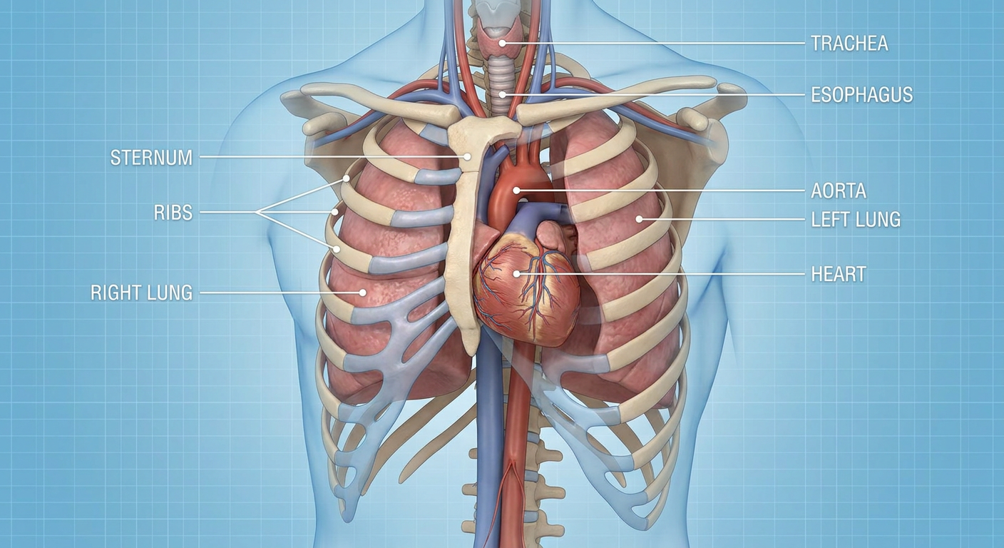
Medical Terms for a "3D Cutaway" of the Chest
Here is what this type of view is called in medical and anatomical contexts:
| Term | Context |
|---|---|
| Cross-sectional anatomy | General anatomy/medical education |
| Transverse (axial) section | Standard anatomical plane - horizontal cut |
| CT scan cross-section | Radiology - Computed Tomography |
| Tomographic slice | Imaging - any modality (CT, MRI) |
| Thoracic dissection view | Gross anatomy / cadaveric study |
| Coronal section | Front-to-back vertical cut |
| Sagittal section | Left-right vertical cut |
| Anatomical cutaway illustration | Medical illustration / textbooks |
Key Structures Visible in a Chest Cutaway:
- Heart (with 4 chambers - right/left atria and ventricles)
- Lungs (right lobe with 3 lobes, left with 2)
- Thoracic aorta - major artery
- Pulmonary vessels - pulmonary trunk, arteries, veins
- Trachea & bronchi - airway tree
- Esophagus - posterior to trachea
- Ribs, sternum, thoracic vertebrae - bony thorax
- Pleural cavity - space around lungs
- Mediastinum - central compartment between lungs
In radiology, the axial (horizontal) CT cut is the most commonly used plane for chest imaging and is the closest clinical equivalent to a "3D cutaway preset."
This is a shared conversation. Sign in to Orris to start your own chat.