
increased hydrostatic pressure leading to edema
Reading File
Increased hydrostatic pressure is one of the main mechanisms of edema formation, acting by pushing fluid out of capillaries into the interstitium. Common causes include heart failure, venous obstruction, and portal hypertension.
Searching sources...
Finding Sources
Finding Sources
Good results. Now let me read the key sections from Frameworks for Internal Medicine and Robbins Pathology in parallel.
Reading File
Reading File
Finding Sources
Great content. Let me now read the Robbins edema section and the key causes section from Frameworks more completely.
Reading File
Reading File
Excellent content. Let me also fetch the key diagrams and read a bit more from Frameworks.
Loading Image
Loading Image
Reading File
I now have comprehensive information from multiple authoritative textbooks. Let me compose the full answer.
Increased Hydrostatic Pressure and Edema
The Starling Forces - Basic Physiology
Fluid movement across the capillary wall is governed by the balance between two opposing forces:
| Force | Direction of Fluid Movement | Normal Value |
|---|---|---|
| Capillary hydrostatic pressure | Out of capillary (filtration) | ~35 mmHg at arteriolar end; ~15 mmHg at venular end |
| Capillary oncotic (colloid osmotic) pressure | Into capillary (reabsorption) | ~25-28 mmHg |
| Interstitial hydrostatic pressure | Into capillary (resists filtration) | ~-3 to 0 mmHg |
| Interstitial oncotic pressure | Out of capillary (opposes reabsorption) | small |
Normally, slight net outflow of fluid at the arteriolar end is nearly balanced by inflow at the venular end. The small residual interstitial fluid is drained by lymphatics back to the circulation, keeping tissues dry.
Edema forms when the lymphatic drainage capacity is exceeded. Because interstitial hydrostatic pressure is normally about -3 mmHg, it must rise ~3 mmHg before significant fluid accumulates - this represents a built-in safety factor against edema. - Guyton and Hall Textbook of Medical Physiology
Mechanism of Hydrostatic Pressure Edema
An increase in capillary hydrostatic pressure opposes capillary oncotic pressure and interstitial hydrostatic pressure, producing net efflux of fluid from capillaries into the interstitial space. When this exceeds lymphatic drainage, edema accumulates. - Frameworks for Internal Medicine
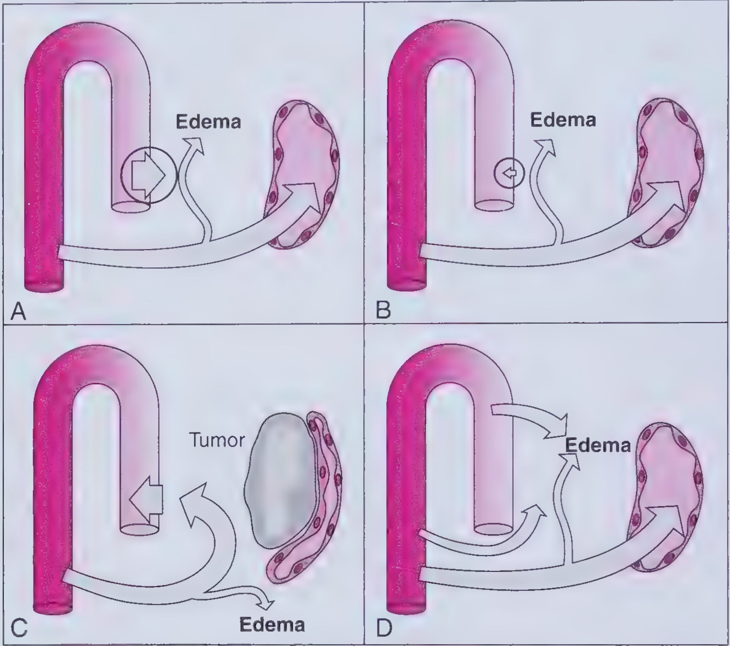
Figure: Mechanisms of edema formation. Panel A shows increased hydrostatic pressure driving fluid out of the capillary into the interstitium.
The fluid produced is a protein-poor transudate (as opposed to the protein-rich exudate of inflammatory edema), because the barrier itself remains intact - only the driving pressure is altered. - Robbins & Kumar Basic Pathology
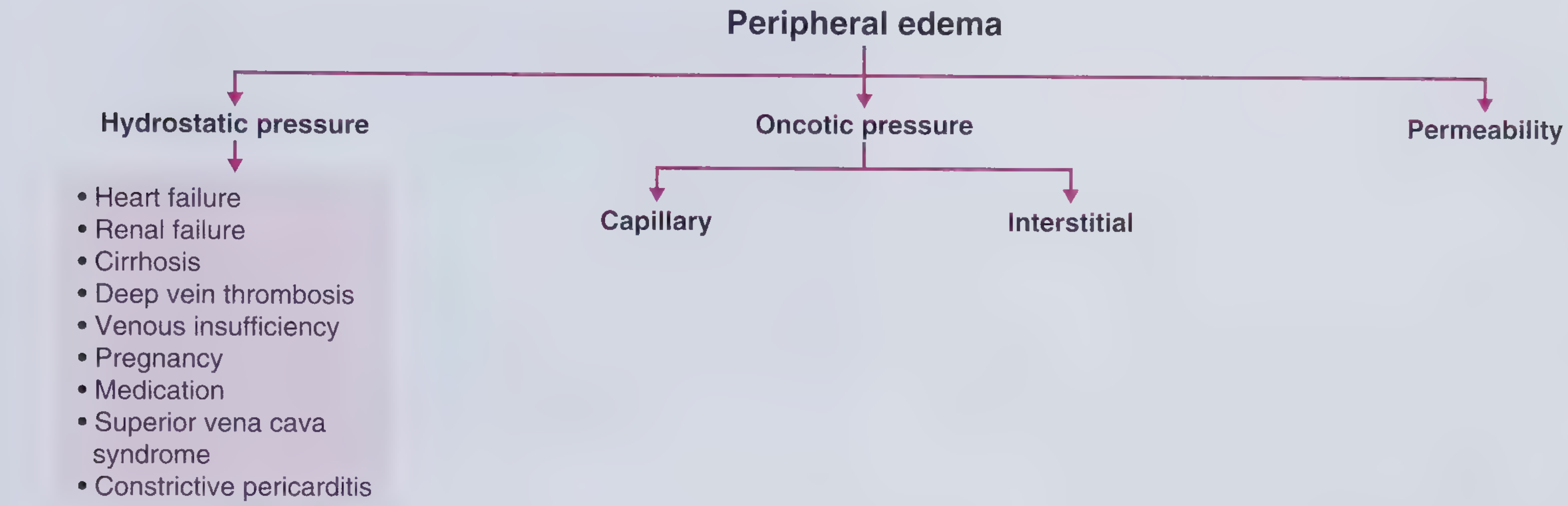
Causes of Increased Hydrostatic Pressure
These are broadly divided into two categories:
1. Impaired Venous Return (most common)
| Cause | Key Features of Edema |
|---|---|
| Congestive heart failure (right-sided) | Bilateral, symmetric, dependent, pitting, gradual onset; may have ascites, elevated JVP |
| Left-sided heart failure | Pulmonary edema / pleural effusions; ± peripheral edema |
| Renal failure | Parallels right-sided HF; fluid removed via dialysis |
| Cirrhosis | Predominantly ascites; bilateral lower limb edema as disease progresses |
| Deep vein thrombosis (DVT) | Unilateral, pitting, acute onset, painful, erythematous |
| Chronic venous insufficiency | Dependent, gradually pitting, may become indurated; hemosiderin pigmentation, ulcers near medial malleoli |
| Constrictive pericarditis | Elevated JVP, Kussmaul's sign, extra diastolic heart sound |
| Superior vena cava (SVC) syndrome | Upper body edema, facial plethora |
| Pregnancy | Lower extremity edema from IVC/iliac vein compression + increased plasma volume |
2. Arteriolar Dilation
- Heat - arteriolar dilation raises downstream capillary pressure
- Medications - dihydropyridine calcium channel blockers (e.g., amlodipine), direct vasodilators (e.g., hydralazine), NSAIDs
Important: Systemic arterial hypertension does NOT cause peripheral edema, because precapillary sphincter autoregulation prevents high arterial pressures from being transmitted to the capillary bed. - Frameworks for Internal Medicine
Characteristics of Hydrostatic Pressure Edema
- Distribution: Localized and dependent (gravity-dependent), usually lower extremities
- Symmetry: Bilateral if systemic (e.g., heart failure, renal failure); unilateral if local (e.g., DVT, venous obstruction)
- Character: Pitting (finger pressure leaves a temporary pit)
- Fluid type: Protein-poor transudate
- Onset: Usually gradual (except DVT, which is acute)
Special Situations
-
Pulmonary edema (cardiogenic): Hemodynamic pulmonary edema is caused by increased hydrostatic pressure, most commonly in left-sided CHF. Edema accumulates initially in the basal regions of the lower lobes (dependent effect). Over time, "heart failure cells" (hemosiderin-laden macrophages) appear in the alveoli. - Robbins, Cotran & Kumar Pathologic Basis of Disease
-
Heart failure cascade: In HF, raised capillary hydrostatic pressure is compounded by ineffective arterial volume, which activates the RAAS, causing renal Na+ and water retention - further increasing total body fluid and worsening edema. - Frameworks for Internal Medicine
-
DVT management: Anticoagulation for proximal DVT; catheter-directed thrombolysis in severe cases. Bilateral lower limb edema should prompt consideration of IVC thrombosis. - Frameworks for Internal Medicine
-
Pregnancy edema: Mechanisms include IVC/iliac vein compression (raising hydrostatic pressure) + increased plasma volume + decreased plasma protein concentration.
Summary Table: Hydrostatic vs. Other Edema Mechanisms
| Mechanism | Fluid Type | Example Causes |
|---|---|---|
| Increased hydrostatic pressure | Transudate (protein-poor) | HF, DVT, cirrhosis, renal failure |
| Decreased oncotic pressure | Transudate (protein-poor) | Nephrotic syndrome, malnutrition, cirrhosis |
| Increased permeability | Exudate (protein-rich) | Inflammation, sepsis, burns, ARDS |
| Lymphatic obstruction | Protein-rich | Malignancy, filariasis, post-surgery |
| Na+ retention | Mixed | Renal insufficiency, RAAS activation |
Sources: Frameworks for Internal Medicine; Robbins & Kumar Basic Pathology; Robbins, Cotran & Kumar Pathologic Basis of Disease; Guyton and Hall Textbook of Medical Physiology
This is a shared conversation. Sign in to Orris to start your own chat.