
First carpometacarpal joint. For 5 mark explain with simple words
first carpometacarpal joint anatomy thumb saddle joint
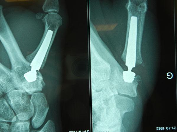
This diagnostic radiograph displays two views of a Moovis carpometacarpal (CMC) joint prosthesis implanted in the thumb. The image illustrates a total joint arthroplasty for the treatment of rhizarthrosis (basal thumb arthritis). The prosthetic system consists of two highly radio-opaque components: a threaded metal cup and a medullary stem. The threaded cup is securely fixed into the trapezium bone, while the elongated, tapered stem is inserted into the proximal medullary canal of the first metacarpal. A ball-and-socket articulation is visible between the stem neck and the trapezial cup, mimicking the natural saddle joint anatomy to restore range of motion and stability. The surrounding cortical bone shows clear contrast against the metallic hardware, and the placement indicates proper alignment within the thumb's CMC joint space. This imaging is characteristic of post-operative orthopaedic assessment to verify component positioning and osseointegration.
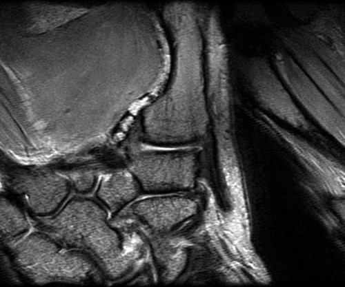
**Modality:** Magnetic Resonance Imaging (MRI), T1-weighted sequence. **Anatomical Region:** Coronal view of the thumb carpometacarpal (CMC) joint, also known as the trapeziometacarpal joint. **Key Landmarks:** The image clearly delineates the articulation between the distal surface of the trapezium and the proximal base of the first metacarpal. Adjacent carpal bones, including the scaphoid and trapezoid, are visible in the proximal and medial fields. **Visual Features:** - **Osseous Structures:** The saddle-shaped morphology of the trapeziometacarpal joint is visualized. Cortical margins appear sharp with high-signal subchondral bone. - **Joint Space:** The articular gap is well-defined, showing the relationship between the reciprocal concave and convex surfaces of the joint. - **Soft Tissues:** Intermediate signal intensity represents the surrounding capsular ligaments and intrinsic musculature of the thenar eminence. - **Texture:** The image demonstrates high-resolution trabecular patterns within the cancellous bone, characteristic of ex vivo cadaveric imaging. **Clinical Significance:** This high-detail anatomical study is utilized for evaluating joint congruivity, detecting subchondral changes, and assessing ligamentous integrity in the context of osteoarthritis or mechanical instability of the first ray.
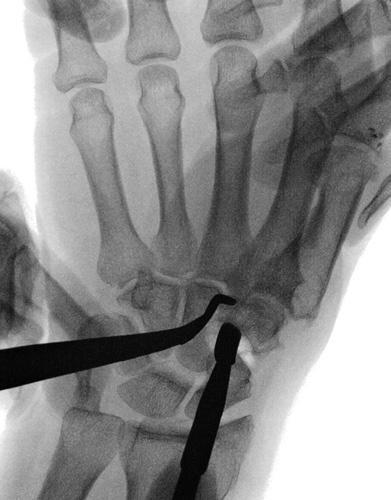
This diagnostic image is an intraoperative fluoroscopic (real-time X-ray) view of the hand and wrist. The field of view focuses on the carpometacarpal region, specifically highlighting the basal joint of the thumb. Visible skeletal anatomy includes the base of the first metacarpal, the trapezium, the scaphoid, and the bases of the second and third metacarpals. Two radio-opaque surgical instruments are present: a curved periosteum elevator positioned superiorly and a straight surgical blade (consistent with a #15 blade) positioned inferiorly. These instruments are shown performing arthrolysis (joint mobilization) of the trapezium. The image demonstrates the spatial relationship between surgical tools and small carpal bones during a trapeziectomy or suspensionplasty procedure for thumb carpometacarpal osteoarthritis. This material is suitable for advanced orthopedic surgical training and demonstrates real-time visualization of joint capsule dissection under fluoroscopic guidance.
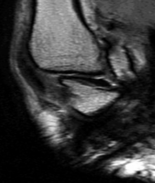
**Imaging Modality:** Magnetic Resonance Imaging (MRI). **Anatomical Region:** Oblique view of the first carpometacarpal (CMC) joint of the hand. **Key Landmarks and Features:** - **Primary Structure:** The anterior oblique ligament (AOL), also known as the beak ligament, is visualized extending from the volar tubercle of the trapezium to the volar base of the first metacarpal. - **Orientation:** The image captures an oblique plane specifically angled to isolate the longitudinal fibers of the AOL. - **Markers:** A high-signal external marker is positioned adjacent to the skin surface, corresponding to the anatomical location of the ligament for localization purposes. - **Joint Characteristics:** The saddle-shaped articulation between the trapezium and the base of the first metacarpal is visible. The ligament appears as a low-signal (hypointense) band, providing essential stability to the CMC joint. **Clinical Significance:** This view is utilized to assess the integrity of the primary stabilizing structures of the thumb base. It is critical for evaluating ligamentous laxity, attenuation, or tearing associated with first CMC joint instability or basal joint osteoarthritis.
First Carpometacarpal (CMC) Joint
1. What It Is (Definition + Bones)
- The base of the 1st metacarpal (the thumb's long bone) above
- The trapezium (a carpal/wrist bone) below
2. Type of Joint - "Saddle Joint"
Simple way to remember: Imagine two saddles placed at right angles to each other, one fitting into the other. Each surface is concave in one direction and convex in the other.
3. Movements
| Movement | Description |
|---|---|
| Flexion & Extension | Thumb folds toward/away from palm |
| Abduction & Adduction | Thumb moves away from/toward fingers |
| Opposition | Thumb tip meets fingertips - the most important movement! |
| Circumduction | A combination of all the above |
Key point: The axes of flexion-extension and abduction-adduction are oblique to each other (not perfectly at 90°), which is why thumb movement feels natural and fluid.
4. Ligaments (Stability)
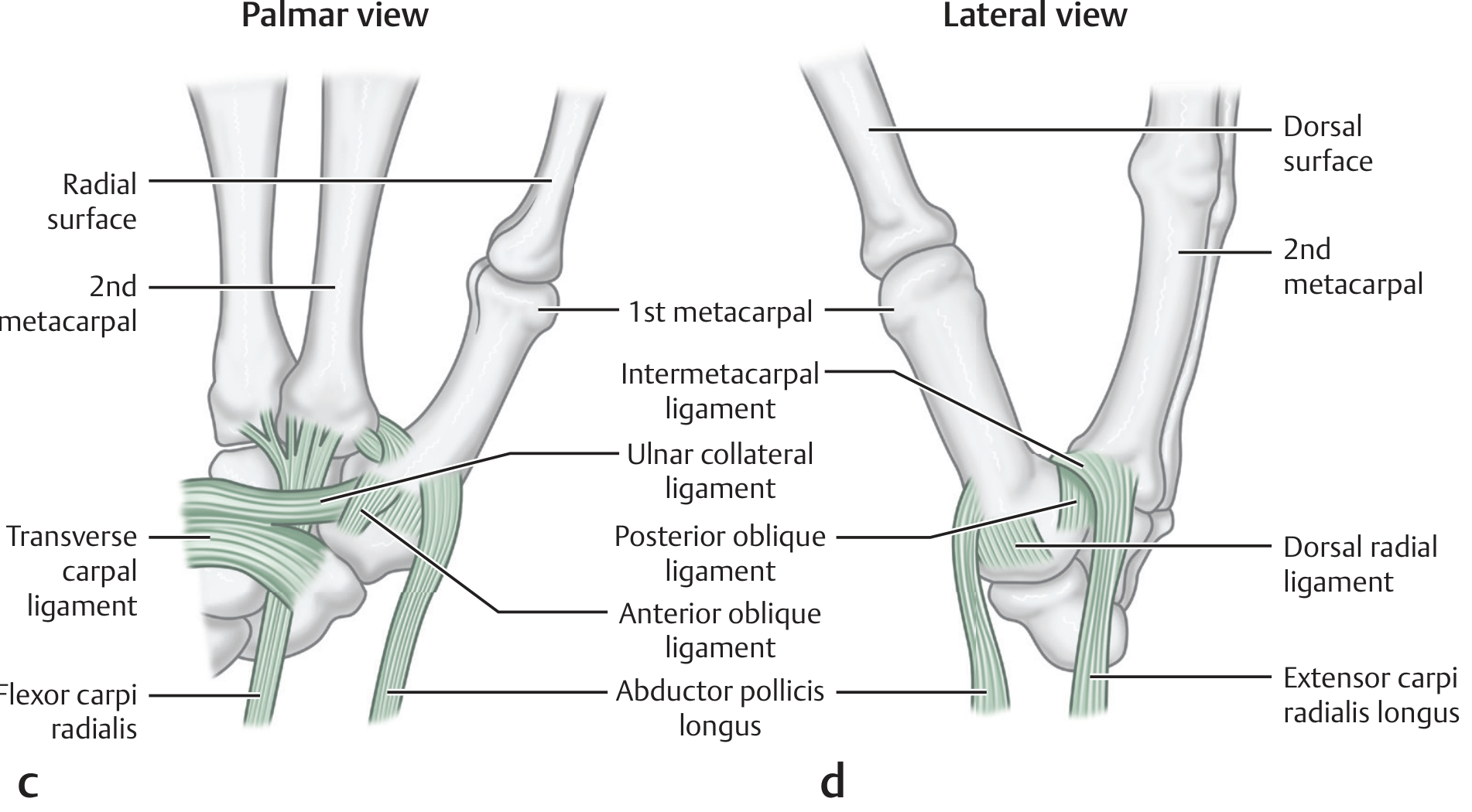
| Ligament | Location | Function |
|---|---|---|
| Dorsal Ligament Complex (Posterior oblique + Dorsal central + Dorsal radial ligaments) | Back (dorsal) side | Main stabilizer of the joint |
| Anterior Oblique Ligament (Beak ligament) | Front (palmar) side | Runs from trapezium to base of 1st metacarpal |
| Ulnar Collateral Ligament | Ulnar/palmar side | Runs from flexor retinaculum to metacarpal |
| Intermetacarpal Ligament | Between 1st and 2nd metacarpals | Adds extra stability |
Simple memory tip: The dorsal complex (back ligaments) is the most important - it keeps the thumb from dislocating.
5. Clinical Importance
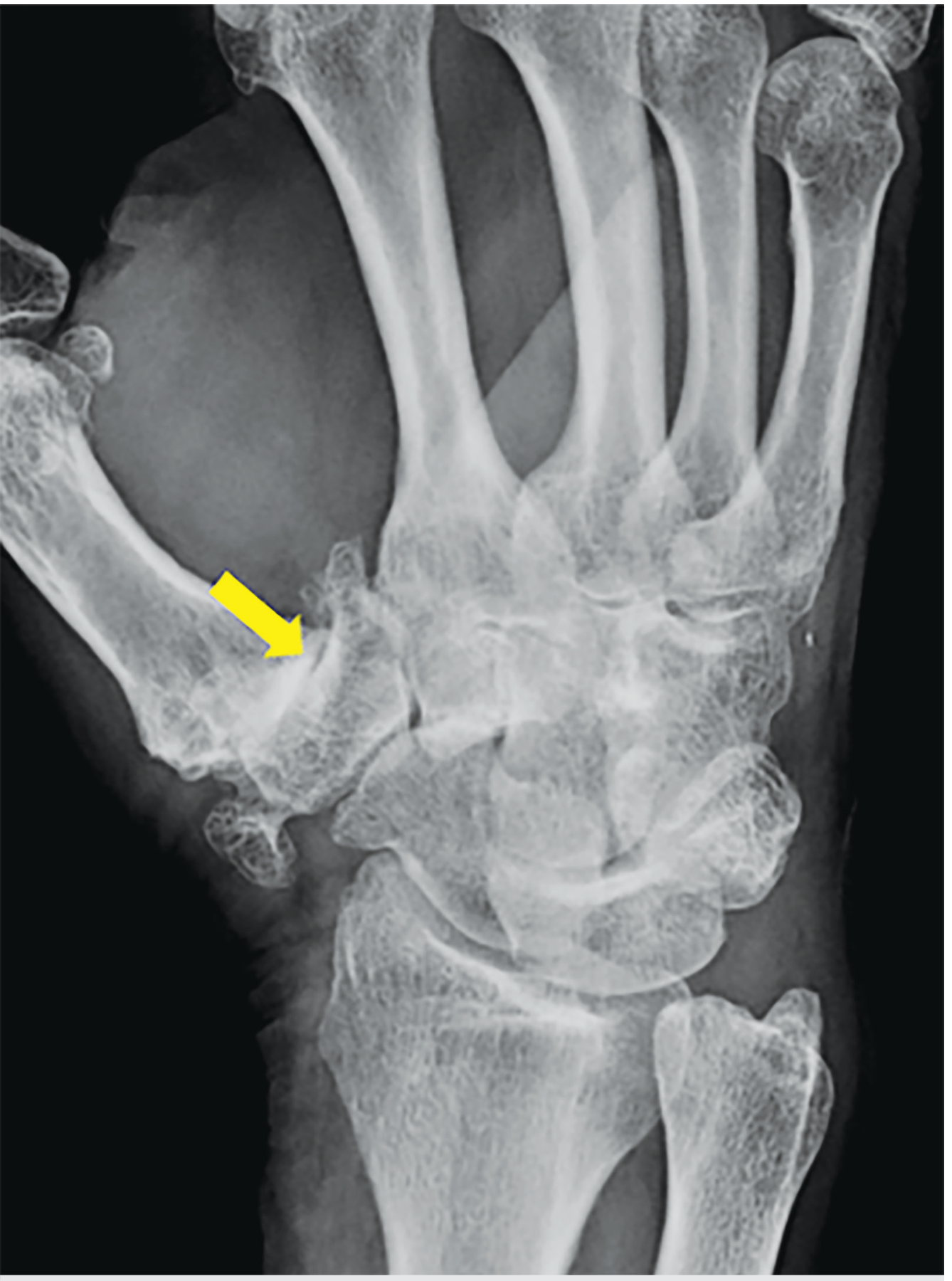
A. Osteoarthritis (OA)
B. Bennett's Fracture
- Intra-articular fracture at the base of the 1st metacarpal
- Unstable because Abductor Pollicis Longus (APL) pulls the main fragment upward/backward
- The small fragment stays held by the ligaments
- Needs surgical fixation
C. Rolando Fracture
- Like a Bennett's but comminuted (broken into many pieces)
- Worse prognosis, almost always needs surgery
Quick Summary Table
| Feature | Detail |
|---|---|
| Bones | 1st Metacarpal + Trapezium |
| Joint type | Saddle (Sellar) joint |
| Movements | Flexion, Extension, Abduction, Adduction, Opposition |
| Main stabilizer | Dorsal ligament complex |
| Most common pathology | Osteoarthritis |
| Classic fracture | Bennett's fracture |
Refer bd chaurasia
Note: BD Chaurasia's Human Anatomy is not available in the textbook library. The answer below is compiled from Gray's Anatomy for Students and the THIEME Atlas of Anatomy (General Anatomy & Musculoskeletal System) - both of which cover the 1st CMC joint in the same depth and style as BD Chaurasia.
First Carpometacarpal (CMC) Joint
1. Type of Joint
- The articular surface of the trapezium is:
- Convex in the dorso-palmar direction
- Concave in the radio-ulnar direction
- The articular surface of the base of 1st metacarpal has the exact opposite curvatures - so the two fit together like two saddles at right angles to each other.
Simple image: Think of a horse's saddle placed upside-down on another saddle. Each is curved both ways.
2. Articulating Bones
| Bone | Part |
|---|---|
| 1st Metacarpal | Base (proximal end) |
| Trapezium | Distal articular surface |
3. Movements & Axes
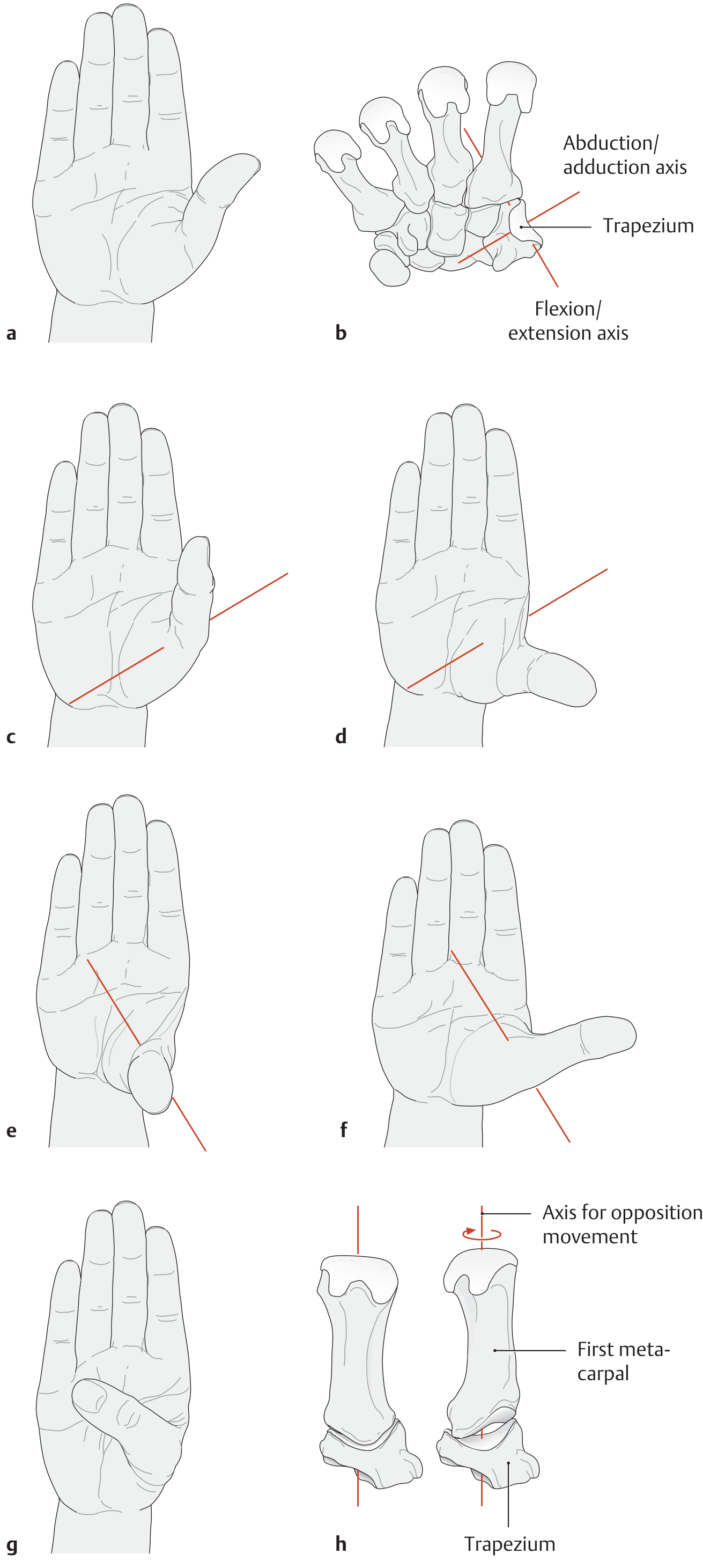
| Axis | Movement |
|---|---|
| Abduction/Adduction axis (runs dorso-palmar) | Thumb moves away from / toward the palm plane |
| Flexion/Extension axis (runs transversely through trapezium) | Thumb folds across palm / straightens |
| Longitudinal axis (3rd degree of freedom) | Rotation - occurs during opposition |
- Abduction
- Flexion
- Medial rotation (about the longitudinal axis of the 1st metacarpal)
In the neutral (0°) position, the thumb metacarpal is already rotated ~60° toward the palm because the trapezium faces radiopalmarward. That is why the thumb naturally points away from the fingers.
4. Ligaments
| Ligament | Origin | Insertion | Importance |
|---|---|---|---|
| Anterior Oblique (Beak) Ligament | Palmar tubercle of trapezium | Volar base of 1st metacarpal | Previously thought to be main stabilizer |
| Dorsal Radial Ligament | Dorsal trapezium | Dorsal 1st metacarpal | Part of dorsal complex |
| Posterior Oblique Ligament | Dorso-ulnar trapezium | Ulnopalmar tubercle of 1st metacarpal | Part of dorsal complex |
| Dorsal Central Ligament | Dorsal trapezium | 1st metacarpal | Part of dorsal complex |
| Ulnar Collateral Ligament | Flexor retinaculum | Volar-ulnar metacarpal | Stabilizes ulnar side |
| Intermetacarpal Ligament | Ulnopalmar 1st metacarpal | Dorsoradial 2nd metacarpal | Links thumb to index |
5. Clinical Importance
A. Osteoarthritis (Most Common)
- The most common site of OA in the hand
- During full opposition, the rotating metacarpal greatly reduces the contact area with the trapezium - concentrating stress on a small area. This repeated stress promotes cartilage wear and OA.
- Seen especially in middle-aged women.
B. Bennett's Fracture
- Intra-articular fracture at the base of the 1st metacarpal (oblique split)
- Unstable - Abductor Pollicis Longus (APL) pulls the main fragment dorsally and proximally
- The small volar fragment stays attached to the trapezium by the anterior oblique ligament
- Requires surgical fixation
C. Rolando's Fracture
- Comminuted (3-part or more) intra-articular fracture of the same base
- Worse prognosis than Bennett's; almost always needs surgery
Summary Box (BD Chaurasia Style)
| Feature | Details |
|---|---|
| Type | Saddle (Sellar) synovial joint |
| Bones | Base of 1st metacarpal + Trapezium |
| Movements | Flexion, Extension, Abduction, Adduction, Opposition, Circumduction |
| Main stabilizer | Dorsal ligament complex |
| Special feature | No deep transverse metacarpal ligament → free thumb movement |
| Common pathology | Osteoarthritis (most common OA in hand) |
| Classic fracture | Bennett's fracture |