
Detailed study stepwise in lap cholecystectomy procedure
laparoscopic cholecystectomy trocar placement and Calot's triangle critical view of safety
laparoscopic cholecystectomy critical view of safety
critical view of safety laparoscopic cholecystectomy Calot triangle
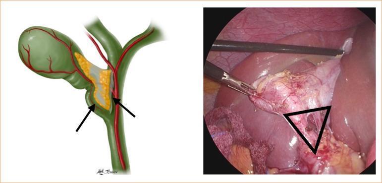
This composite image consists of an anatomical illustration and a corresponding endoscopic photograph demonstrating the landmarks of Calot's triangle (hepatocystic triangle) during a laparoscopic cholecystectomy. The anatomical diagram on the left illustrates the gallbladder, cystic duct, and common hepatic duct (marked with arrows), with the cystic artery passing through the triangle. The laparoscopic view on the right displays the surgical field with laparoscopic graspers retracting a distended, erythematous gallbladder. A black triangular overlay identifies Calot’s triangle, bounded superiorly by the inferior margin of the liver, laterally by the cystic duct, and medially by the common hepatic duct. The operative photograph shows evidence of inflammatory changes, including tissue hypervascularity and edema, which can obscure clear identification of biliary structures. This visual is designed to teach the 'critical view of safety' by emphasizing the anatomical boundaries required to prevent iatrogenic bile duct injuries during minimally invasive hepatobiliary surgery.
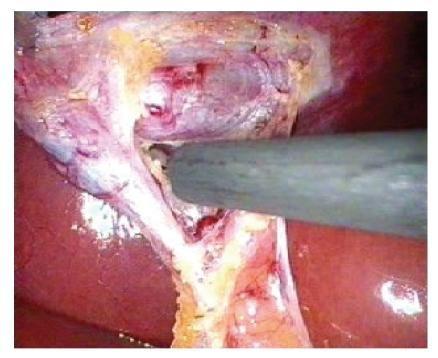
Laparoscopic intraoperative photograph showing the dissection of the Triangle of Safety during a cholecystectomy. The image displays the inferior surface of the liver (reddish-brown parenchyma) and the retracted gallbladder. A laparoscopic dissecting instrument is actively manipulating and dividing connective tissues within the triangle, which is bordered by the cystic duct, the cystic artery/gallbladder wall, and the liver bed. Visible features include glistening peritoneal surfaces, yellow fatty tissue along the gallbladder wall, and the initial exposure of the cystic duct-infundibular junction. This clinical visual demonstrates the 'top-down' or fundus-first dissection technique where the surgeon clears tissues close to the gallbladder wall to achieve the critical view of safety, thereby avoiding injury to the common bile duct and other vital structures in Calot's triangle.
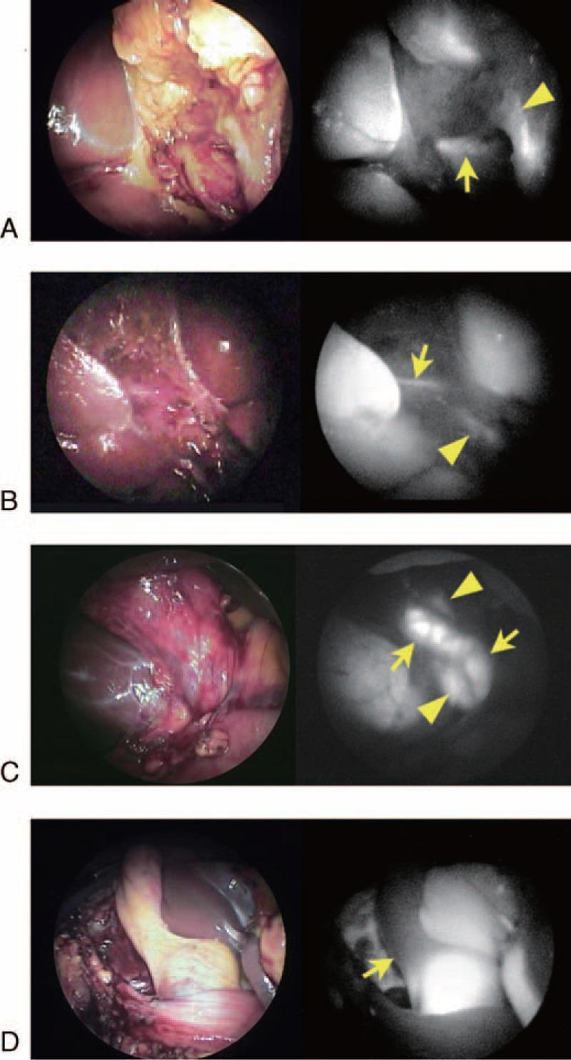
This medical image displays four pairs of laparoscopic views (A–D) comparing standard white-light endoscopy with indocyanine green (ICG) fluorescence cholangiography (FC) for intraoperative bile duct identification. (A) Shows an accessory hepatic duct (arrow) draining the right lateral liver sector into the common hepatic duct (arrowhead) near Rouviere's sulcus after dissection of Calot's triangle. (B) Illustrates a thin hepatic duct (arrow) running along the gallbladder bed, likely draining Couinaud's segment V, with the common hepatic duct marked by an arrowhead. (C) Visualizes the cystic duct (arrows) draining into the common hepatic duct (arrowheads) from the left side before Calot's triangle dissection, demonstrating spiral-type anatomy. (D) Highlights a fluorescence defect (arrow) within the cystic duct, indicating the presence of impacted gallstones. The series demonstrates the clinical utility of FC in mapping biliary variants and identifying pathology like cholecystolithiasis during laparoscopic cholecystectomy, enhancing the 'critical view of safety' by delineating extrahepatic biliary structures.
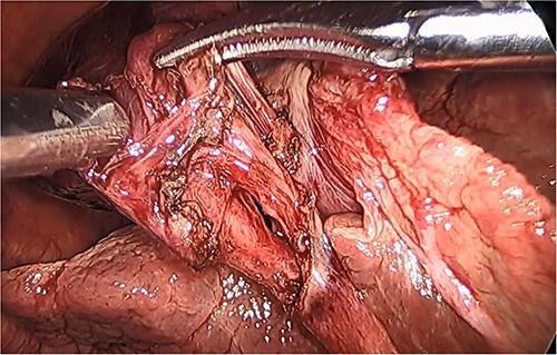
This laparoscopic intraoperative clinical photograph illustrates the achievement of the 'Critical View of Safety' during a cholecystectomy for gallbladder volvulus. The field shows the hepatobiliary triangle (Calot's triangle) following surgical dissection. Visible structures include the cystic duct and cystic artery, which have been isolated and cleared of surrounding fibrofatty tissue. The gallbladder appears distended with darkened, hemorrhagic walls consistent with gangrenous necrosis due to torsion. The liver surface is visible in the background, showing the lower third of the cystic plate cleared to ensure anatomical identification. Metal surgical graspers are positioned to retract the gallbladder and facilitate exposure of the ductal and vascular structures. This image serves as an educational example of proper surgical identification techniques to prevent bile duct injury during complex cholecystectomy procedures involving anomalous gallbladder positions and vascular compromise.
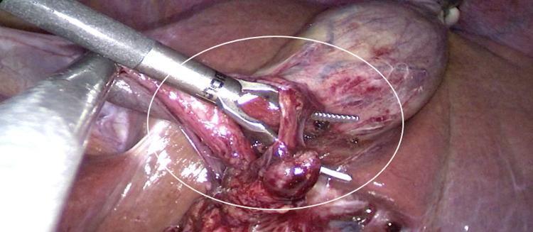
Laparoscopic surgical view demonstrating the dissection of the hepatocystic triangle (Calot's triangle) during a cholecystectomy in a patient with situs inversus totalis. The image shows the gallbladder, which appears erythematous with visible vascularity on its surface, positioned in the left hypochondrium (anatomical left). Within the white circle, a surgical grasper is manipulating the cystic duct and cystic artery to achieve the 'critical view of safety.' A white surgical clip is visible, having been applied to a tubular structure, presumably the cystic artery or duct, for ligation. The surgical field contains evidence of minor bleeding consistent with active tissue dissection. This image illustrates the 'mirror-image' surgical approach required for laparoscopic procedures in patients with total visceral transposition, where gallbladder removal is performed on the patient's left side rather than the standard right side.
Laparoscopic Cholecystectomy: Stepwise Study
Overview
Step 1: Anesthesia and Positioning
- General anesthesia with muscle relaxation is mandatory (a contraindication is inability to tolerate GA).
- Patient supine; one or both arms may be tucked (to allow intraoperative cholangiogram if needed).
- Foley catheter if a long operation or critical illness is anticipated; orogastric tube to decompress the stomach for a better upper-abdominal view.
- Surgeon stands to the patient's left, first assistant to the right (American technique); in the French technique the patient's legs are abducted and the surgeon stands between them.
- Sabiston Textbook of Surgery; Maingot's Abdominal Operations, p. 1018
Step 2: Pneumoperitoneum
- A working space is created with CO2 insufflation (non-combustible, rapidly absorbed, but can cause hypercarbia in cardiopulmonary disease).
- Entry is via the umbilicus, either:
- Closed technique: Veress needle insufflation, then blind port placement, or
- Open (Hasson) technique: direct-vision port placement through a small subumbilical incision - preferred by many as the initial camera port.
- Maingot's Abdominal Operations, p. 1018; Bailey and Love's Short Practice of Surgery
Step 3: Port Placement
- Umbilical port (10-12 mm) - camera, often used later for specimen extraction.
- Epigastric/subxiphoid port (10 mm) - surgeon's primary working instrument.
- Right subcostal (midclavicular) port (5 mm) - grasps and retracts the infundibulum/Hartmann's pouch.
- Right anterior axillary line port (5 mm) - elevates the gallbladder fundus toward the right shoulder.
Step 4: Exposure and Retraction
- The fundus is grasped and retracted cephalad/toward the right shoulder.
- The infundibulum is retracted laterally and inferiorly (toward the right iliac fossa), which distracts the cystic duct away from the common bile duct (CBD) and opens the hepatocystic triangle into a trapezoid shape.
- Adhesions to omentum, duodenum, or hepatic flexure are lysed bluntly, taking care to avoid electrocautery injury to unvisualized structures like the duodenum or CBD.
Step 5: Dissection of the Hepatocystic Triangle (Calot's Triangle)
- Using fine Maryland dissecting forceps, dissection starts from a known structure (the gallbladder wall), never in an unknown space, and proceeds in a "top-down"/fundus-first fashion.
- The peritoneum overlying the triangle is scored and fibroareolar/fatty tissue is swept away, exposing the cystic duct and cystic artery.
- The Calot's lymph node is a reliable landmark - it typically overlies the cystic artery; dissection is kept on the gallbladder side of this node to protect the CBD.
- Sabiston Textbook of Surgery, p. 1838; Maingot's Abdominal Operations, p. 1019
Step 6: Achieving the Critical View of Safety (CVS)
- The hepatocystic triangle is completely cleared of fat and fibrous tissue.
- The lower third of the gallbladder is dissected off the liver bed to expose the cystic plate.
- Only two structures (the cystic duct and cystic artery) are seen entering the gallbladder.
Step 7: Clipping and Division
- Once CVS is confirmed, the cystic artery and cystic duct are each doubly clipped (proximal and distal to the intended division site) and divided between the clips.
- It is unnecessary and potentially harmful to dissect the cystic duct all the way down to the CBD junction.
- If the cystic artery crosses anterior to the duct, it is usually taken first to improve exposure.
Step 8: Dissecting the Gallbladder off the Liver Bed
- Using electrocautery (hook or spatula), the gallbladder is dissected free from the cystic plate/liver bed, working from the divided cystic duct/artery up toward the fundus.
- Meticulous hemostasis is essential since gallbladder venous drainage empties directly into the liver bed via small venules.
- Clips on the cystic duct/artery stumps are re-inspected once fundic traction has improved exposure of the porta hepatis, before final detachment.
Step 9: Specimen Extraction
- The gallbladder is placed in a retrieval bag and withdrawn through the umbilical (or epigastric) port, most commonly.
- Bile or stone spillage should be controlled and irrigated; the liver bed and porta are irrigated and inspected for hemostasis and bile leak.
Step 10: Closure
- Ports are removed under vision to confirm no bleeding from port sites.
- Fascial closure at 10-12 mm ports (to prevent port-site hernia); skin closed with subcuticular sutures or adhesive strips.
Key Complications to Anticipate
- Bile duct injury (most feared - historically from mistaking the CBD or an aberrant right hepatic duct for the cystic duct)
- Cystic artery/hepatic artery bleeding
- Bile leak from the cystic duct stump or an accessory duct of Luschka
- Bowel or vascular injury at port entry
- Conversion to open surgery, more likely in acute cholecystitis (longer operative time, higher conversion rate)