
Cervical neck conditions
cervical spine anatomy diagram
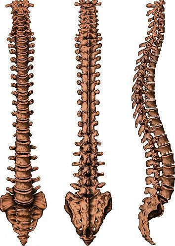
This anatomical diagram presents a comprehensive three-view study of the human vertebral column, including anterior (frontal), posterior (rear), and lateral (side) perspectives. The illustration highlights the structural organization of the spine, starting from the cervical vertebrae down through the thoracic and lumbar regions, terminating at the fused sacrum and the coccyx. Each vertebra is depicted with distinct anatomical features: the anterior view emphasizes the vertebral bodies and intervertebral disc spaces; the posterior view showcases the spinous and transverse processes; and the lateral view clearly demonstrates the four physiological curvatures—cervical lordosis, thoracic kyphosis, lumbar lordosis, and sacral kyphosis. This 'S' shaped alignment is shown as essential for weight-bearing, flexibility, and protecting the spinal cord. The diagram serves as an educational resource for studying osteology, spinal biomechanics, and regional anatomy, illustrating the progressive increase in vertebral size to accommodate increasing gravitational loads.
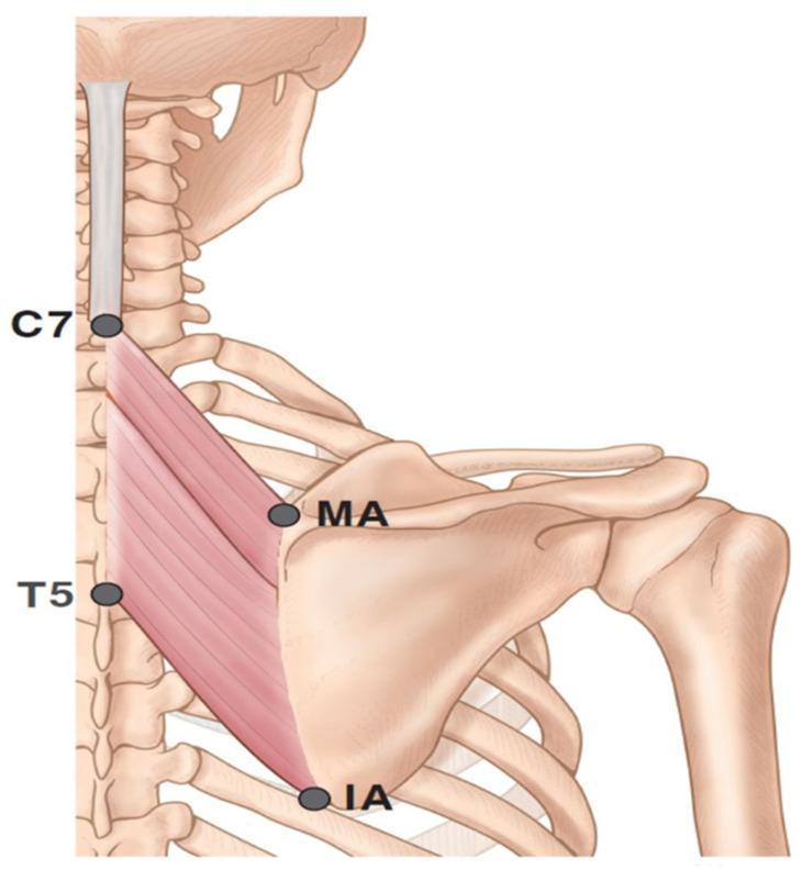
This anatomical diagram illustrates the superficial and deep musculature of the posterior thoracic region, specifically focusing on the left rhomboid muscles. The illustration depicts the skeletal framework including the cervical and thoracic spine, the scapula, and the proximal humerus. Four key anatomical landmarks are identified with markers: the spinous process of the seventh cervical vertebra (C7), the spinous process of the fifth thoracic vertebra (T5), the medial angle (MA) of the scapula, and the inferior angle (IA) of the scapula. The rhomboid muscles are shown with their characteristic oblique fiber orientation, originating from the midline nuchal ligament and spinous processes (ranging from C7 to T5) and inserting along the medial border of the scapula. This diagram serves as a clinical reference for understanding musculoskeletal anatomy, surface landmarks for physical examination, and the localization of intramuscular targets for therapeutic interventions such as botulinum toxin injections or electromyography.
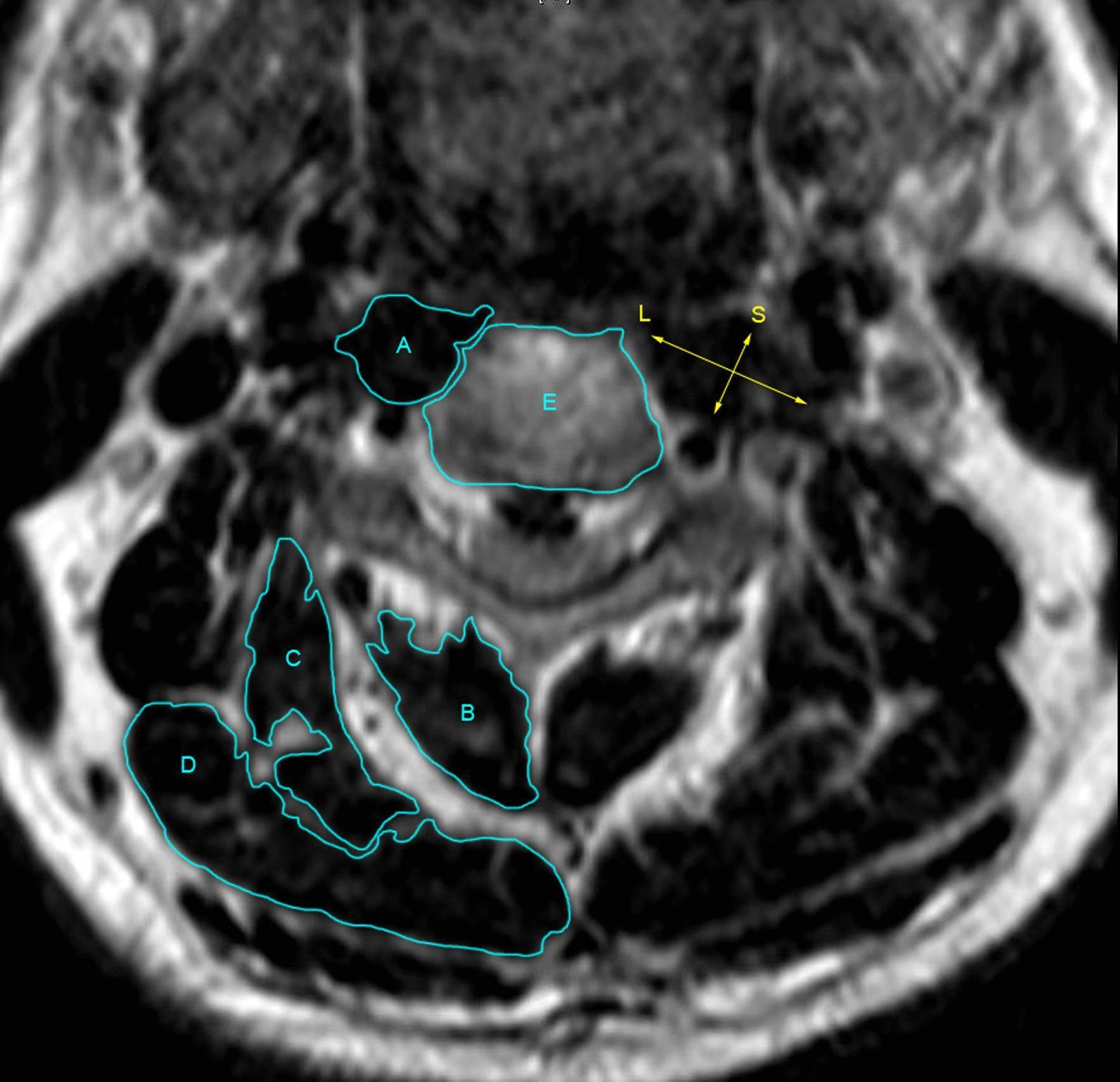
This diagnostic image is an axial T2-weighted MRI scan of the cervical spine, serving as a measurement diagram for cervical musculature. The central vertebral body is labeled 'E'. To its anterior and medial aspect, the cervical longus muscle is outlined and labeled 'A'. Posterior to the vertebral column, the cervical extensor muscles are delineated: 'B' identifies the combined multifidus and semispinalis cervicis muscles; 'C' denotes the splenius cervicis and semispinalis capitis; and 'D' represents the splenius capitis. Yellow arrows illustrate the methodology for calculating the Ratio of Long and Short diameter line (RLS) of the cervical longus muscle. The 'L' arrow represents the longest diameter, while the 'S' arrow represents the shortest diameter, intersecting to perform a 'dichotomy cutting' of the muscle's cross-sectional area. This imaging is clinically significant for evaluating muscle morphology and atrophy, particularly in the context of preoperative and postoperative assessments for anterior cervical surgery. The target audience includes radiology and orthopedics specialists studying cervical spinal anatomy and surgical outcomes.
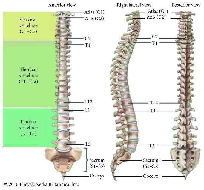
This anatomical diagram presents three perspectives of the human vertebral column: anterior view, right lateral view, and posterior view. The illustration categorizes the 33 vertebrae into five distinct regions, color-coded for clarity: cervical (C1–C7), thoracic (T1–T12), lumbar (L1–L5), the fused sacrum (S1–S5), and the coccyx. Key anatomical landmarks are labeled, including the Atlas (C1) and Axis (C2). The anterior and posterior views demonstrate the vertical alignment and central symmetry of the spine. The right lateral view illustrates the physiological curvatures of the column, showing the cervical lordosis (inward curve), thoracic kyphosis (outward curve), lumbar lordosis (inward curve), and the sacrococcygeal curve. This comparison highlights the structural organization of individual vertebrae, the intervertebral discs, and the overall S-shaped profile necessary for balance and shock absorption. The diagram serves as a foundational educational tool for understanding spinal anatomy, regional segmentation, and normal postural curvature in clinical and musculoskeletal medicine.
cervical disc herniation radiculopathy MRI
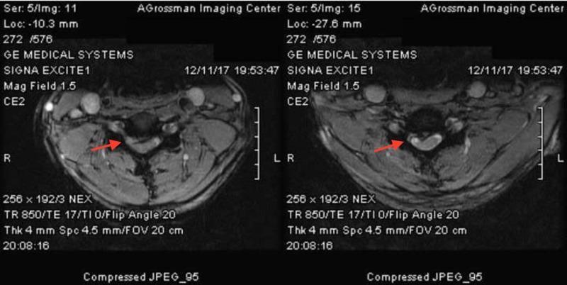
Diagnostic axial MRI images of the cervical spine illustrating varying degrees of intervertebral disc pathology and resulting spinal canal stenosis. The left image demonstrates a 5 mm right paracentral disc herniation that extends toward the neural foramen, causing moderate encroachment. The right image shows a more severe right paracentral disc extrusion, marked by a focal mass effect that significantly compresses and displaces the right anterior aspect of the spinal cord, resulting in visible cord deformity. Both images highlight the spatial relationship between the herniated disc material (indicated by red arrows) and the hyperintense cerebrospinal fluid surrounding the spinal cord. These findings are clinically significant for diagnosing cervical radiculopathy and myelopathy, typically associated with degenerative disc disease. The images serve as an educational comparison between simple herniation and more aggressive extrusion causing mechanical cord compression.
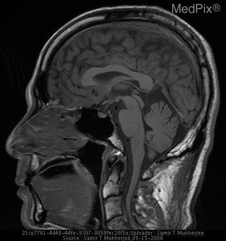
**Imaging Modality:** Magnetic Resonance Imaging (MRI), T1-weighted sequence.
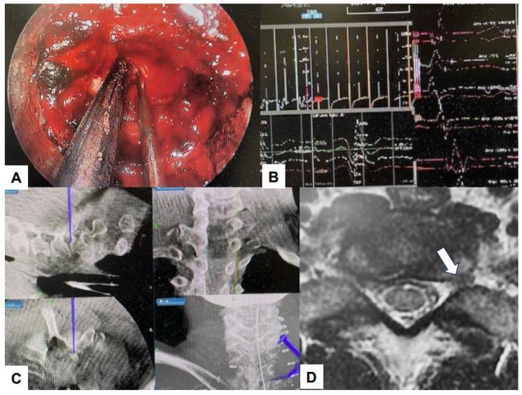
A multi-panel medical illustration showcasing a cervical endoscopic keyhole foraminotomy procedure. Panel (A) provides an intraoperative endoscopic view showing surgical instruments, including a dissector and a suction-irrigation cannula, positioned within the operative field against paraspinal muscle and soft tissue. Panel (B) demonstrates real-time intraoperative neurophysiological monitoring (IONM) waveforms used to ensure nerve root integrity. Panel (C) displays a surgical navigation monitor showing multi-planar fluoroscopic or CT-based reconstructions of the cervical spine with a blue virtual probe indicating the surgical trajectory. Panel (D) is an axial T2-weighted MRI image of the cervical spine, where a white arrow highlights a paracentral disc herniation impinging on the neural foramen and spinal cord. This composite image illustrates the integration of minimally invasive endoscopic techniques, navigation, and neuromonitoring for treating cervical radiculopathy and disc herniation.
cervical spondylotic myelopathy cord compression
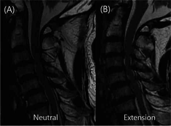
This diagnostic image consists of side-by-side sagittal T2-weighted magnetic resonance imaging (MRI) scans of the cervical spine, demonstrating dynamic changes in spinal cord compression. Image A shows the spine in a neutral position, where focal spinal cord compression (Cervical Spondylotic Myelopathy) is primarily visible at the C3–C4 level. Image B shows the same patient in an extension posture, which reveals significant multi-level dynamic compression extending from C3 through C7. Key visual features include the buckling of the ligamentum flavum posteriorly and disc-osteophyte complexes anteriorly that narrow the spinal canal more severely during extension than in the neutral state. The image serves as an educational tool for dynamic cervical MRI (dMRI), illustrating how neutral imaging can underestimate the extent of myelopathy and how extension maneuvers can identify additional levels of pathological cord impingement, critical for surgical planning in Cervical Spondylotic Myelopathy (CSM).
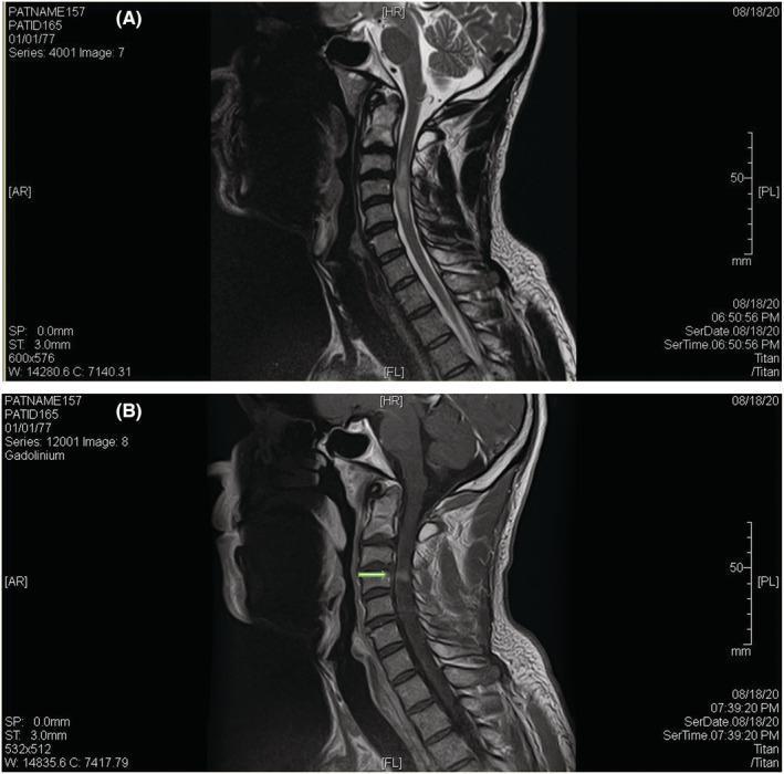
This diagnostic image consists of two sagittal MRI views of the cervical spine illustrating spondylotic myelopathy. Image A is a T2-weighted sagittal MRI showing a long-segment hyperintense signal within the cervical spinal cord, extending from approximately C2-C3 to the C5 level, consistent with cord edema. A degenerative disc bulge at the C3-C4 level is seen mildly indenting the anterior spinal cord. Image B is a T1-weighted sagittal MRI with gadolinium enhancement, which reveals a distinct, linear, transverse band of contrast uptake within the spinal cord just below the level of maximal compression. This finding is known as the 'pancake enhancement pattern' and is a characteristic radiological sign of chronic compressive myelopathy. The imaging highlights key indicators for diagnosing cervical spondylotic myelopathy (CSM) and distinguishing it from other intramedullary pathologies like multiple sclerosis or tumors.
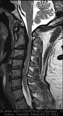
**Imaging Modality:** Sagittal T2-weighted Magnetic Resonance Imaging (MRI) of the cervical spine.
whiplash injury cervical spine trauma
Cervical Neck Conditions
Overview
Anatomy & Joints Involved
- Intervertebral disc
- Two uncovertebral (Luschka) joints
- Two facet joints (zygoapophyseal joints)
Classification of Cervical Conditions
1. Cervical Degenerative Disc Disease (Cervical Spondylosis)
| Entity | Description |
|---|---|
| Discogenic neck pain | Axial pain from disc degeneration |
| Radiculopathy | Nerve root compression |
| Myelopathy | Spinal cord compression |
| Myeloradiculopathy | Combined cord + root involvement |
2. Discogenic Neck Pain (Axial Pain)
- Secondary to intervertebral disc degeneration without instability, fracture, or neural compression
- Presents as axial neck pain, often with occipital headache
- Exam: Normal motor/sensory/reflexes; reduced ROM from pain
- Imaging: MRI shows decreased T2 disc signal ("dark disc"), possible annular tear or high-intensity zone (HIZ); X-rays may show disc space narrowing
- Treatment: NSAIDs, symptomatic care, patient education (self-limiting); surgical options are generally avoided for pure discogenic pain
3. Cervical Radiculopathy
- Cervical nerve roots exit above their corresponding vertebrae (C5 exits at C4-C5 foramen)
- Disc herniation at C5-6 compresses the C6 root
- Disc herniation at C6-7 compresses the C7 root
- Spurling test - rotation + lateral bending + axial compression reproduces radicular pain
- Shoulder abduction sign - relief of pain when patient's hand is placed on top of the head
| Level | Motor | Sensory | Reflex |
|---|---|---|---|
| C5 | Deltoid, biceps | Lateral arm | Biceps |
| C6 | Wrist extensors | Thumb/index finger | Brachioradialis |
| C7 | Triceps, wrist flexors | Middle finger | Triceps |
| C8 | Hand intrinsics | Ring/little finger | - |
4. Cervical Myelopathy (Degenerative Cervical Myelopathy)
- Normal: ≥14 mm
- Relative stenosis: 10-13 mm
- Absolute stenosis: <10 mm
- Torg (Pavlov) ratio <0.8 suggests risk for neurologic involvement
- Clumsiness of hands (difficulty with buttons, writing)
- Unsteady/wide-based gait
- Bladder/bowel dysfunction (late)
5. Inflammatory / Autoimmune Conditions
- Rheumatoid Arthritis: ~50% of RA patients develop atlantoaxial subluxation (C1-C2 instability), which can lead to cord compression and neurologic deterioration. - Harrison's 22E, p. 175
- Ankylosing Spondylitis: Causes neck pain and stiffness, typically worse with rest/inactivity; may lead to cervical fusion and fracture risk.
- Polymyalgia Rheumatica: Pain and stiffness in the neck, shoulders, and pelvic girdle, with systemic symptoms (elevated ESR/CRP).
- Crowned Dens Syndrome: Calcium pyrophosphate crystal deposition around the dens (C2); presents as severe acute neck pain and stiffness, mimicking meningitis.
6. Infectious Causes
- Cervical Epidural Abscess: Severe neck pain + progressive neurologic deterioration from cord compression; associated with IV drug use, immunocompromise, diabetes. Urgent surgical decompression typically required.
- Discitis/Osteomyelitis: Fever, elevated inflammatory markers, severe localized pain; often hematogenous seeding.
- Meningitis: Neck pain + fever + headache + meningismus.
7. Vascular Causes
- Vertebral / Carotid Artery Dissection: Sudden-onset neck pain + headache; can cause ischemic stroke. Consider in young patients with neck trauma.
- Cervical Epidural Hematoma: Sudden neck pain + rapid neurologic deficit; can be mistaken for stroke. Pain at the spinous process on palpation is a distinguishing sign.
- Acute Coronary Syndrome: Can present as referred neck pain (important red flag).
8. Congenital / Structural
- Chiari Type 1 Malformation: Headache and neck pain aggravated by cough/Valsalva; cerebellar tonsils herniate below the foramen magnum.
- Klippel-Feil Syndrome: Congenital fusion of cervical vertebrae; presents with short neck, limited motion, low hairline.
- Congenital Canal Stenosis: Predisposes to myelopathy even with minor degenerative changes.
9. Endocrine / Metabolic
- Paget's Disease: Cervical involvement less common but causes rest pain; abnormal bone remodeling.
- Osteoporosis: Vertebral compression fractures possible; can be precipitated by hyperparathyroidism or glucocorticoid use.
10. Malignant Causes
- Cervical Metastases: Osteolytic lesions (breast, lung, prostate, renal) compressing the spinal cord; presents with progressive pain and neurologic deficit. Requires urgent investigation with CT/MRI.
- Primary spinal tumors (rare): meningioma, neurofibroma, ependymoma.
Red Flags - When to Investigate Urgently
| History | Physical Exam |
|---|---|
| History of cancer | Fever |
| Recent head/neck trauma | Severe neck pain and tenderness |
| Signs or symptoms of infection | Torticollis |
| History of inflammatory arthritis | Motor weakness in limbs |
| Unexplained weight loss | Upper motor neuron signs (Hoffman, Babinski, clonus, hyperreflexia) |
| Progressive neurologic deficits | Unsteady gait |
| Bladder or bowel incontinence | Sensory changes in extremities |
| Saddle anesthesia | Difficulty with tandem walk |
Diagnostic Approach
- Plain X-rays - 5 vertebral lines on lateral view; disc space narrowing, osteophytes, canal diameter
- MRI - Gold standard for soft tissue, disc, cord signal change; T2 hyperintensity in cord = myelopathy
- CT - Better for bony detail, fractures, foraminal stenosis
- EMG/NCS - Localizes radiculopathy, differentiates from peripheral nerve disease
- Labs - ESR, CRP, RF, anti-CCP, HLA-B27 when inflammatory cause suspected; blood cultures if infection likely
- Provocative injections (fluoroscopic) - Diagnostic and therapeutic for facet-mediated and discogenic pain
Treatment Summary
| Condition | Conservative | Interventional / Surgical |
|---|---|---|
| Discogenic pain | NSAIDs, physiotherapy, activity modification | Avoid surgery unless clear instability |
| Radiculopathy | NSAIDs, cervical traction, physical therapy, cervical collar (short-term), epidural steroids | ACDF or posterior foraminotomy if refractory |
| Myelopathy | Surgery generally indicated for moderate/severe; conservative for mild stable cases | Anterior (ACDF, corpectomy) or posterior (laminoplasty, laminectomy) decompression |
| RA atlantoaxial subluxation | Cervical collar, DMARDs | Surgical fusion (C1-C2) if neurologic risk |
| Epidural abscess | IV antibiotics | Urgent surgical decompression |
| Facet-mediated pain | Physical therapy, NSAIDs | Medial branch blocks, radiofrequency ablation |
Sources
- Harrison's Principles of Internal Medicine 22E (2025) - Chapter 19: Neck Pain (pp. 173-176)
- Miller's Review of Orthopaedics 9th Edition - Chapter 8: Cervical Spine (pp. 758-761)
- Firestein & Kelley's Textbook of Rheumatology - Axial Neck Pain (p. 865)