plantar warts

Reading File
Finding Sources
Finding Sources
Searching Images

plantar wart verruca plantaris foot

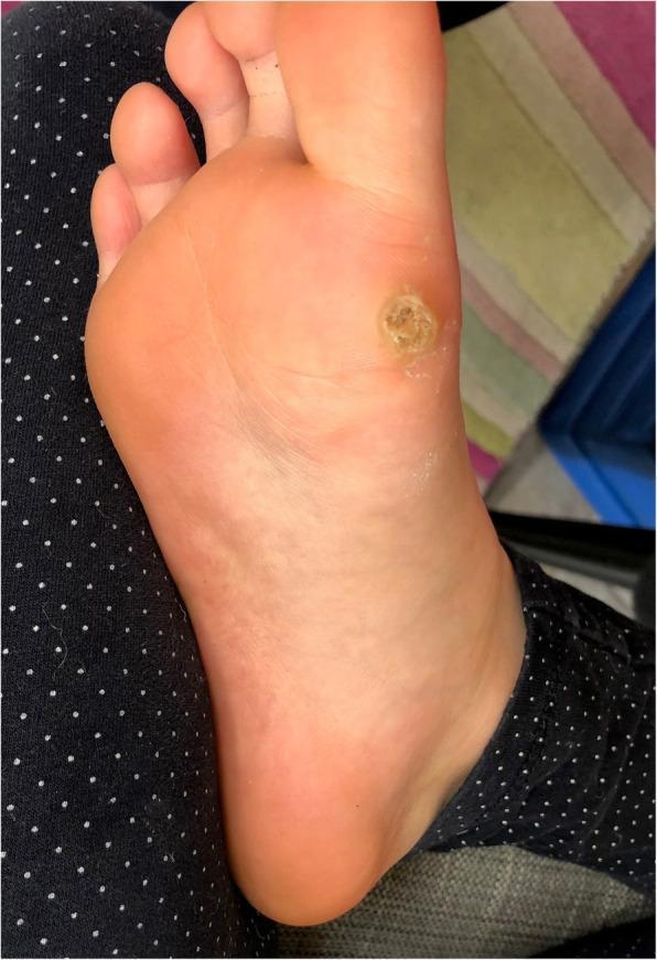
This clinical photograph displays a solitary, deep plantar wart (verruca plantaris) located on the weight-bearing plantar surface of the human foot, specifically over the metatarsal region. The lesion is well-circumscribed and approximately circular, characterized by a central area of significant hyperkeratosis. The surface texture is rough, verrucous, and uneven, exhibiting a beige to yellowish-tan discoloration that contrasts with the surrounding healthy pink skin. A slightly raised, indurated outer rim demarcates the lesion from the adjacent tissue. The visual findings are consistent with a myrmecia-type wart typically associated with Human Papillomavirus type 1 (HPV 1). Key educational features include the disruption of normal dermatoglyphics and the thick keratotic plug, which are hallmark diagnostic signs of plantar verrucae in dermatology.
This clinical photograph displays a solitary, deep plantar wart (verruca plantaris) located on the weight-bearing plantar surface of the human foot, specifically over the metatarsal region. The lesion is well-circumscribed and approximately circular, characterized by a central area of significant hyperkeratosis. The surface texture is rough, verrucous, and uneven, exhibiting a beige to yellowish-tan discoloration that contrasts with the surrounding healthy pink skin. A slightly raised, indurated outer rim demarcates the lesion from the adjacent tissue. The visual findings are consistent with a myrmecia-type wart typically associated with Human Papillomavirus type 1 (HPV 1). Key educational features include the disruption of normal dermatoglyphics and the thick keratotic plug, which are hallmark diagnostic signs of plantar verrucae in dermatology.

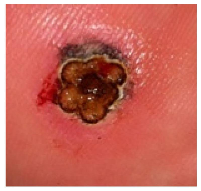
This clinical photograph displays a large plantar wart (verruca plantaris) measuring over 4 mm in diameter on the weight-bearing surface of the foot. The lesion is characterized by a raised, hyperkeratotic appearance with a clustered, multi-lobulated morphology resembling a 'mosaic' pattern. The central tissue exhibits dark brown to black discoloration, likely due to the application of India ink used in laser therapy protocols to enhance thermal absorption. A distinct, pale rim or halo surrounds the lobules, followed by a wider zone of erythematous, inflamed perilesional skin. Minor punctate bleeding is visible on the lateral margin, typical of exposed capillaries (thrombosed capillaries) following the removal of overlying hyperkeratosis. This image serves as a clinical example of a recalcitrant plantar wart prepared for laser treatment, illustrating the anatomical challenges of treating thick epidermal lesions on the plantar surface.
This clinical photograph displays a large plantar wart (verruca plantaris) measuring over 4 mm in diameter on the weight-bearing surface of the foot. The lesion is characterized by a raised, hyperkeratotic appearance with a clustered, multi-lobulated morphology resembling a 'mosaic' pattern. The central tissue exhibits dark brown to black discoloration, likely due to the application of India ink used in laser therapy protocols to enhance thermal absorption. A distinct, pale rim or halo surrounds the lobules, followed by a wider zone of erythematous, inflamed perilesional skin. Minor punctate bleeding is visible on the lateral margin, typical of exposed capillaries (thrombosed capillaries) following the removal of overlying hyperkeratosis. This image serves as a clinical example of a recalcitrant plantar wart prepared for laser treatment, illustrating the anatomical challenges of treating thick epidermal lesions on the plantar surface.

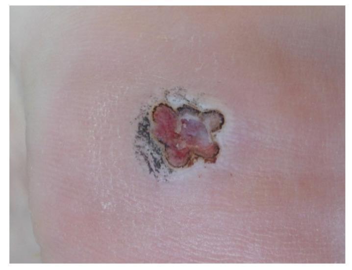
This clinical photograph displays a follow-up view of a plantar wart (Verruca plantaris) on the foot, one week post-laser treatment using an India ink chromophore. The lesion, which is greater than 4 mm in diameter, shows a central zone of reddish-purple and pinkish granulation tissue or inflammation. This central area is bordered by a thin, dark, almost black ring, likely representing remnants of the India ink or charred tissue from the laser irradiation. The overall morphology is roughly circular with irregular, non-uniform borders. The immediate periphery of the lesion exhibits a lighter, whitish-pink hue indicative of localized inflammation or re-epithelialization, transitioning into the normal surrounding skin tone. The skin texture at the site appears slightly raised and uneven, typical of a healing wound after the removal of hyperkeratotic tissue. This image illustrates the clinical progression and tissue reaction during the healing phase of laser-treated recalcitrant plantar warts.
This clinical photograph displays a follow-up view of a plantar wart (Verruca plantaris) on the foot, one week post-laser treatment using an India ink chromophore. The lesion, which is greater than 4 mm in diameter, shows a central zone of reddish-purple and pinkish granulation tissue or inflammation. This central area is bordered by a thin, dark, almost black ring, likely representing remnants of the India ink or charred tissue from the laser irradiation. The overall morphology is roughly circular with irregular, non-uniform borders. The immediate periphery of the lesion exhibits a lighter, whitish-pink hue indicative of localized inflammation or re-epithelialization, transitioning into the normal surrounding skin tone. The skin texture at the site appears slightly raised and uneven, typical of a healing wound after the removal of hyperkeratotic tissue. This image illustrates the clinical progression and tissue reaction during the healing phase of laser-treated recalcitrant plantar warts.

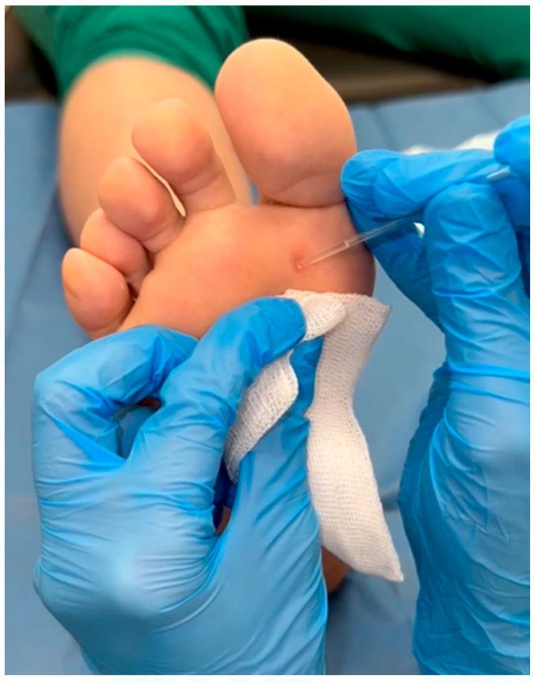
A close-up clinical photograph demonstrating a dermatological procedure on the plantar surface of a human foot. The image shows a small, localized, hyperkeratotic lesion—identified as a plantar wart (verruca plantaris)—located on the forefoot near the base of the first metatarsal head. The lesion is approximately 2-3 mm in diameter, circular, and slightly darker than the surrounding healthy skin. A healthcare professional wearing blue nitrile gloves is shown performing a topical treatment application. A thin, clear plastic capillary applicator is being used to precisely apply a liquid medicinal substance (nitric-zinc complex) directly onto the surface of the wart. A piece of sterile white gauze is held beneath the treatment site to catch excess fluid and protect the surrounding skin. The clinical focus is on targeted chemical cauterization or topical therapy for common viral warts in a primary care or podiatric setting.

A close-up clinical photograph demonstrating a dermatological procedure on the plantar surface of a human foot. The image shows a small, localized, hyperkeratotic lesion—identified as a plantar wart (verruca plantaris)—located on the forefoot near the base of the first metatarsal head. The lesion is approximately 2-3 mm in diameter, circular, and slightly darker than the surrounding healthy skin. A healthcare professional wearing blue nitrile gloves is shown performing a topical treatment application. A thin, clear plastic capillary applicator is being used to precisely apply a liquid medicinal substance (nitric-zinc complex) directly onto the surface of the wart. A piece of sterile white gauze is held beneath the treatment site to catch excess fluid and protect the surrounding skin. The clinical focus is on targeted chemical cauterization or topical therapy for common viral warts in a primary care or podiatric setting.

Searching PubMed

plantar wart treatment

Reading File
Reading File
Reading File
Reading File
Reading File
Loading Image
Excellent. I now have comprehensive information from multiple authoritative sources. Here is the full answer:

Plantar Warts (Verruca Plantaris)

Definition and Etiology

Plantar warts are endophytic, hyperkeratotic lesions caused by human papillomavirus (HPV) infection of the plantar surface of the foot. The term "verruca plantaris" is used interchangeably with plantar wart. While over 100 HPV subtypes exist, HPV-1 is classically associated with the deep "myrmecia" type. Other HPV types (2, 4, 60, 63) can also cause plantar warts. - Harrison's Principles of Internal Medicine, 22E, p. 1815

Clinical Features

  • Endophytic growth: Unlike common warts on other skin surfaces, plantar warts grow inward due to the pressure of weight-bearing. They appear flat or slightly raised, often flush with the skin surface.
  • Thick hyperkeratosis: Covered by a dense keratin layer that can be mistaken for a callus.
  • Punctate hemorrhages: Paring the keratotic surface reveals a central core of keratinized debris and punctate bleeding points (thrombosed capillaries) - a hallmark diagnostic sign that distinguishes a wart from a callus.
  • Disruption of dermatoglyphics: Normal skin lines are interrupted across the lesion.
  • Mosaic warts: Clusters of multiple smaller warts that fuse together in a plaque-like pattern; particularly resistant to treatment.
  • Pain: Often painful on weight-bearing due to inward pressure.
Here is a typical clinical appearance:
Plantar wart on the metatarsal region showing hyperkeratosis and disrupted dermatoglyphics

Diagnosis

The diagnosis is primarily clinical. Key differentiating features:
FeaturePlantar WartCallus/Corn
Skin linesInterruptedPreserved
Paring revealsPunctate black/red dots, bleedingLayers of yellowish keratin, no bleeding
TendernessLateral (pinching)Direct pressure
HPV etiologyYesNo
The differential diagnosis also includes: punctate palmoplantar keratoderma, punctate porokeratosis, arsenical keratoses, eccrine poromas/poromatosis, and - importantly - amelanotic melanoma, which should be suspected if a persistent lesion shows erosion or subtle pigmentation. - Dermatology 2-Volume Set, 5e, p. 422

Natural History

Most warts in immunocompetent individuals resolve spontaneously within 1-2 years. Approximately 60% resolve spontaneously over time. Recurrence rates after any treatment are 30% or higher. These facts should guide the decision to treat and the choice of modality. - Pfenninger and Fowler's Procedures for Primary Care, p. 4165; Harrison's 22E

Treatment

There is no single universally effective therapy. Treatment choice depends on the number and location of warts, patient age and immune status, and patient preference.

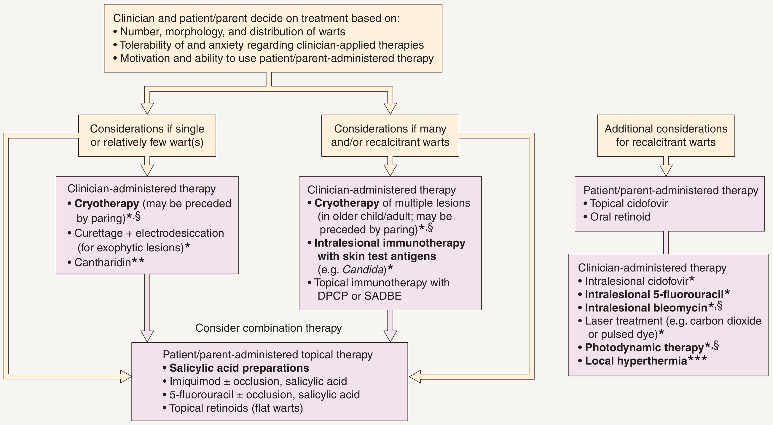
Treatment Algorithm

Flowchart for treatment of common warts showing clinician- and patient-administered options for single/few warts, multiple warts, and recalcitrant warts
From Dermatology 2-Volume Set, 5e - therapies in bold are supported by controlled trials.

First-Line Approaches

1. Salicylic Acid (patient-administered)
  • 17% solution (e.g., Compound W) or 40% salicylic acid pads (Mediplast, Trans-Ver-Sal)
  • Patient soaks and cleans the area, applies the preparation daily
  • After 2 weeks, the soft white keratin layer can be peeled away, revealing the wart base
  • Keratolysis improves efficacy of subsequent in-office procedures
  • Equally effective to cryotherapy for non-genital warts but requires higher patient compliance
2. Cryotherapy (clinician-administered)
  • Liquid nitrogen is the most effective and convenient in-office modality
  • Plantar warts have dense keratin and are very resistant to cryosurgery; pretreatment with salicylic acid for 2 weeks significantly improves outcomes
  • Keratin can also be pared with a no. 10 or 15 scalpel blade until the first punctate vasculature (verruca) is seen, then frozen
  • Does not result in scarring (important on the plantar surface - see below)
  • Pfenninger and Fowler's, p. 5573

Avoid Surgical Excision on the Plantar Surface

"Physicians should avoid surgical excisions on the bottom of the feet because the scar tissue often remains painful after healing. A patient may suffer with the irritated scar, which produces an effect not unlike a pebble in a shoe."
  • Pfenninger and Fowler's, p. 4167

For Multiple or Recalcitrant Warts

  • Intralesional immunotherapy (Candida antigen): Has become the first-line approach for all but the simplest cases. Cost-effective, least traumatic, virtually no residual scarring. - Pfenninger and Fowler's, p. 4167
  • Intralesional bleomycin: Effective in controlled trials
  • Intralesional 5-fluorouracil
  • Topical immunotherapy: DPCP (diphenylcyclopropenone) or SADBE (squaric acid dibutyl ester)
  • Laser therapy: CO2 or pulsed dye laser for recalcitrant lesions
  • Photodynamic therapy
  • Local hyperthermia: 44°C/111°F for 30 minutes on days 1, 2, 3, 17, and 18
  • Topical/intralesional cidofovir: Reserved for refractory cases (evidence from case reports including HPV-66 type)

Immunocompromised Patients

In patients with cellular immunodeficiencies (solid organ transplant recipients, HIV/AIDS, hematologic malignancies), warts are more numerous, more persistent, and more resistant to treatment. They should also be monitored for development of squamous cell carcinoma, as HPV-associated SCC is increased in long-term immunosuppressed patients. - Harrison's 22E, p. 1821; Dermatology 5e, p. 424

HPV Subtypes and Wart Types (Summary)

Wart TypeLocationHPV Types
Verruca plantarisPlantar foot1, 2, 4, 60, 63
Verruca vulgarisHands, any skin2, 4, 7, 27, 57
Verruca plana (flat wart)Face, arms, legs3, 10, 28
Condyloma acuminatumAnogenital6, 11
High-risk (oncogenic)Cervix, anus16, 18
From Pfenninger and Fowler's Procedures for Primary Care, p. 480

Recent evidence note: A 2026 systematic review and meta-analysis (PMID 40873359) evaluated ionic contra-viral therapy (digoxin-furosemide combination) for multiple cutaneous warts - an emerging immunomodulatory approach. This is newer territory and should be weighed against the established modalities above.

so having plantar warts means that the person had hpv?

Finding Sources
Reading File
Yes - and here are the important nuances to understand:

Plantar Warts = HPV Infection (But Context Matters a Lot)

Yes, definitively: Plantar warts are caused exclusively by HPV. There is no other mechanism. The virus infects the keratinocytes of the plantar skin epithelium and drives their proliferation. - Goldman-Cecil Medicine, p. 3597

But here's what's important to understand:

1. This is NOT the same HPV as the sexually transmitted kind.
The HPV types that cause plantar warts (types 1, 2, 4, 60, 63) are entirely different from the high-risk genital HPV types (16, 18) or the genital wart types (6, 11). Having a plantar wart says nothing about a person's sexual history.
2. How plantar warts are actually transmitted:
  • Fomite transmission from moist surfaces is the primary route - walking barefoot on wet floors (locker rooms, pools, showers, public bathrooms)
  • Direct skin-to-skin contact
  • Autoinoculation - spreading from one area of your own skin to another
  • Children are particularly susceptible: annual incidence rates of cutaneous warts in children have been reported at up to 30% - it is one of the most common childhood skin infections
3. Most HPV infections are subclinical.
HPV can infect skin without producing a visible wart. The virus can also persist in the tissue for a period before a wart becomes visible. So a person could technically have had HPV on their foot without knowing it, and only develop a visible wart later.
4. The infection is localized.
Plantar HPV infection is confined to the skin of the foot. It does not reflect systemic HPV infection and carries no cancer risk. The oncogenic HPV types responsible for cervical cancer, oropharyngeal cancer, and anal cancer are completely different subtypes.

Bottom line: Plantar warts mean the person has/had a localized HPV infection on their foot skin - acquired almost certainly from a contaminated surface, not sexually. It is extremely common, affects up to 1 in 3 children at some point, and is clinically unrelated to "HPV" in the sense most people mean when they hear that term.

is it latent?

Finding Sources
Finding Sources
Reading File
Finding Sources
This is a nuanced and well-answered question. Here is a precise answer:

Is HPV in Plantar Warts "Latent"?

Short answer: not in the traditional sense - but it does persist in a unique way.

What "latent" means in virology

True latency (as in herpesviruses like HSV or VZV) means the virus goes completely silent in nerve ganglia, produces no proteins, causes no symptoms, and can reactivate later. HPV does not work that way.

How HPV actually persists - the episomal mechanism

The molecular biology is well understood. From Goldman-Cecil Medicine:
"In the basal layer, the viral genome becomes established in the nucleus as an episome that replicates in tandem with cellular replication, thus maintaining a stable copy number of viral genomes. As basal cells migrate up and differentiate in the superficial layers of the epithelium, full vegetative viral DNA replication and expression of structural proteins occur, with assembly of infectious virions in the most superficial layer of the epithelium."
So in a wart:
  • HPV sits in the basal keratinocytes as an episome (a circular piece of DNA separate from the chromosomes)
  • It replicates quietly alongside the cell with low-level gene expression in the basal layer
  • Full viral replication and infectious virus production happen in the upper, differentiating layers
  • Virions are shed from the surface as dead cells slough off
This low-level basal presence is sometimes called "latent" or "subclinical" infection, but it is not true latency - the virus is still actively (if quietly) maintained.

Why warts recur after treatment

This episomal persistence explains the 30%+ recurrence rate after all treatments. When you destroy the visible wart, you eliminate the upper keratotic layers and the bulk of viral replication - but basal keratinocytes at the margins may still carry the episome. If those cells are not eliminated, the wart regrows.

What happens when a wart "resolves spontaneously"

When the immune system clears a wart (which happens in 60% of cases within 1-2 years), cell-mediated immunity eliminates the infected basal cells. Current understanding is that viral clearance can be complete - unlike herpesviruses, there is no permanent reservoir in a ganglion. However, whether truly every last viral copy is eliminated is hard to prove, and low-level subclinical HPV DNA has been detected in perilesional skin even after clinical resolution.

Summary

ConceptHPV in plantar wart
Visible wartActive, productive infection
After treatment/resolutionLikely cleared, but basal episomal persistence can cause recurrence
True latency (like herpes)No
Permanent reservoirNo established permanent reservoir
Reactivation years laterPossible but not a defining feature the way herpes reactivation is
The practical takeaway: a resolved plantar wart is most likely gone, but the high recurrence rate in treated warts reflects how deeply the episome can embed in basal skin cells before complete clearance. - Goldman-Cecil Medicine, p. 3597 (pathobiology section)
This is a shared conversation. Sign in to Orris to start your own chat.