
Verrucus epidermal nevus based on iadvl rooks fitzpatrick and Bolognia textbook of dermatology
verrucous epidermal nevus linear Blaschko lines
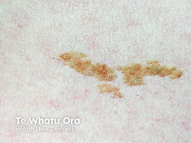
Clinical photography of a linear epidermal nevus. This image captures a single, elongated, hyperpigmented plaque with a slightly verrucous surface and mild keratotic scaling, arranged in a linear/serpiginous pattern along the skin. The lesion appears well circumscribed, non-inflammatory, and located on the cutaneous surface without obvious regional lymphadenopathy or ulceration. The distribution follows Blaschko's lines, a hallmark feature of epidermal nevi, indicating a mosaic somatic mutation in keratinocytes. The color ranges from light brown to tan with focal darker hyperpigmented conglomerates; surface texture is rough with papillomatous outgrowths at the margins. The image is a high-resolution clinical photograph (macroscopic close-up) without dermoscopic magnification or contrast enhancement. In suspected cases, histopathology would typically show hyperkeratosis, acanthosis, and slight papillomatosis with elongation of rete ridges, consistent with an epidermal nevus. Clinically, linear epidermal nevi are usually benign but can be associated with epidermal nevus syndrome if extensive or multisystemic. Differential considerations include inflammatory linear verrucous epidermal nevus (ILVEN), linear porokeratosis, and epidermal keratinocytic nevi of other distributions. The image is valuable for dermatology education, pattern recognition, genetic counseling discussion, and patient counseling about cosmetic management or laser/ablative therapy options. Proper documentation supports diagnosis, prognosis assessment, and multidisciplinary treatment planning for patient care.
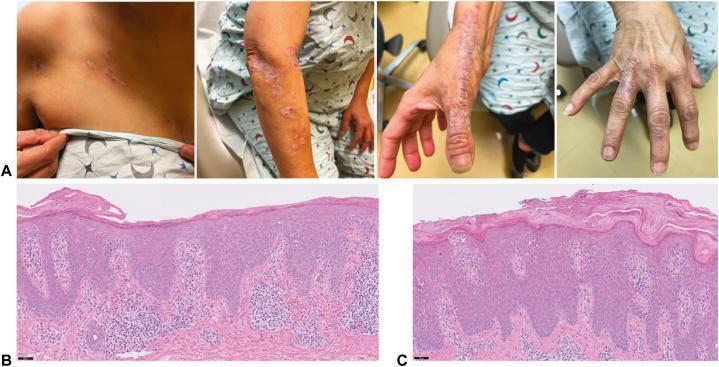
This composite educational image illustrates the clinical and histopathological features of Inflammatory Linear Verrucous Epidermal Nevus (ILVEN). Panel A presents clinical photographs showing erythematous to violaceous, scaly papules and plaques. These lesions exhibit a distinct linear arrangement following the Lines of Blaschko, extending from the right pectoral region down the upper extremity to the dorsal aspect of the fingers. Panels B and C are high-power photomicrographs (H&E stain) of skin biopsies from the chest and wrist. Pathological findings include marked epidermal acanthosis and spongiosis. A key diagnostic feature shown is the characteristic horizontally alternating pattern of orthokeratosis and parakeratosis within the stratum corneum. The underlying papillary dermis reveals a superficial perivascular lymphocytic infiltrate. This presentation is a classic example used in dermatology to differentiate ILVEN from other Blaschkoid dermatoses such as linear psoriasis or lichen striatus based on chronicity and specific histomorphological patterns.
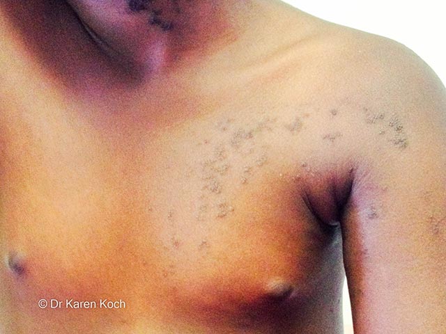
This is a high-resolution clinical photograph of a cutaneous epidermal nevus located along the left shoulder and anterior upper chest. Modality: clinical photography; technique: standard color image, with even lighting to depict surface texture and pigmentation; no dermoscopy or adjunct contrast used. The lesion demonstrates a classic Blaschko-linear distribution, a left-to-right, anterior-lateral cascade that follows embryologic skin cleavage lines. The primary subject is an epidermal nevus arising from keratinocyte proliferation within the epidermis, presenting as hyperpigmented, verrucous plaques that are well demarcated and mildly raised. In skin of colour, the pigment intensity may be accentuated, producing dark brown to black plaques with subtle scale and ridging; surface may be minimally hyperkeratotic. The pattern is typically unilateral and unilateral to the trunk and proximal limbs, with linear arrays aligning with Blaschko lines. Clinically, these lesions are generally benign but can be cosmetically disfiguring; differential considerations include inflammatory linear verrucous epidermal nevus (ILVEN), linear porokeratosis, and other pigmentary mosaicisms. Pathology, if pursued, would show acanthosis, hyperkeratosis, and mild papillomatosis without significant atypia. This image is useful for dermatology education, mosaicism concepts, and digital documentation in genodermatoses clinics. Helpful for patient counseling, telemedicine assessment, and research on Blaschko-linear cutaneous disorders in diverse populations.
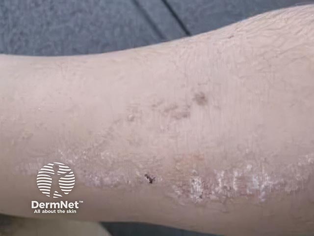
Clinical photography of an inflamed, linear, verrucous skin plaque consistent with inflammatory linear verrucous epidermal nevus (ILVEN) located on the forearm. The lesion appears as a long, narrow band of scaly, hyperkeratotic plaques with erythematous border and mild crusting. The linear distribution follows Blaschko lines, a characteristic pattern for epidermal nevi. Visual features include raised, rough surface texture, intermittent excoriations, and focal remodeling of surrounding epidermis. Inflammation manifests as surrounding erythema and subtle edema with secondary flaking. The lesion length spans several centimeters with variable width, tapering at the ends. There is no evidence of systemic involvement. The image is a standard clinical dermatology photograph without dermoscopic magnification or histologic confirmation. ILVEN typically presents in infancy or early childhood but can be seen later; it is often intensely pruritic and recalcitrant to routine emollients, requiring targeted therapy. Histopathology, if obtained, would show alternating areas of hypergranulosis and hypogranulosis with psoriasiform epidermal hyperplasia and inflammatory infiltrate, supporting an ILVEN diagnosis. This image supports clinical recognition, differential diagnosis against linear psoriasis, verrucous epidermal nevus, and lichen striatus, and can aid educational discussions, patient counseling, and treatment planning including topical steroids, retinoids, or laser therapy.
IADVL textbook verrucous epidermal nevus classification treatment India
Note on sources: The library contains Fitzpatrick's Dermatology (9th ed.), Rook's Dermatology (Dermatology 2-Volume Set 5e), and Andrews' Diseases of the Skin. IADVL's Textbook of Dermatology is not in this digital library, but its published chapters (e.g. IADVL Dermatopathology Ch.31 Epidermal Nevi) follow the same classification. The synthesis below integrates all three available references with IADVL-consistent framing.
Verrucous Epidermal Nevus (VEN)
1. Definition and Terminology
-
Linear epidermal nevus
-
Hard nevus of Unna / soft epidermal nevus
-
Nevus verrucosus
-
Nevus unius lateris (unilateral extensive variant)
-
Ichthyosis hystrix (bilateral extensive variant)
-
Fitzpatrick's Dermatology, 9th ed., Ch. 108 | Andrews' Diseases of the Skin, Ch. 29
2. Epidemiology
-
Incidence: 1 in 1000 live births; no sex predilection
-
Most cases sporadic; rare familial cases documented
-
80% present at birth or within the first year of life; the remainder manifest by adolescence
-
Adult-onset cases are thought to represent subclinical lesions that became clinically apparent with growth
-
Fitzpatrick's Dermatology, p. 1836
3. Pathogenesis and Genetics
Key Mutations:
| Gene | Pathway | Notes |
|---|---|---|
| HRAS (most common ~40%) | RAS/MAPK | Mosaic RASopathy |
| KRAS | RAS/MAPK | |
| FGFR3 (~40%) | FGFR/PIK3CA/AKT | Also found in seborrheic keratoses |
| PIK3CA | PI3K/AKT | |
| KRT1, KRT10, KRT2 | Keratin | Epidermolytic variant |
| KRT16 | Keratin | Palmoplantar non-epidermolytic |
- RAS/MAPK pathway (mosaic RASopathies)
- FGFR/PIK3CA/AKT1 pathway (negatively regulated by PTEN)
- Rook's Dermatology 5e, p. 1219-1220 | Fitzpatrick's, p. 1837 | Andrews', p. 736
4. Clinical Features
- Skin-colored to hyperpigmented verrucous or velvety papules coalescing into plaques
- Follow Blaschko's lines - linear on limbs, S-shaped/whorled on trunk
- May appear macerated/whitish-pink at birth, becoming more verrucous/pigmented over time
- Lesions tend to be more pronounced in flexural skin (intertriginous accentuation) - can resemble acanthosis nigricans in skin folds
- Rarely: pale/hypopigmented streaks in dark skin types
- Most common on neck, trunk, and extremities
- Intertriginous: softer, less hyperkeratotic
| Variant | Features |
|---|---|
| Nevus unius lateris | Unilateral systematized epidermal nevus |
| Ichthyosis hystrix | Bilateral, widespread involvement |
| ILVEN | Inflammatory variant - erythematous, pruritic, psoriasiform plaques |
| RAVEN | Rounded and velvety epidermal nevus; arranged in linear pattern (FGFR2/3 mutations) |
| PENS | Papular epidermal nevus with "skyline" basal cell layer; sporadic or familial |
- Rook's Dermatology 5e, p. 1218-1219 | Fitzpatrick's, p. 1836-1837 | Andrews', p. 735
5. Histopathology
Non-epidermolytic (most common, ~62%):
- Hyperkeratosis + acanthosis + papillomatosis (the classic triad)
- Elongation of rete ridges
- No granular/vacuolar degeneration
Epidermolytic (~16%):
- Marked orthokeratosis with vacuolization and coarse keratohyalin granule deposition in granular and spinous layers - epidermolytic hyperkeratosis
- Caused by KRT1/KRT10/KRT2 mutations
Other histologic patterns (less common):
- Psoriasiform type
- Acrokeratosis verruciformis-like type
- Darier disease-like (acantholytic dyskeratotic)
ILVEN histology:
- Psoriasiform epidermal hyperplasia
- Alternating bands of orthokeratosis and parakeratosis (absent granular layer under parakeratotic areas)
- Chronic dermal lymphocytic infiltrate
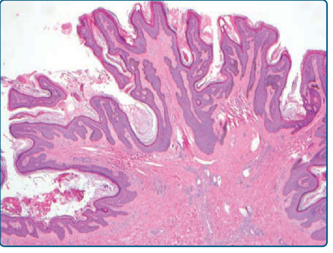
- Fitzpatrick's, p. 1837-1838 | Andrews', p. 735
6. Differential Diagnosis
| Condition | Distinguishing features |
|---|---|
| Seborrheic keratosis (SK) | Individual papules mimic SK; not linear |
| Verruca vulgaris | HPV; not linear/blaschkoid |
| Nevus sebaceous | Head/neck; waxy yellow-orange; hair loss |
| Lichen striatus | Inflammatory; follows Blaschko; self-limited (usually resolves) |
| ILVEN | Pruritic; psoriasiform; recalcitrant; distinguishable histologically |
| Linear psoriasis | Family history; more confluent parakeratosis; responds to treatment |
| Linear porokeratosis | Cornoid lamellae; keratotic ridge; SCC risk |
| Linear Darier disease | Acantholytic dyskeratosis histologically |
| Incontinentia pigmenti stage 2 | Female predominance; vesicular stage preceding |
| Linear and whorled nevoid hypermelanosis | Macular, no verrucous component |
| Acanthosis nigricans | Flexural, diffuse; associated metabolic syndrome |
- Fitzpatrick's, p. 1838 | Rook's 5e, p. 1219
7. Epidermal Nevus Syndrome (ENS)
- CNS abnormalities (more common with head/neck lesions): intellectual disability, seizures, hemimegalencephaly
- Skeletal abnormalities (more common with trunk/extremity lesions)
- Ocular anomalies
-
Schimmelpenning syndrome (nevus sebaceous + CNS/ocular/skeletal)
-
CHILD syndrome (congenital hemidysplasia, ichthyosiform erythroderma, limb defects) - NSDHL mutation
-
Proteus syndrome - mosaic AKT1 mutation; cerebriform plantar nevi, asymmetric overgrowth
-
CLOVES syndrome - PIK3CA; congenital lipomatous overgrowth, vascular malformations
-
Phakomatosis pigmentokeratotica - mosaic HRAS/KRAS
-
SOLAMEN syndrome - mosaic PTEN; verrucous EN + arteriovenous malformation + lipomatosis
-
FGFR3-ENS - widespread epidermal nevus + developmental brain defects + vitamin D-resistant hypophosphatemic rickets (FGF-23 excess)
-
Rook's 5e, p. 1221-1222 | Andrews', p. 736 | Fitzpatrick's, p. 1838
8. Course and Complications
-
Congenital lesions tend to be quiescent; those developing postnatally may enlarge before stabilizing at puberty
-
Intertriginous lesions: maceration, secondary infection
-
Malignant transformation is rare but documented - BCC and SCC can arise within VEN during adulthood; any rapidly enlarging or ulcerated nodule within a nevus requires biopsy
-
Epidermolytic variant: risk of offspring with epidermolytic ichthyosis (gonadal mosaicism)
-
Fitzpatrick's, p. 1837-1838
9. ILVEN (Inflammatory Linear Verrucous Epidermal Nevus) - Special Variant
- Linear erythematous, scaly, intensely pruritic plaques following Blaschko's lines
- Usually appears in childhood; occasionally at birth or adult life
- Extends over months to years; rarely remits spontaneously
- Postzygotic mutations in GJA1 (connexin 43; mosaic erythrokeratodermia variabilis)
- Postzygotic mutations in CARD14
- Germline + postzygotic mutations in ABCA12 (mosaic autosomal recessive congenital ichthyosis)
- Some ILVEN-phenotype patients have forme fruste of CHILD syndrome (NSDHL mutation) - termed PEN/PENCIL syndrome
10. Treatment
Definitive treatment:
- Full-thickness surgical excision - most definitive; low recurrence because it removes the superficial dermal component. Best for small, localized lesions.
Ablative/destructive modalities:
- CO₂ laser / Er:YAG laser - effective for larger or cosmetically sensitive areas
- Cryotherapy - can be effective with good cosmetic results
- Dermabrasion / electrofulguration - partial clearance; recurrence common if superficial only
- Medium- to full-depth chemical peels
Topical therapies (partial/temporary benefit):
- 5-FU 5% + tretinoin 0.1% (once daily, may enhance with occlusion) - Andrews' recommends this combination
- Calcipotriene (calcipotriol) - useful particularly in ILVEN and epidermolytic variants
- Topical corticosteroids - for ILVEN
- Corticosteroid + calcipotriene combination - for ILVEN
ILVEN-specific (pathogenesis-directed):
- Topical statin/cholesterol cream (for CHILD-related/NSDHL mutation variant)
- Topical retinoids - limited benefit
Key principle: Any treatment that does not extend into the dermis will likely result in recurrence.
- Fitzpatrick's, p. 1838 | Andrews', p. 736 | Rook's 5e, p. 1221-1222
Summary Table
| Feature | Details |
|---|---|
| Type | Benign hamartoma (keratinocytic) |
| Mechanism | Postzygotic somatic mutation → cutaneous mosaicism |
| Key genes | HRAS, FGFR3, PIK3CA (~80%); KRT1/KRT10 (epidermolytic) |
| Distribution | Blaschko's lines (linear on limbs, whorled on trunk) |
| Incidence | 1 in 1000 live births |
| Onset | 80% at birth/first year; rest by adolescence |
| Histology | Acanthosis + papillomatosis + hyperkeratosis (non-EH); EH variant: vacuolization + coarse keratohyalin granules |
| ILVEN | Pruritic, psoriasiform; alternating ortho/parakeratosis; GJA1, CARD14, ABCA12 mutations |
| Malignant risk | Rare BCC/SCC in adults |
| EH genetic risk | Offspring risk epidermolytic ichthyosis (gonadal mosaicism) |
| Definitive treatment | Full-thickness excision |
| Alternatives | CO₂/Er:YAG laser, cryotherapy, 5-FU + tretinoin, calcipotriol |
Verrucous Epidermal Nevus - Comprehensive Review
Definition
Epidemiology
- Incidence: 1 in 1000 live births; no sex predilection
- Onset: 80% present at birth or by 1 year of age; remainder by adolescence
- Most cases sporadic; rare familial cases exist
- Adult "onset" usually represents subclinical lesions becoming evident - Fitzpatrick's, p. 1836
Pathogenesis and Genetics
Key Genes and Pathways:
| Gene | Pathway | Germline Counterpart |
|---|---|---|
| HRAS (~40%) | RAS/MAPK | Costello syndrome |
| KRAS | RAS/MAPK | |
| FGFR3 | FGFR/PI3K/AKT | Thanatophoric dysplasia |
| PIK3CA | PI3K/AKT | CLOVES/Megalencephaly syndromes |
| KRT1, KRT10, KRT2 | Keratin | Epidermolytic ichthyosis |
| FGFR2 | FGFR/PI3K/AKT | Apert syndrome (germline) |
- RAS/MAPK (mosaic RASopathies)
- FGFR/PIK3CA/AKT1, negatively regulated by PTEN
Critical gonadal mosaicism point (Fitzpatrick's): Patients with epidermolytic histology (KRT1/KRT10 mutations) who have concomitant gonadal mosaicism can have offspring with full epidermolytic ichthyosis. Prenatal counseling is essential for this subtype.
Clinical Features
- Skin-colored to hyperpigmented verrucous or velvety papules coalescing into serpiginous plaques
- Linear on limbs; S-shaped/whorled on trunk
- More pronounced in flexural (intertriginous) skin - may resemble acanthosis nigricans
- May appear whitish-pink and macerated at birth, becoming hyperkeratotic with age
| Variant | Description |
|---|---|
| Nevus unius lateris | Unilateral systematized form |
| Ichthyosis hystrix | Bilateral, widespread |
| ILVEN | Inflammatory, pruritic, psoriasiform; recalcitrant |
| RAVEN | Rounded/velvety; linear; FGFR2/3 mutations |
| PENS | Papular; "skyline" basal cells on histology |
Histopathology
| Pattern | Features | Frequency |
|---|---|---|
| Non-epidermolytic | Hyperkeratosis + acanthosis + papillomatosis | ~62% |
| Epidermolytic (EH) | Vacuolization + coarse keratohyalin granules in spinous/granular layers | ~16% |
| Psoriasiform | Psoriasis-like epidermal changes | Rare |
| Acrokeratosis verruciformis-like | - | Rare |
| Darier disease-like | Acantholytic dyskeratosis | Rare |
Differential Diagnosis
| Condition | Key differentiating point |
|---|---|
| Seborrheic keratosis | Not linear/blaschkoid |
| Verruca vulgaris | HPV; dermoscopy shows thrombosed capillaries |
| Nevus sebaceous | Head/neck; waxy yellow-orange; alopecia |
| Lichen striatus | Self-limited; inflammatory; resolves spontaneously |
| ILVEN | Pruritic; psoriasiform; recalcitrant; characteristic histology |
| Linear psoriasis | Family history; responds to therapy; confluent parakeratosis |
| Linear porokeratosis | Keratotic ridge with cornoid lamellae; SCC risk |
| Linear Darier disease | Acantholytic dyskeratosis on biopsy |
| Incontinentia pigmenti | Female; vesicular stage 1 precedes verrucous stage 2 |
| Linear hyperpigmentation | Macular only; no verrucous texture |
Epidermal Nevus Syndrome (ENS)
- Head/neck lesions → CNS anomalies (seizures, intellectual disability, hemimegalencephaly)
- Trunk/extremity lesions → Skeletal anomalies
- Schimmelpenning - nevus sebaceous + CNS + ocular
- CHILD - NSDHL mutation; ipsilateral hemidysplasia
- Proteus - mosaic AKT1; cerebriform plantar nevi; VTE risk
- CLOVES - PIK3CA; congenital lipomatous overgrowth
- FGFR3-ENS - widespread EN + brain defects + hypophosphatemic rickets
- SOLAMEN - mosaic PTEN; verrucous EN + AVM + lipomatosis
- Phakomatosis pigmentokeratotica - mosaic HRAS/KRAS
Course and Complications
- Congenital lesions: generally quiescent
- Postnatal lesions: may enlarge, then stabilize at puberty
- Intertriginous lesions: maceration, secondary infection
- Malignant transformation (rare): BCC and SCC in adults within VEN - any new nodule warrants biopsy
- Gonadal mosaicism risk (epidermolytic subtype)
ILVEN - Special Focus
- Represents a spectrum of inflammatory mosaic disorders - not a single entity
- Characteristically pruritic, erythematous, scaly linear plaques
- Appears in childhood; extends over months to years; rarely remits spontaneously
- Responds temporarily (if at all) to topical anti-inflammatories
- GJA1 mutations (connexin 43)
- CARD14 mutations
- ABCA12 mutations
- NSDHL mutations (CHILD/PEN/PENCIL variant)
Treatment
First-line (Definitive):
- Full-thickness surgical excision - most effective; removes superficial dermal component, hence low recurrence. Best for small/localized lesions.
Ablative options:
- CO₂ laser - effective; preferred for larger lesions or cosmetically sensitive areas (commonly used in Indian practice per IADVL guidelines)
- Er:YAG laser - alternative
- Cryotherapy - effective with good cosmetic results (Andrews')
- Dermabrasion / electrofulguration - partial; recurrence if superficial only
- Chemical peels (medium to full depth)
Topical therapies:
- 5-FU 5% + tretinoin 0.1% once daily (± occlusion) - Andrews'
- Calcipotriene alone or with corticosteroids
- Topical retinoids - limited benefit
- Topical corticosteroids - for ILVEN
ILVEN / CHILD-targeted (Rook's 5e):
- Cholesterol 2%/lovastatin 2% cream - for NSDHL-mutation CHILD/PEN lesions
- Topical statin/cholesterol combinations
- Miransertib (AKT inhibitor) - for Proteus syndrome-associated lesions in clinical trials
Key principle: Treatments that do not reach the dermis invariably recur - Fitzpatrick's, p. 1838; Andrews', p. 736
- Fitzpatrick's Dermatology, 9th ed., Ch. 108, pp. 1835-1838
- Rook's Dermatology (Dermatology 2-Volume Set 5e), Ch. 62, pp. 1218-1222
- Andrews' Diseases of the Skin, Ch. 29, pp. 734-736
- IADVL Textbook of Dermatopathology, Ch. 31 (Epidermal Nevi) - Jaypee Digital
Genetics involved in Verrucus epidermal nevus
"epidermal nevus" AND "somatic mutation" AND genetics
"epidermal nevus" AND "HRAS" AND mosaicism
RAS MAPK signaling pathway epidermal nevus mosaicism diagram
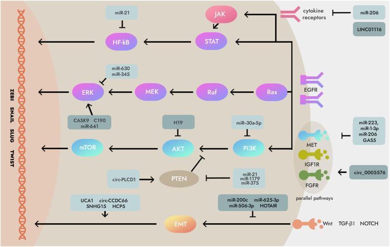
This pathophysiology diagram illustrates the complex regulatory network of non-coding RNAs (ncRNAs) on essential intracellular signaling pathways in human cancers, specifically gastric cancer. The visual details the downstream and parallel signaling cascades of the Epidermal Growth Factor Receptor (EGFR) and other transmembrane receptors like cytokine receptors, MET, IGF1R, FGFR, and Wnt/TGF-β1/NOTCH. Key signaling axes depicted include: 1. The MAPK/ERK pathway: Ras triggers a Raf-MEK-ERK phosphorylation cascade, regulated by miR-630, miR-345 (inhibitors), and CASK9, C190, miR-641 (activators). 2. The PI3K/AKT/mTOR axis: Regulated by miR-30a-5p (PI3K inhibitor), H19 (AKT inhibitor), and the PTEN tumor suppressor. PTEN is modulated by circ-PLCD1 and various miRNAs like miR-21. 3. The JAK/STAT/NF-κB pathway: Showing cytokine-mediated activation and miRNA inhibition. All pathways culminate in nuclear translocation and regulation of Epithelial-Mesenchymal Transition (EMT) transcription factors (ZEB1, SNAIL, SLUG, TWIST), driving oncogenic progression. This schematic highlights the therapeutic potential of targeting ncRNAs like miRNAs, lncRNAs, and circRNAs to modulate cancer cell behavior and radiosensitivity.
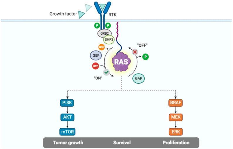
This pathophysiology diagram illustrates the Receptor Tyrosine Kinase (RTK) and RAS signaling pathway, highlighting mechanisms of intracellular signal transduction. The process initiates at the plasma membrane where 'Growth factors' bind to the extracellular domain of an RTK. This triggers intracellular autophosphorylation, represented by green 'P' markers, which recruits adapter proteins GRB2 and SHP2. The central component is the RAS molecular switch, shown cycling between an 'ON' state (GTP-bound) and an 'OFF' state (GDP-bound). Transition to the active 'ON' state is facilitated by Guanine Nucleotide Exchange Factors (GEF), while inactivation is mediated by GTPase-activating proteins (GAP). Activated RAS triggers two primary downstream effector cascades: the PI3K/AKT/mTOR pathway and the BRAF/MEK/ERK (MAPK) pathway. The diagram links these pathways to critical biological outcomes including 'Tumor growth', 'Survival', and 'Proliferation'. This educational visual is designed to explain oncogenic signaling and cellular regulation at a level suitable for medical and biomedical students.
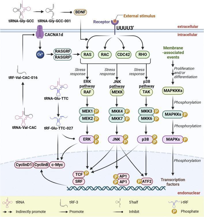
This pathophysiology diagram illustrates the Mitogen-Activated Protein Kinase (MAPK) signaling pathways and their modulation by tRNA-derived small RNAs (tsRNAs). The diagram is organized into extracellular, membrane-associated, and intracellular (cytoplasmic and nuclear) compartments. It depicts three primary cascades: the ERK pathway (RAF to MEK1/2 to ERK), the JNK pathway (MEKK to MKK4/7 to JNK), and the p38 pathway (TAK to MKK3/6 to p38). These pathways are activated by external stimuli via receptors and GTPases including RAS, RAC, CDC42, and RHO. Terminal events show MAPKs phosphorylating nuclear transcription factors such as TCF/SRF, AP1, and ATF2 to regulate genes like CyclinD1, CyclinB, and c-Myc. The graphic highlights the inhibitory roles of specific tsRNAs: tiRNA-Gly-GCC-001 inhibits BDNF; tRF-Val-CAC-016 inhibits CACNA1d (blocking calcium-mediated RASGRF/RASGRP activation); and tRF-Glu-TTC-027 inhibits ERK and p38 phosphorylation. This educational visual explains how tRNA fragments act as post-transcriptional regulators of cell proliferation, differentiation, and stress responses.
Genetics of Verrucous Epidermal Nevus
Fundamental Concept: Cutaneous Mosaicism
"Epidermal nevi are a product of cutaneous mosaicism, defined as a postzygotic genetic mutation or alteration occurring at the level of skin development."
- Fitzpatrick's Dermatology, 9th ed., p. 1836
Two Master Signaling Pathways
Growth Factor
     ↓
  RAS (HRAS/KRAS/NRAS)
     ↓
  RAF → MEK → ERK       ← RAS/MAPK pathway

  PI3K → AKT → mTOR     ← FGFR/PIK3CA/AKT pathway
            ↑
          PTEN (negative regulator)
Gene-by-Gene Breakdown
1. Non-Epidermolytic (Classic) VEN
| Gene | Protein | Pathway | Frequency | Notes |
|---|---|---|---|---|
| HRAS | H-Ras GTPase | RAS/MAPK | ~40% of all VEN | Most common single gene; germline HRAS mutation causes Costello syndrome |
| KRAS | K-Ras GTPase | RAS/MAPK | Part of the ~40% RAS group | Germline causes Noonan syndrome; mosaic KRAS also seen in Schimmelpenning |
| FGFR3 | Fibroblast growth factor receptor 3 | FGFR/PI3K/AKT | Together with PIK3CA ~50% | Germline FGFR3 mutations cause thanatophoric dysplasia/achondroplasia; the FGFR3-mutant EN mimics acanthosis nigricans in flexures |
| PIK3CA | Phosphatidylinositol-3-kinase catalytic subunit α | PI3K/AKT | Part of ~50% FGFR group | Downstream of FGFR signaling; germline causes CLOVES/megalencephaly syndromes |
| FGFR2 | Fibroblast growth factor receptor 2 | FGFR/PI3K/AKT | Less common | Mainly in RAVEN variant and Munro acne nevus; germline causes Apert syndrome |
| NRAS | N-Ras GTPase | RAS/MAPK | Rare | Associated with cutaneous skeletal hypophosphatemia syndrome |
| AKT1 | AKT serine/threonine kinase | PI3K/AKT | Syndromic (Proteus) | Germline mutation is lethal; only survives as mosaic |
| PTEN | Phosphatase and tensin homolog | PI3K/AKT (negative reg.) | Syndromic (SOLAMEN) | Loss-of-function mosaic "second hit" in setting of germline PTEN mutation |
| BRAF | B-Raf kinase | RAS/MAPK | Rare subset | Mainly in linear syringocystadenoma papilliferum and woolly hair nevi |
- Rook's 5e, p. 1219-1220 | Fitzpatrick's, p. 1837 | Andrews', p. 735-736
2. Epidermolytic VEN (~16% of VEN)
| Gene | Protein | Germline Counterpart | Clinical Implication |
|---|---|---|---|
| KRT1 | Keratin 1 | Epidermolytic ichthyosis (bullous congenital ichthyosiform erythroderma) | Gonadal mosaicism risk |
| KRT10 | Keratin 10 | Epidermolytic ichthyosis | Gonadal mosaicism risk |
| KRT2 | Keratin 2 | Ichthyosis bullosa of Siemens | Less common |
| KRT16 | Keratin 16 | Pachyonychia congenita, palmoplantar keratoderma | Palmoplantar non-epidermolytic variant |
Critical Gonadal Mosaicism Warning (Fitzpatrick's, p. 1837-1838): If a patient with VEN showing EH on biopsy also has a KRT1 or KRT10 mutation in gonadal tissue, their offspring are at risk of full-blown epidermolytic ichthyosis. Prenatal genetic counseling is mandatory for this subtype.
3. ILVEN Genetics (Updated - Rook's 5e)
| Gene | Protein | Condition | Treatment implication |
|---|---|---|---|
| GJA1 | Connexin 43 | Mosaic erythrokeratodermia variabilis et progressiva | - |
| CARD14 | Caspase recruitment domain family member 14 | Mosaic psoriasiform disorder | Ustekinumab (IL-12/23 inhibitor) |
| ABCA12 | ATP-binding cassette transporter A12 | Mosaic autosomal recessive congenital ichthyosis | - |
| NSDHL (germline) | NAD(P)H steroid dehydrogenase-like | CHILD syndrome / PEN/PENCIL variant | Topical cholesterol/statin |
| HRAS or KRT10 (postzygotic) | Ras / Keratin 10 | Secondary inflammation within standard VEN | Treat underlying nevus |
| PMVK (germline + somatic second hit) | Phosphomevalonate kinase | Linear porokeratosis mimicking ILVEN | Topical cholesterol/lovastatin |
- Rook's 5e, p. 1221-1222
Mosaicism Types Relevant to VEN
| Type | Mechanism | Example |
|---|---|---|
| Type 1 (simple segmental) | Postzygotic heterozygous mutation on wild-type background | Non-epidermolytic VEN (HRAS, FGFR3) |
| Type 1 (EH) | Postzygotic KRT1/KRT10 mutation on wild-type background | Epidermolytic VEN |
| Type 2 (superimposed/second hit) | Postzygotic "second hit" in patient with heterozygous germline mutation | SOLAMEN (germline PTEN + somatic second hit) |
| Gonadal mosaicism | Mutation also present in germline/gonadal tissue | EH-VEN → offspring with epidermolytic ichthyosis |
- Rook's 5e, p. 1055 | Fitzpatrick's, p. 1837
Germline vs. Mosaic: The Key Concept
| Gene | Mosaic → VEN type | Germline → Syndrome |
|---|---|---|
| HRAS | Keratinocytic VEN, nevus sebaceous | Costello syndrome |
| KRAS | Keratinocytic VEN | Noonan syndrome |
| FGFR3 | Non-epidermolytic VEN (acanthosis nigricans-like) | Thanatophoric dysplasia, achondroplasia |
| PIK3CA | Keratinocytic VEN | CLOVES, megalencephaly-capillary malformation |
| AKT1 | Keratinocytic VEN (in Proteus syndrome) | Lethal germline - not seen |
| KRT1/KRT10 | Epidermolytic VEN | Epidermolytic ichthyosis |
If the germline mutation is lethal (e.g. Proteus/AKT1), only a postzygotic mosaic form exists - there is no germline correlate. - Rook's 5e, p. 1219
Genetics of Associated Epidermal Nevus Syndromes
| Syndrome | Gene | Pathway | Key Features |
|---|---|---|---|
| Schimmelpenning | HRAS, KRAS (mosaic) | RAS/MAPK | Nevus sebaceous + CNS/ocular/skeletal |
| Phakomatosis pigmentokeratotica | HRAS, KRAS (mosaic) | RAS/MAPK | Nevus spilus + nevus sebaceus |
| CHILD syndrome | NSDHL (X-linked germline) | Cholesterol biosynthesis | Unilateral ichthyosiform EN + limb defects; females only |
| Proteus syndrome | AKT1 (mosaic) | PI3K/AKT | Cerebriform plantar nevi, asymmetric overgrowth; VTE risk |
| CLOVES syndrome | PIK3CA (mosaic) | PI3K/AKT | Congenital lipomatous overgrowth + vascular malformations + EN |
| SOLAMEN syndrome | PTEN (germline + somatic second hit) | PI3K/AKT | EN + AVM + lipomatosis + macrocephaly; thicker verrucous nevi |
| Cutaneous skeletal hypophosphatemia | HRAS, NRAS (mosaic) | RAS/MAPK → FGF-23 excess | EN + vitamin D-resistant rickets; FGF-23 overproduction |
| FGFR3-ENS | FGFR3 (mosaic) | FGFR/PI3K/AKT | Widespread EN + brain defects + hypophosphatemic rickets |
- Rook's 5e, pp. 1130-1134 | Andrews', p. 736
FOXN1 Expression
Pathogenesis-Directed Therapy (Genetics → Treatment)
| Mutation | Targeted therapy |
|---|---|
| CARD14 (ILVEN) | Ustekinumab (IL-12/23 blocker) |
| NSDHL/PMVK (CHILD/linear porokeratosis ILVEN) | Topical cholesterol 2% + lovastatin 2% |
| AKT1 (Proteus/PROS) | Miransertib (AKT inhibitor - clinical trials) |
| PIK3CA (PROS/CLOVES) | Alpelisib (PI3K inhibitor) |
| HRAS/NRAS (cutaneous skeletal hypophosphatemia) | Burosumab (FGF23 inhibitor) or trametinib (MEK inhibitor) |
- Rook's 5e, pp. 1138-1139, 1175 | Recent reviews: Mosaic RASopathies PMID 38290824 | RASopathies Part I PMID 40518121
Summary Diagram
POSTZYGOTIC SOMATIC MUTATION
          ↓
    Cutaneous Mosaicism
          ↓
  ┌───────────────────────────────────┐
  │     KERATINOCYTIC (VEN)           │
  ├───────────────┬───────────────────┤
  │ Non-EH (~84%) │ Epidermolytic     │
  │               │ (~16%)            │
  │ HRAS (~40%)   │ KRT1              │
  │ KRAS          │ KRT10  ← Gonadal  │
  │ FGFR3 (~50%) │ KRT2    mosaicism │
  │ PIK3CA        │ KRT16   risk!     │
  │ FGFR2         │                   │
  └───────────────┴───────────────────┘
          ↓                ↓
    RAS/MAPK pathway   Keratin structural
    FGFR/PI3K/AKT      disruption → EH

          ↓
    ILVEN (inflammatory) variant:
    GJA1, CARD14, ABCA12, NSDHL
- Rook's Dermatology (Dermatology 2-Volume Set 5e), Ch. 62, pp. 1219-1223
- Fitzpatrick's Dermatology 9th ed., Ch. 108, pp. 1836-1838
- Andrews' Diseases of the Skin, Ch. 29, pp. 734-736
- Mosaic RASopathies review - Morren et al. J Med Genet 2024 - PMID 38290824
- RASopathies Part I - Jaeger et al. JAAD 2025 - PMID 40518121
Alopecia areata based on iadvl rooks bolognia main points
alopecia areata exclamation point hairs dermoscopy trichoscopy yellow dots
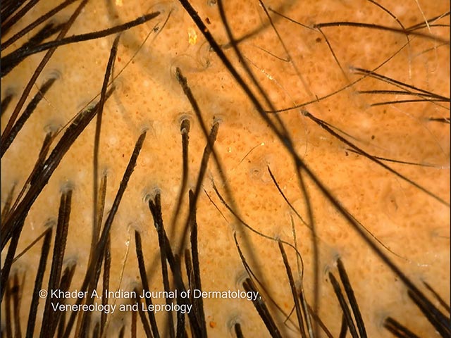
Trichoscopic (scalp dermoscopy) image of a PLACK syndrome patient presents close-up visualization of scalp hair and surrounding epidermis during noninvasive examination. The study field shows a beige-orange scalp background with multiple dark hair shafts projecting over the surface, oriented obliquely and overlying miniature follicles. This pattern is characteristic of alopecia areata in which non-scarring hair loss occurs; dermoscopic features commonly described for alopecia areata include yellow dots representing keratin within follicular infundibula, exclamation-mark hairs indicating fragility, black dots corresponding to follicular ruptures, and short broken or regrowing vellus hairs. In this image, the dense distribution of terminal hair shafts and the relatively intact epidermis suggest an early or patchy alopecia is occurring rather than a scarring process. The presence of many hair shafts crossing at various angles across a sparse follicular opening may reflect ongoing follicular activity with shedding and re-growth; such patterns are useful for longitudinal assessment and therapeutic response. In the clinical context of PLACK syndrome, alopecia areata patterns observed on trichoscopy can aid differential diagnosis from other etiologies such as traction alopecia, tinea capitis, and androgenetic-type hair loss. Overall, this trichoscopic snapshot provides rapid, noninvasive confirmation of alopecia areata involvement in PLACK syndrome. Correlative clinical assessment is recommended.
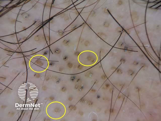
Low-power dermoscopic evaluation of the scalp in alopecia areata reveals diagnostic yellow dots and dark perifollicular dots. In this in vivo dermoscopy image, numerous yellow dots indicate keratin-filled follicular infundibulae, where the hair shafts are absent or completely regressed. Superimposed black dots reflect pigmented, broken hair shafts lodged at follicular openings. The surrounding skin shows relatively preserved texture without extensive scarring or scale, consistent with non-scarring alopecia. Follicular ostia are occasionally dilated and plug-filled by keratinous material, while visible hair shafts within these follicles are lacking, producing the signature AA pattern. The lesions appear as discrete, patchy alopecic areas on the scalp; the appearance is typical for early to mid-stage alopecia areata, though exclamation mark hairs may be absent in some images. Dermoscopy is performed in vivo, using polarized and non-polarized light modes, at approximately 10–20x magnification, enabling visualization of follicular openings and dot patterns. Clinically, these features correlate with autoimmune-mediated hair cycle disruption and a favorable response to intralesional or topical therapies. This image is diagnostic when correlated with history of sudden patchy hair loss and supports monitoring treatment response and disease activity in AA patients. These findings strengthen diagnostic confidence and guide therapeutic strategy for individualized AA management.
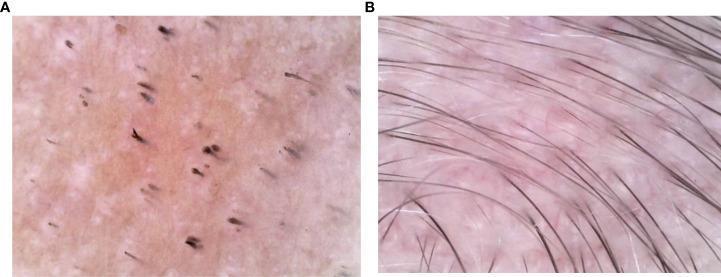
This comparative dermoscopy image displays two clinical states of alopecia areata on the scalp. Image A (baseline) illustrates the characteristic diagnostic signs of active disease: prominent 'yellow dots' representing follicular infundibula filled with sebum and keratinous debris, 'black dots' indicating hairs broken at the skin surface, and several 'exclamation mark hairs' (short hairs with distal fraying and proximal tapering). The surrounding skin surface is pinkish-tan and shows a significant reduction in hair density. Image B (post-treatment) demonstrates clinical resolution after a 6-month therapeutic course. The previously noted pathological markers (yellow and black dots) have disappeared. The follicle density has restored, showing 1-2 healthy, terminal hairs emerging from each follicle. The hairs appear thicker, more uniform in pigment, and display normal growth patterns across the field of view. This comparison serves as an educational tool for identifying trichoscopic features of alopecia areata and evaluating clinical response to microneedling and corticosteroid therapy in pediatric dermatology.
IADVL alopecia areata management guidelines India 2024
Alopecia Areata - Main Points
Note: IADVL's Textbook of Dermatology (4th ed.) is not in the digital library. The content below integrates Fitzpatrick's (9th ed.), Rook's/Dermatology 2-Volume Set 5e, and Andrews', which align closely with IADVL's approach. IADVL-specific clinical pearls are noted where applicable based on published Indian dermatology practice.
Definition
Epidemiology
| Parameter | Data |
|---|---|
| Prevalence | ~0.1-0.2% of population at any time |
| Lifetime risk | ~1.7-2% |
| Sex | Equal in both sexes |
| Age | Any age; most common alopecia in children |
| Family history | 15-25% positive (up to 42% in early-onset disease) |
| AT risk | 5% of AA patients progress to totalis |
| AU risk | 1% progress to universalis |
- Fitzpatrick's, p. 1547 | Rook's 5e, p. 1399
Pathogenesis - The Core Mechanism
1. Loss of Hair Follicle Immune Privilege
- Normal anagen hair bulbs express low MHC Class I and II antigens - "immune privilege"
- AA results from collapse of this immune privilege - upregulation of MHC on follicular epithelium exposes follicular antigens to autoreactive T cells
2. CD8+ NKG2D+ T Cell Attack
- CD8+ cytotoxic T cells and NK cells infiltrate the hair bulb (peri-bulbar "swarm of bees" infiltrate)
- IFN-γ is the dominant driver cytokine - Th1/IFN-γ skewed response
- IL-15 is upregulated in affected follicles → activates IFN-γ-producing CD8+NKG2D+ cells → positive feedback loop → sustained autoimmune attack
- CD4+ CD25- helper T cells promote disease; CD4+ CD25+ Tregs suppress it
- Result: premature catagen transition → hair loss
3. JAK-STAT Pathway
- IFN-γ and IL-15 signaling depend on JAK1/JAK2/JAK3-STAT pathway
- This explains the therapeutic response to JAK inhibitors (the most important mechanistic insight for modern treatment)
4. Follicular Melanocytes as Target
-
Follicular melanocytes appear to substitute as antigen - explains why:
- White hair is spared (lacks melanocytes)
- Regrowing hair is often initially depigmented
- Sudden "whitening" (canities subita) can occur when pigmented hairs selectively fall out
-
Rook's 5e, p. 1399-1400 | Fitzpatrick's, p. 229 | Andrews', p. 864
Genetics
- Polygenic/complex disease with multiple susceptibility loci
- HLA associations: HLA-DQB1*03 (important susceptibility marker); HLA-DR4 in early-onset familial clustering
- ULBP gene cluster (chromosome 6q25) - encodes activating ligands for NKG2D receptor on cytotoxic T cells - strong association
- Shared loci with other autoimmune diseases: CTLA-4, IL-2/IL-21, IL-2RA (explains co-occurrence with thyroid disease, RA)
- PTPN22 (lymphoid protein tyrosine phosphatase - normally suppresses T cell proliferation) and IL-1 cluster (IL-1RA) associated with severe AA
- Rook's 5e, p. 1400
Clinical Features
Morphological Types / Patterns
| Pattern | Description |
|---|---|
| Patchy AA | Single or multiple round/oval well-circumscribed smooth bald patches, 1-5 cm |
| Alopecia Totalis (AT) | Complete loss of all scalp hair |
| Alopecia Universalis (AU) | Complete loss of all body hair including scalp |
| Ophiasis | Band-like loss along temporal and occipital scalp periphery - poor prognosis |
| Sisaipho (Ophiasis inversus) | Spares temporal/occipital; diffuse involvement elsewhere |
| Reticular | Recurrent patches - hair loss in one area while regrowth in another |
| Diffuse AA | Widespread thinning mimicking AGA or telogen effluvium - diagnosis challenge |
| AA incognita | Acute diffuse loss without visible patches; positive pull test; many yellow dots |
Cardinal Signs
- Smooth scalp surface within patches (vs. scarring alopecia)
- Exclamation point hairs - proximal tapering, blunt distal end; at active patch margins - pathognomonic
- Black dots (cadaver hairs) - broken hairs visible at follicular ostia
- Yellow dots - empty follicles filled with keratotic material and sebum
- Sparing of white/grey hairs (early disease)
- Canities subita - apparent sudden whitening when all pigmented hairs fall out simultaneously
Nail Changes (in ~10%, more in severe/extensive disease)
- Nail pitting (most common) - geometric, grid-like pattern
- Trachyonychia - sandpaper-like roughness, longitudinal ridging
- Brittle nails, onycholysis, koilonychia, onychomadesis, red lunulae
Trichoscopy (Dermoscopy) - Key Feature
| Trichoscopic Feature | Meaning | Activity |
|---|---|---|
| Yellow dots | Empty follicles + keratotic/sebaceous plugs | Active/chronic |
| Black dots | Broken hair shafts at ostia | Active |
| Exclamation point hairs | Proximal thinning due to arrested mitosis | Active (moderate) |
| Tapered hairs | Very long exclamation point hairs | Active (moderate) |
| Pigtail/circle hairs | Regrowing hairs curling | Regrowth |
| Upright regrowing hairs | Short stubble emerging | Regrowth |
Histopathology
Acute/Early/Progressive Phase:
- Normal total hair count
- Peribulbar mononuclear infiltrate - predominantly CD4+ and CD8+ T cells + NK cells around anagen/catagen bulbs - "swarm of bees"
- Occasional eosinophils, mast cells
- Degenerative changes in hair matrix
- Increased catagen/telogen hairs
- Trichomalacia, pigment casts
Chronic/Longstanding Phase:
- Miniaturization of most follicles - "nanogen hairs" (arrested, rapidly cycling miniaturized hairs)
- Mild peribulbar infiltrate around nanogen bulbs
- Follicular dropout possible in very long-standing disease (>10 years)
Key histology pearl: AA should be considered whenever high percentages of telogen or miniaturized hairs are found even without peribulbar inflammation (as the infiltrate may be absent in chronic disease). - Fitzpatrick's, p. 1548
- Rook's 5e, p. 1400-1401
Associated Conditions / Comorbidities
| Association | Odds Ratio / Note |
|---|---|
| Thyroid disease (Hashimoto's) | OR 4.4 |
| SLE | OR 3.5 |
| Atopic dermatitis | OR 2.4 |
| Vitiligo | Common co-occurrence |
| Down syndrome | Increased incidence |
| APECED syndrome | AA in 1/3 of patients |
| Vitamin D-dependent rickets type II | Familial association |
| Depression and anxiety | Psychological comorbidity |
| Metabolic syndrome | Reported |
Differential Diagnosis
| Condition | Differentiating Feature |
|---|---|
| Tinea capitis | Broken hairs, scaling, fluorescence, KOH positive |
| Trichotillomania | Irregular shape, varying hair lengths, hook/coiled hairs on trichoscopy, no inflammation |
| Androgenetic alopecia | Gradual onset, androgenic pattern, no exclamation point hairs |
| Telogen effluvium | Diffuse, pull test positive but no yellow/black dots |
| Secondary syphilis | "Moth-eaten" alopecia; plasma cells on biopsy |
| Temporal triangular alopecia | Congenital; triangular patch; vellus hairs on trichoscopy |
| Early scarring alopecia | Follicular ostia lost; inflammation; biopsy diagnostic |
| Loose anagen syndrome | Hair pulls easily without taper |
| Alopecia neoplastica | Metastatic deposits; scalp induration |
Prognosis
- Single small patch
- Short disease duration
- Postpubertal onset
- No nail changes
- No family history
- Ophiasis pattern
- Onset in childhood
- Extensive involvement (>50% scalp)
- Nail changes present
- Duration >5 years (may be irreversible >10 years)
- Atopic dermatitis comorbidity
- Involvement of occiput/hairline
-
~60% partial regrowth by 1 year
-
~25% have only a single episode
-
~40% relapse within first year
-
Fitzpatrick's, p. 1548 | Andrews', p. 869
Treatment
Treatment Algorithm by Extent
Limited disease (< 50% scalp / SALT ≤ 20)
    ↓
Intralesional triamcinolone ± topical steroids
    ↓
No response → topical immunotherapy (DPCP/SADBE)

Extensive disease (> 50% scalp / SALT > 20)
    ↓
Systemic: Oral JAK inhibitors (first choice now)
          OR pulse corticosteroids
          OR cyclosporine / methotrexate
1. Intralesional Corticosteroids (IL-CS) - First line for patchy AA
- Triamcinolone acetonide 2.5-10 mg/mL (Rook's recommends 2.5-5 mg/mL)
- 0.1 mL per injection site, 1 cm apart, into mid-deep dermis
- Maximum dose per session: 10-20 mg (Rook's 5e)
- Repeat every 3-4 weeks
- Risk: skin atrophy with high concentrations; retinal artery embolization with high pressure injection (Andrews')
2. Topical Corticosteroids
- Class I (ultrapotent) corticosteroids - first-line topical
- Less reliable than intralesional; suitable as first-line in children
- Safer than injections
3. Oral/Pulse Corticosteroids (for rapidly progressive or widespread disease)
| Regimen | Protocol |
|---|---|
| Daily prednisolone | 0.4-0.6 mg/kg/day, taper over ≥12 weeks |
| Pulse dexamethasone | 0.1 mg/kg twice weekly |
| Prednisolone pulse | 200 mg weekly (adults); 5 mg/kg monthly |
| Methylprednisolone | 500 mg/day × 3 days monthly (Kaposi-type pulse) |
- Ophiasis does not respond to pulse steroids
- Predictors of response to pulse steroids: duration <6 months, onset <10 years, multifocal disease
4. JAK Inhibitors - Current Standard for Severe AA
| Drug | Type | Status | Notes |
|---|---|---|---|
| Baricitinib | Oral JAK1/2 inhibitor | FDA-approved for severe AA in adults | First approved |
| Ritlecitinib | Oral JAK3/TEC inhibitor | FDA-approved for AA + extensive hair loss ≥12 years | Approved for adolescents too |
| Tofacitinib | Oral JAK1/3 inhibitor | Off-label | Good evidence |
| Ruxolitinib (oral) | Oral JAK1/2 inhibitor | Off-label | |
| Topical ruxolitinib 1.5% | Topical | Less effective than oral | |
| Topical tofacitinib 2% | Topical | Less effective than oral |
Maintenance therapy is necessary to avoid relapse after stopping JAK inhibitors. - Rook's 5e, p. 1402
Low-dose oral minoxidil can be added as supplemental therapy alongside any primary treatment. - Rook's 5e
5. Contact Immunotherapy (for refractory / widespread / children with severe disease)
- Diphencyprone (DCP/DPCP) - most used in practice
- Squaric acid dibutyl ester (SADBE)
- Mechanism: induces contact hypersensitivity → shifts T cell response away from follicle
- Note: avoid concurrent systemic immunosuppressants as they blunt the desired contact reaction
6. Steroid-Sparing Systemic Agents
| Drug | Dose | Notes |
|---|---|---|
| Cyclosporine | 3-5 mg/kg/day | Can combine with PUVA |
| Methotrexate | 15-20 mg/week (adults); 0.4 mg/kg/week (children) | |
| Mycophenolate mofetil | - | Limited data |
| Azathioprine | - | Limited data |
| Sulfasalazine | Up to 1.5 g three times daily |
7. Other Therapies
- PUVA (topical or oral) - for refractory/widespread lesions
- Topical anthralin 1% (short contact 15-20 min, then shampooed off)
- Topical minoxidil - as adjunct
- Excimer laser (308 nm) - 300-2300 mJ/cm² per session; 11-12 sessions over 9-11 weeks
- Bimatoprost / latanoprost - for eyelash/eyebrow disease (prostaglandin analogues); note periocular pigmentation risk
- Dupilumab - successful when AA occurs with comorbid atopic dermatitis
Treatment for Children
- Topical Class I steroids first
- Contact immunotherapy (DPCP) before systemic options
- Pulse corticosteroids: prednisolone 5 mg/kg (up to 300 mg) monthly
- JAK inhibitors in children: off-label; ritlecitinib approved ≥12 years; max baricitinib 0.625 mg (<20 kg) to 1.25 mg (20-40 kg)
Scoring Tools
- SALT score (Severity of Alopecia Tool): Most widely used; SALT >20 = indication for systemic therapy
- AAPS (Alopecia Areata Predictive Score): Based on trichoscopy; estimates regrowth probability
Psychological Aspect (IADVL emphasis)
- Disease process and natural course
- High rate of spontaneous remission
- Cosmetic alternatives (wigs)
- Support organizations (National Alopecia Areata Foundation)
Summary of Key Points (High-Yield)
| Feature | Key Point |
|---|---|
| Type | Non-scarring, autoimmune |
| Target | Anagen hair bulb (NOT bulge/stem cells) |
| Key cell | CD8+NKG2D+ cytotoxic T cells |
| Key cytokine | IFN-γ (Th1 driven) |
| Pathway | JAK-STAT (drives IFN-γ/IL-15 signaling) |
| Hallmark sign | Exclamation point hairs |
| Trichoscopy triad | Yellow dots + Black dots + Exclamation point hairs |
| Histology | Peribulbar "swarm of bees" infiltrate |
| White hair spared? | Yes (melanocytes are the target) |
| Nail change | Nail pitting (grid-like) most common |
| Best prognosis | Single patch, short duration, postpubertal |
| Worst prognosis | Ophiasis, childhood onset, atopy, nails involved |
| FDA-approved treatments | Baricitinib (adults), Ritlecitinib (≥12 yrs) |
| First-line limited disease | Intralesional triamcinolone |
| First-line severe disease | Oral JAK inhibitors |
| Contact allergens used | DPCP, SADBE, DNCB |
- Fitzpatrick's Dermatology 9th ed., Ch. 13 (p. 229) and Ch. 87 (pp. 1546-1550)
- Rook's Dermatology 5e (Dermatology 2-Volume Set), Ch. 69 (pp. 1399-1403)
- Andrews' Diseases of the Skin, Ch. 33 (pp. 864-870)
- IADVL approach: consistent with above; pulse steroids and DPCP are pillars of Indian practice (IJDVL)