
GOLD STANDARD DIAGNOSTIC TEST FOR DENDUE
gold standard diagnostic test dengue fever NS1 RT-PCR serology
Gold Standard Diagnostic Tests for Dengue (DENV)
The Key Principle: Phase-Dependent Testing
1. Viral Isolation - Historical Gold Standard
-
Requires specimens from the first 6 days of illness
-
Takes >1 week for results
-
Is only available in specialized/reference laboratories
-
Is not practical for routine clinical use
-
Brenner and Rector's The Kidney and Park's Textbook of Preventive and Social Medicine
2. RT-PCR (Reverse Transcriptase Polymerase Chain Reaction) - Current Practical Gold Standard
-
Detects dengue viral RNA from day 0 to day 7-10 of illness
-
Higher specificity than virus isolation
-
Can identify and differentiate all 4 serotypes (DENV-1 to DENV-4)
-
Much faster turnaround than viral culture
-
Used on serum, plasma, urine, or blood samples
-
Cited as gold standard reference method against which all other tests (NS1, IgM ELISA) are evaluated
-
Park's Textbook of Preventive and Social Medicine; Tietz Textbook of Laboratory Medicine, 7th Ed.
3. NS1 Antigen Detection - Best Early Clinical Test
-
Detectable from day 1 up to day 9-14 of illness (sometimes day 18)
-
Detected in both primary and secondary infections
-
Available as ELISA or rapid immunochromatographic kits
-
Does not differentiate between serotypes
-
NS1 ELISA showed sensitivity ~96.8% using RT-PCR as reference
-
Park's PSM; Red Book 2021 (AAP); ROSEN's Emergency Medicine
4. Serological Tests (MAC-ELISA / IgM & IgG)
| Test | Window | Details |
|---|---|---|
| IgM capture ELISA (MAC-ELISA) | Day 3-5 onwards; 99% positive by day 10 | Standard for late/convalescent phase |
| IgG ELISA | Persists for life | Useful for secondary infections; can cross-react with other flaviviruses |
| Haemagglutination Inhibition (HI) Assay | Convalescent | Classically called "gold standard" for serology; 4-fold rise confirms infection |
| Complement Fixation (CF) / Neutralization Test | Convalescent | Less commonly used now |
- Brenner and Rector's The Kidney; Red Book 2021
- IgM can cross-react with Zika virus and other flaviviruses
- A 4-fold or greater rise in IgG between acute (≤5 days) and convalescent (>15 days) specimens confirms recent infection
5. WHO-Recommended Optimal Approach
Combine NS1 antigen + IgM ELISA on a single serum specimen taken in the first 10 days - this identifies ≥90% of both primary and secondary dengue cases.
Summary Table
| Phase | Days of Illness | Gold Standard Test |
|---|---|---|
| Early febrile | Days 1-5 | RT-PCR (gold standard) + NS1 Ag detection |
| Late febrile / early defervescence | Days 5-10 | NS1 + IgM ELISA |
| Convalescent | >Day 10 | IgM ELISA (MAC-ELISA); 4-fold IgG rise (HI assay) |
| Confirmation / reference standard | Any time | Viral isolation (historical); RT-PCR (practical) |
For Exam Purposes
- Overall gold standard: Virus isolation (classical) / RT-PCR (practical, modern)
- Gold standard for serology: Haemagglutination Inhibition (HI) Assay
- Best early rapid test: NS1 antigen detection (ELISA or RDT)
- Best late test: IgM capture ELISA (MAC-ELISA)
- Best combined approach: NS1 + IgM ELISA on a single specimen (days 1-10)
WHAT IS IgM ELISA
IgM ELISA MAC-ELISA principle dengue diagnosis explained
https://www.cdc.gov/dengue/hcp/diagnosis-testing/serologic-t…
ELISA principle sandwich IgM antibody detection diagram
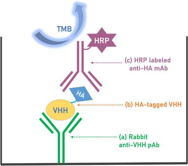
This medical laboratory diagram illustrates a sandwich Enzyme-Linked Immunosorbent Assay (ELISA) technique used for the detection of VHH (variable heavy-chain antibodies). The schematic depicts the spatial arrangement of the assay components within a microtiter plate well from bottom to top: (a) A capture layer consisting of Rabbit anti-VHH polyclonal antibodies (pAb), which are passively adsorbed to the bottom of the well. (b) The analyte, represented as an HA-tagged VHH molecule, where the VHH core binds to the capture antibody and an HA (hemagglutinin) tag is available for detection. (c) The detection layer featuring an HRP-labeled anti-HA monoclonal antibody (mAb). This antibody targets the HA tag of the analyte and is conjugated to Horseradish Peroxidase (HRP), shown as a purple star-shaped molecule. At the top, a blue curved arrow labeled TMB (3,3′,5,5′-Tetramethylbenzidine) represents the chromogenic substrate added to the well. The diagram demonstrates the biochemical principle of signal transduction where HRP catalyzes the oxidation of TMB to produce a measurable colorimetric signal, indicating the presence and concentration of the VHH analyte.
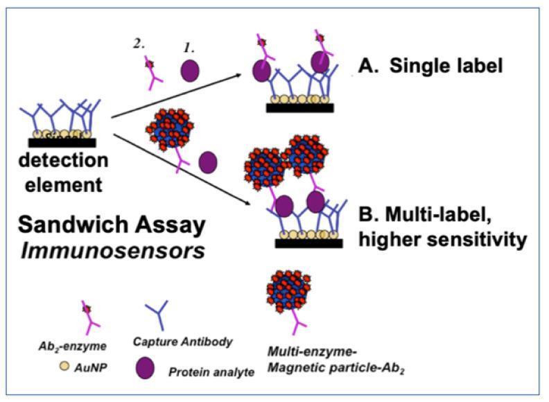
This pathophysiology diagram illustrates the principles of ELISA-like sandwich immunosensors for protein detection. The schematic compares two diagnostic methods: (A) Single label and (B) Multi-label, higher sensitivity. Both assays utilize a solid-phase 'detection element' (black base) containing gold nanoparticles (AuNP) and immobilized Y-shaped 'Capture Antibodies' (blue). In method A, a single target 'Protein analyte' (purple circle) is sandwiched between the capture antibody and a 'Detection Antibody' (Ab2) conjugated to a single enzyme label. In method B, the detection antibody is part of a 'Multi-enzyme-magnetic particle' complex, featuring a large spherical carrier coated with numerous enzymatic labels (red dots). This configuration facilitates signal amplification for increased sensitivity. The diagram highlights key biochemical components including antibody-antigen binding dynamics and labeling strategies for optical or electrochemical clinical assays used in biomarker detection, such as cancer diagnostics. Labels identify key elements: Ab2-enzyme, AuNP, Capture Antibody, Protein analyte, and Multi-enzyme-Magnetic particle-Ab2.
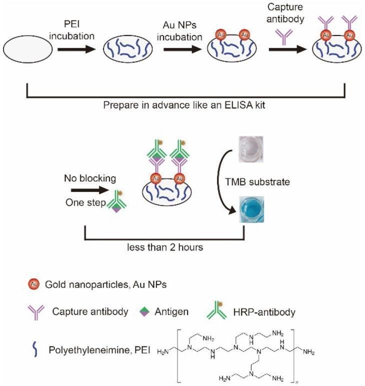
This schematic diagram illustrates a specialized, blocking-free 'sandwich' Enzyme-Linked Immunosorbent Assay (ELISA) utilizing a polyethyleneimine (PEI) and gold nanoparticle (Au NP) coated microwell plate. The process is divided into two phases: preparation and detection. In the preparation phase, a microwell undergoes PEI incubation followed by Au NP incubation to form a functionalized base layer; capture antibodies are then immobilized onto the Au NPs. In the detection phase, labeled as a 'No blocking - One step' procedure, the antigen and horseradish peroxidase (HRP)-conjugated antibody are introduced. The final step shows the addition of TMB substrate, which undergoes a visual transition from a colorless liquid to a blue-colored solution, indicating successful antigen detection. The diagram highlights that this modified protocol simplifies traditional ELISA by omitting the 2-hour blocking step, reducing total assay time to less than 2 hours. Chemical structures for PEI and icons for antigens, Au NPs, and various antibodies are included to clarify the molecular binding sequence and surface modifications.
IgM ELISA (Enzyme-Linked Immunosorbent Assay)
What is ELISA?
| Word | Meaning |
|---|---|
| Enzyme-Linked | An enzyme is attached to an antibody as a label/reporter |
| Immunosorbent | Antibodies or antigens are "adsorbed" (bound) onto a solid surface (microplate) |
| Assay | A test or measurement |
Basic Principle of ELISA
- Coat the plate - A capture antibody or antigen is fixed to the wells of a microtiter plate
- Add the patient sample - The target antibody (IgM) or antigen from the patient's serum binds to the coated surface
- Add detector antibody - A secondary antibody conjugated to an enzyme (usually Horseradish Peroxidase / HRP) is added and binds to the captured molecule
- Add substrate (TMB) - A chromogenic substrate (Tetramethylbenzidine / TMB) is added; the HRP enzyme converts it, producing a color change (blue → yellow)
- Read result - The intensity of color is measured by a spectrophotometer. More color = more IgM = positive result
IgM ELISA Specifically
MAC-ELISA (IgM Antibody Capture ELISA)
How MAC-ELISA Works (Step by Step):
| Step | What Happens |
|---|---|
| 1. Capture | Microplate wells are coated with anti-human IgM antibody (captures all IgM from the patient sample) |
| 2. Patient sample added | Diluted patient serum is added - all IgM (including dengue-specific IgM) is captured onto the plate |
| 3. Dengue antigen added | Dengue viral antigen is added - it binds ONLY to dengue-specific IgM captured on the plate |
| 4. Enzyme-conjugated antibody | An anti-dengue antibody linked to HRP enzyme is added - it binds the dengue antigen |
| 5. Substrate added | TMB substrate is added - HRP converts it producing a yellow color |
| 6. Read result | Absorbance measured at 450 nm; positive = color change above cutoff |
Why capture IgM first?
IgM ELISA in Dengue Diagnosis
| Feature | Details |
|---|---|
| When does IgM appear? | 3-5 days after onset of fever |
| When does it peak? | ~2 weeks after onset |
| When does it decline? | Falls to undetectable over 2-3 months |
| 99% patients IgM positive by... | Day 10 of illness |
| Sensitivity (days 5-10) | ~47-80% (varies by study) |
| Best combined strategy | NS1 + IgM MAC-ELISA on a single specimen (days 1-10) - detects ≥90% of cases |
- Red Book 2021 (AAP); Park's Textbook of Preventive and Social Medicine
Primary vs. Secondary Dengue Infection
| Primary Infection | Secondary Infection | |
|---|---|---|
| IgM | High levels, appears day 3-5 | Lower levels, may be undetectable |
| IgG | Appears later (day 14+), low titers | Rapidly rises to very high titers |
| IgM/IgG ratio | High (>1.2) | Low (<1.2) |
| Clinical significance | Milder disease usually | Higher risk of dengue hemorrhagic fever (DHF) |
Limitations of IgM ELISA
- Not useful in first 3-5 days of illness (IgM not yet detectable)
- Cross-reactivity with other flaviviruses - Zika, West Nile, Japanese Encephalitis, Yellow Fever (can give false positives)
- Lower sensitivity in secondary infections
- Positive IgM results should ideally be confirmed by PRNT (Plaque Reduction Neutralization Test) - the gold standard confirmatory serology test
- Cannot differentiate between dengue serotypes (DENV-1 to 4)
Summary: Where IgM ELISA Fits in Dengue Diagnosis
Day 0-1:  Fever onset
Day 1-5:  RT-PCR + NS1 Ag  ← Use these (IgM negative)
Day 3-5:  IgM begins to appear
Day 5-10: NS1 + IgM ELISA  ← Best window for combined testing
Day 10+:  IgM ELISA alone (NS1 may be declining)
Months:   IgG only (IgM gone, IgG persists for life)
- Park's Textbook of Preventive and Social Medicine (Table 2)
- CDC Dengue Serologic Tests
GOLD STANDARD DIAGNOSTIC TEST FOR ANRMIA WHICH OCCURS DURING DENGUE INFECTIONS
gold standard diagnostic test anemia dengue infection hemolysis bone marrow
Anemia in Dengue Infection - Types & Gold Standard Diagnostics
Why This Question Needs Context First
| Mechanism | Type of Anemia | Primary Cause |
|---|---|---|
| Bone marrow suppression | Hypoproliferative/aplastic-like | Direct viral invasion of bone marrow precursors |
| Hemolysis | Hemolytic anemia | Autoimmune (polyclonal antibodies vs RBC antigens) + complement activation |
| Hemorrhage (DHF/DSS) | Acute blood loss anemia | Plasma leakage, thrombocytopenia, mucosal bleeding |
| Hemodilution | Dilutional anemia | Plasma leakage causes hemoconcentration (opposite effect = raised Hct) |
Type 1: Hemolytic Anemia in Dengue
Gold Standard Investigation: Direct Antiglobulin Test (DAT) / Direct Coombs Test
- Detects antibodies or complement (C3) coating the surface of the patient's RBCs
- A positive DAT confirms autoimmune hemolytic anemia (AIHA) is occurring
- In dengue, DAT may be positive due to the transient polyclonal autoantibodies formed
"The direct antiglobulin test (DAT) or Coombs test is useful in revealing cells coated with antibody or complement and can aid in diagnosis. A positive DAT alone, however, does not define AIHA and must be supported by clinical context and evidence of hemolysis on laboratory evaluation."
- ROSEN's Emergency Medicine
Supporting Tests for Hemolysis (All Required Together):
| Test | What It Shows | Finding in Hemolysis |
|---|---|---|
| Peripheral blood smear | Morphology of RBCs | Spherocytes, schistocytes, fragmented RBCs |
| Reticulocyte count | Bone marrow compensatory response | Elevated (reticulocytosis) = RBCs being destroyed, marrow trying to compensate |
| LDH (Lactate Dehydrogenase) | Released from destroyed RBCs | Elevated |
| Haptoglobin | Binds free hemoglobin | Decreased (consumed by free Hb) |
| Indirect bilirubin | Breakdown product of heme | Elevated |
| Urine hemosiderin / hemoglobinuria | Intravascular hemolysis marker | Present in severe intravascular hemolysis |
- Goldman-Cecil Medicine; ROSEN's Emergency Medicine
Type 2: Bone Marrow Suppression Anemia
Gold Standard: Bone Marrow Aspiration and Biopsy
- Reveals hypocellular marrow with suppressed erythroid precursors
- Confirms hemophagocytosis (macrophages engulfing blood cells) which can occur in severe dengue
- Also confirms reactive changes including dyserythropoiesis
- This is the definitive test when bone marrow suppression is suspected
Type 3: Acute Blood Loss Anemia (Dengue Hemorrhagic Fever / DSS)
Gold Standard: Complete Blood Count (CBC) + Hematocrit
Standard haematological parameters such as platelet count and haematocrit are important and are part of the diagnosis of dengue infection and should be closely monitored.
- In dengue, paradoxically, rising hematocrit (hemoconcentration ≥20% above baseline) signals plasma leakage (not true anemia)
- Falling Hb/Hct = actual blood loss anemia in hemorrhagic dengue
Practical Diagnostic Algorithm for Anemia in Dengue
Step 1: CBC + Reticulocyte count + Peripheral Blood Smear
        ↓
Step 2: LDH + Haptoglobin + Indirect Bilirubin
        ↓
        Evidence of hemolysis?
        YES → Direct Coombs Test (DAT)  ← GOLD STANDARD for immune hemolysis
        NO  → Check bone marrow / blood loss
        ↓
Step 3 (if bone marrow suppression suspected):
        Bone Marrow Aspiration + Biopsy  ← GOLD STANDARD for BM suppression
Summary: Gold Standard by Anemia Type
| Type of Anemia in Dengue | Gold Standard Test |
|---|---|
| Immune hemolytic anemia (most common) | Direct Antiglobulin Test (DAT / Direct Coombs) |
| Bone marrow suppression | Bone Marrow Aspiration and Biopsy |
| Blood loss anemia (DHF/DSS) | CBC + Hematocrit (with clinical context) |
| Overall initial screening | Complete Blood Count (CBC) + Peripheral Blood Smear |
For Exam Purposes
-
If asked simply "gold standard for anemia in dengue" in a short answer context: Peripheral blood smear + Direct Coombs Test (DAT) for hemolytic anemia (the most clinically relevant type)
-
If the question emphasizes bone marrow involvement: Bone marrow aspiration/biopsy
-
The CBC with reticulocyte count and peripheral smear is always the essential first step in evaluating any anemia
-
ROSEN's Emergency Medicine, p. 2447; Park's Textbook of Preventive and Social Medicine, p. 294; Goldman-Cecil Medicine