
Hey
Hey! How can I help you today?
Tell me about mycosis fungoides
Reading File
Searching sources...
Finding Sources
Searching PubMed
"mycosis fungoides"[MeSH Terms]
Good results. Now let me read the most comprehensive sections in parallel.
Reading File
Reading File
The Andrews' textbook has very rich content. Let me read more of it, including the histology, variants, and treatment sections.
Reading File
Excellent - I have very rich content. Let me fetch the key clinical images and read the treatment/histology section.
Loading Image
Reading File
I now have very comprehensive content. Let me compile the full answer.
Mycosis Fungoides (MF)
What Is It?
Mycosis fungoides is the most common primary cutaneous T-cell lymphoma (CTCL). It is a malignant neoplasm of T-lymphocytes - almost always a memory CD4+ T-helper (Th) cell that homes to the skin. It is twice as common in men as in women, affects all races (Black individuals in the US are relatively more often affected), and has an incidence of approximately 1 in 300,000/year, which has been increasing.
Clinical Stages
MF classically progresses through three clinical stages:
1. Patch Stage
- Flat, macular, or slightly elevated patches, usually ≥5 cm
- Favor the "bathing suit area" - lower abdomen, buttocks, upper thighs, breasts in women
- Resemble eczema; pruritus (sometimes severe) is common
- Variants at this stage include poikiloderma vasculare atrophicans, large-plaque parapsoriasis, and "invisible MF" (histologically proven but visually subtle)
- Biopsy may not be diagnostic early on
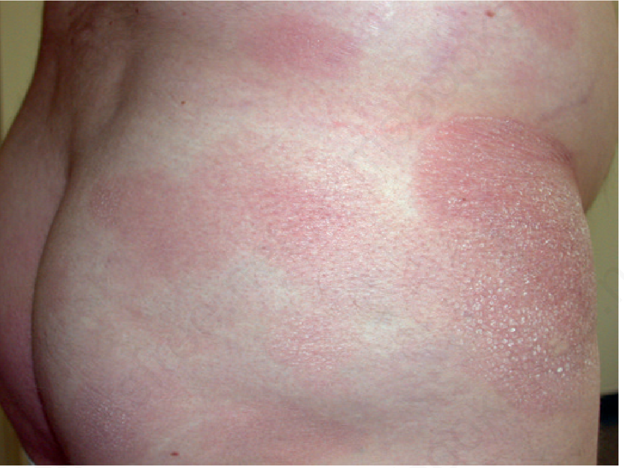
2. Plaque Stage
- Lesions become more infiltrated, may resemble psoriasis or granulomatous dermatitis
- Plaques can reach up to 30 cm
- Palms and soles may be involved with hyperkeratotic, fissuring plaques
- Diagnosis is usually confirmed at this stage with biopsy
3. Tumor Stage
- Large nodules arise on infiltrated plaques
- Ulceration may occur
- Lymph node enlargement typically develops (nontender, firm, freely movable)
- A rare d'emblée form presents with tumors from the outset with no prior patches/plaques
Some patients progress to erythroderma (generalized skin involvement). Advanced disease may involve lymph nodes, peripheral blood, and visceral organs - most patients die of septicemia.
Variants
- Folliculotropic MF - tumor cells invade hair follicles; can resemble lichen nitidus or lichen spinulosus
- Syringotropic MF - involves sweat glands
- Hypopigmented MF - predominates in dark-skinned children
- Granulomatous MF - poorer prognosis and poorer response to skin-directed therapy
- CD8+ MF - rare suppressor cell variant; can behave indolently or aggressively
- Pagetoid reticulosis (Woringer-Kolopp) - solitary lesion, excellent prognosis
- Vesicular/bullous, verrucous/hyperkeratotic, and pigmented purpura-like forms also exist
Histopathology
- Neoplastic T cells infiltrate the epidermis and upper dermis
- Epidermotropism - lymphocytes migrating into the epidermis is the hallmark
- Pautrier microabscesses - collections of atypical lymphocytes within the epidermis (highly characteristic but not always present)
- Tumor cells have cerebriform nuclei - hyperconvoluted, deeply indented nuclear membranes
- Dermal infiltrate is band-like in the upper dermis in patch stage
- CD4+, CD45RO+ (memory T-cell) immunophenotype in most cases
Pathogenesis
- MF cells express cutaneous lymphocyte antigen (CLA), which binds E-selectin on inflamed skin endothelium, enabling skin homing
- CCR4 (a chemokine receptor) is also expressed, with its ligand on basal keratinocytes
- Early MF: Th1-dominant environment with CD8+ tumor-infiltrating lymphocytes (TILs) that may suppress the malignant clone. Patients with >20% CD8+ cells survive longer
- Advanced MF/Sézary syndrome: shift to Th2 environment (via IL-4, IL-10), which downregulates suppressor cell function and allows clonal expansion
- Common chromosomal changes: gain of 7q36 and 7q21-22, loss of 5q13 and 9p21 (distinct from Sézary syndrome)
- Circulating malignant cells can be identified by flow cytometry as CD4+/CD7- or CD4+/CD26-
Staging (TNMB System)
| Stage | Skin (T) | Nodes (N) | Viscera (M) |
|---|---|---|---|
| IA | <10% BSA (T1) | N0 | M0 |
| IB | >10% BSA (T2) | N0 | M0 |
| IIA | T1-T2 | N1 (palpable, not MF) | M0 |
| IIB | Tumors (T3) | N0-N1 | M0 |
| IIIA | Erythroderma (T4) | N0 | M0 |
| IIIB | T4 | N1 | M0 |
| IVA | T1-T4 | N2-N3 | M0 |
| IVB | T1-T4 | Any N | M1 (visceral) |
Blood involvement (B) is separately noted but does not alter formal staging.
Prognosis
- Stage IA: Life expectancy identical to general population; only 8-9% progress; only 2% die of disease
- Stage IB (T2): Median survival 11.7-15.6 years; 24% progress
- T3 (tumor stage): Median survival 3.2-8.4 years
- T4 (erythroderma): Median survival 1.8-3.7 years
- Palpable adenopathy: median survival 7.7 years vs. 21.8 years without
- Key negative prognostic factors: lymphadenopathy, tumors, cutaneous ulceration (any one reduces survival; all three together = median 1-year survival)
- Worse prognosis in non-Caucasian patients with early-onset MF, especially African American women
- Robbins Pathology notes median survival of ~10 years; transformation to aggressive T-cell lymphoma occasionally occurs as a terminal event
Relationship to Sézary Syndrome
Both MF and Sézary syndrome are CD4+ Th-cell tumors homing to skin via CLA and CCR4. Key differences:
- MF: progressive patch → plaque → tumor stages; extracutaneous disease only late
- Sézary syndrome: presents with generalized erythroderma + leukemia of cerebriform "Sézary cells" + generalized lymphadenopathy; skin rarely forms tumors; worse prognosis (minority survive 5 years)
Treatment (Skin-Directed)
Early-stage (patch/plaque) disease is managed with skin-directed therapies:
- Topical corticosteroids (especially stage IA)
- Topical nitrogen mustard (mechlorethamine)
- Topical carmustine (BCNU)
- Phototherapy: Narrowband UVB (patch stage), PUVA (psoralen + UVA) for deeper plaques
- Total skin electron beam therapy (TSEBT)
- Topical retinoids (bexarotene gel)
Advanced/systemic disease:
- Systemic retinoids (bexarotene), interferon-alpha, extracorporeal photopheresis
- Targeted therapies: romidepsin, vorinostat (HDAC inhibitors), mogamulizumab (anti-CCR4)
- Systemic chemotherapy is reserved for refractory/advanced disease; early aggressive chemotherapy is not indicated given excessive morbidity and lack of curative benefit
Sources: Andrews' Diseases of the Skin, 13th ed.; Robbins, Cotran & Kumar Pathologic Basis of Disease; Goldman-Cecil Medicine
This is a shared conversation. Sign in to Orris to start your own chat.