
Generate a short answer on this question for MBBS biochemistry exam based on Vasudevan with simple cycle(easily understandable) with simple diagram
Which biochemistry topic/question do you want a short answer for?
maple syrup urine disease
Simple biochemistry diagram for MBBS exam showing Maple Syrup Urine Disease (MSUD) pathway. Show: Leucine, Isoleucine, Valine (branched-chain amino acids) at the top with arrows going down through Transamination step to produce branched-chain alpha-keto acids (alpha-ketoisocaproic acid, alpha-keto-beta-methylvaleric acid, alpha-ketoisovaleric acid). Then a large red X block at the enzyme "Branched-Chain Alpha-Keto Acid Dehydrogenase (BCKD) - requires TPP, FAD, NAD+, CoA, Lipoic acid" to show enzyme deficiency. Below the block show accumulation with upward arrows. Label the consequences: accumulation in blood and urine, maple syrup odor in urine. Show downstream products (blocked): Acetyl-CoA, Succinyl-CoA. Clean white background, bold text, color-coded arrows, simple exam-style diagram.
Maple Syrup Urine Disease (MSUD)
Definition
Pathway & Diagram
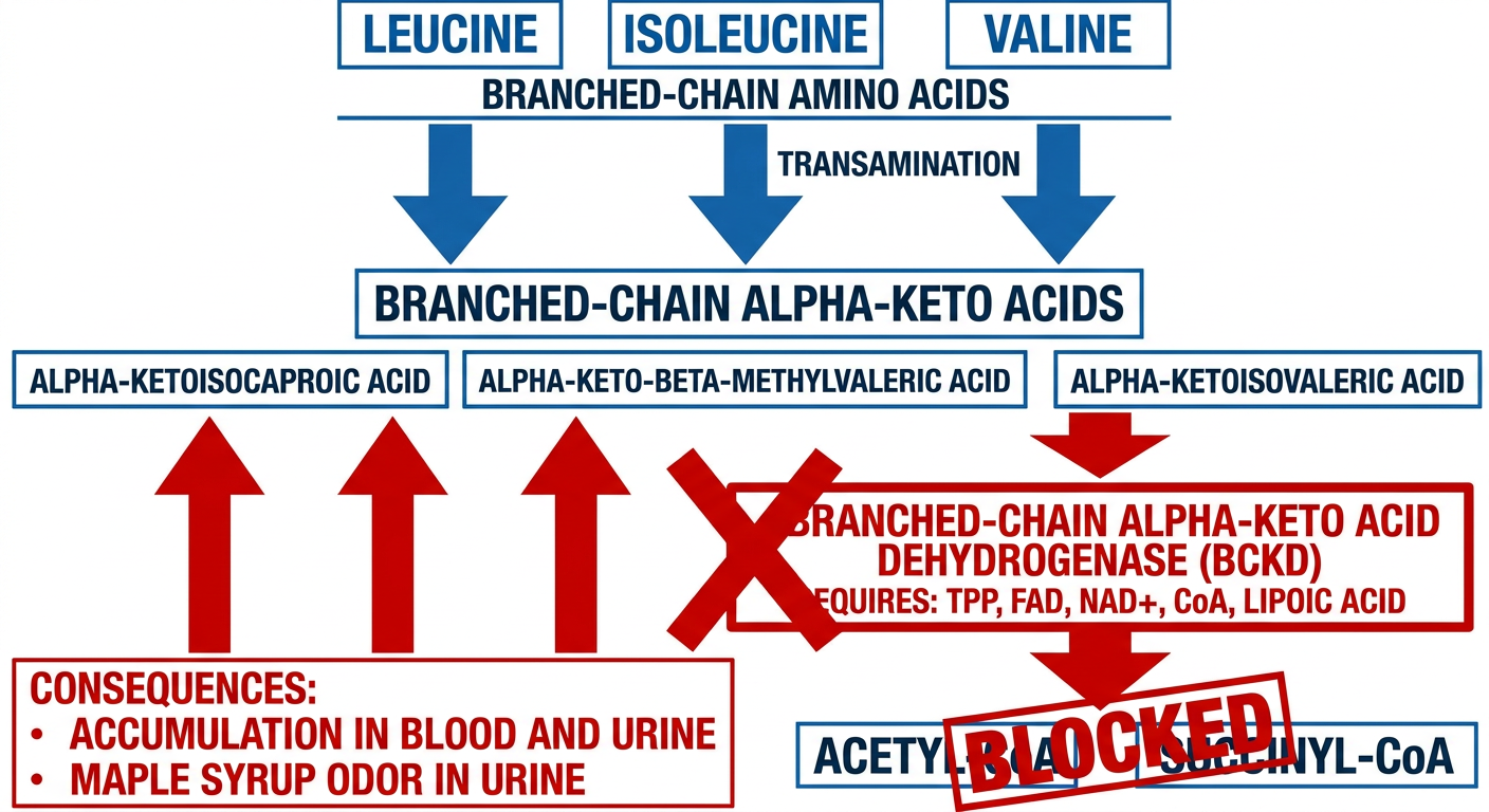
Simple Cycle (Step by Step):
Leucine, Isoleucine, Valine  (Branched-Chain Amino Acids - BCAAs)
          |
          ↓  (Transamination - by BCAA transaminase)
          |
Branched-Chain α-Keto Acids
  • α-Ketoisocaproic acid       (from Leucine)
  • α-Keto-β-methylvaleric acid (from Isoleucine)
  • α-Ketoisovaleric acid       (from Valine)
          |
          ↓  ← ❌ BLOCK HERE ❌
          |
  BCKD Complex (Deficient)
  [Requires: TPP, FAD, NAD+, Lipoic acid, CoA]
          |
          ↓  (Normally produces)
          |
  Acetyl-CoA / Succinyl-CoA → TCA Cycle (BLOCKED in MSUD)
Enzyme Deficient
- Cofactors required: TPP, FAD, NAD+, Lipoic acid, CoA-SH
Key Features (Clinical Points)
| Feature | Detail |
|---|---|
| Inheritance | Autosomal Recessive |
| Enzyme defect | BCKD complex |
| Amino acids accumulated | Leucine, Isoleucine, Valine |
| Characteristic sign | Maple syrup/burnt sugar odor of urine (due to isoleucine) |
| Onset | First few days of life (neonatal) |
| Main toxic amino acid | Leucine (causes neurological damage) |
Clinical Features
- Feeding difficulty, vomiting
- Ketoacidosis (high anion gap)
- Neurological deterioration: seizures, hypotonia, coma
- Characteristic maple syrup smell in urine
- Fatal if untreated in neonates
Diagnosis
- Newborn screening (blood spot test)
- Elevated plasma BCAAs (Leucine, Isoleucine, Valine)
- Elevated urinary branched-chain keto acids
- Urine dinitrophenylhydrazine (DNPH) test - positive
Treatment
- Dietary restriction of BCAAs (specially formulated BCAA-free formula)
- Supplement small, controlled amounts of Leucine, Isoleucine, Valine (for growth)
- Thiamine (B1) supplements - helps in thiamine-responsive variant (some MSUD patients)
- Monitor leucine levels closely (leucine is the neurotoxic agent)
Mnemonic
"LIV in Maple Trees"
- Leucine, Isoleucine, Valine accumulate
- BCKD is the blocked enzyme
- Maple syrup odor in urine
Distribution of Water & Electrolytes
Simple MBBS biochemistry exam diagram showing Distribution of Body Water and Electrolytes. Top section: Human body outline showing Total Body Water = 60% of body weight (42L in 70kg man) Split into two main compartments below: LEFT BOX (blue): Intracellular Fluid (ICF) = 40% body weight = 28L (2/3 of TBW). Major electrolytes inside: K+ (156 mmol/L) HIGH, Mg2+, HPO4 2-, Proteins HIGH. Na+ LOW, Cl- LOW. RIGHT BOX (yellow/orange): Extracellular Fluid (ECF) = 20% body weight = 14L (1/3 of TBW). Divided into two sub-boxes: - Interstitial Fluid: 10.5L (15% body weight) - Plasma: 3.5L (5% body weight) Major electrolytes: Na+ (140 mmol/L) HIGH, Cl- HIGH, HCO3- HIGH. K+ LOW. Separating membranes labeled: "Cell Membrane" between ICF and ECF. "Capillary Wall" between Plasma and Interstitial fluid. Bottom: Mnemonic box: "ICF = K+, Mg2+, Phosphate | ECF = Na+, Cl-, HCO3-" Clean white background, bold labels, color-coded compartments, simple arrows, exam-ready style.
Distribution of Body Water & Electrolytes
Total Body Water (TBW)
- TBW = 60% of body weight (in an average 70 kg adult male = 42 litres)
- In females: ~50% (due to higher fat content)
- In infants: ~75% (higher water content)
- Fat is nearly water-free, so obese individuals have lower % TBW
Diagram - Compartmental Distribution
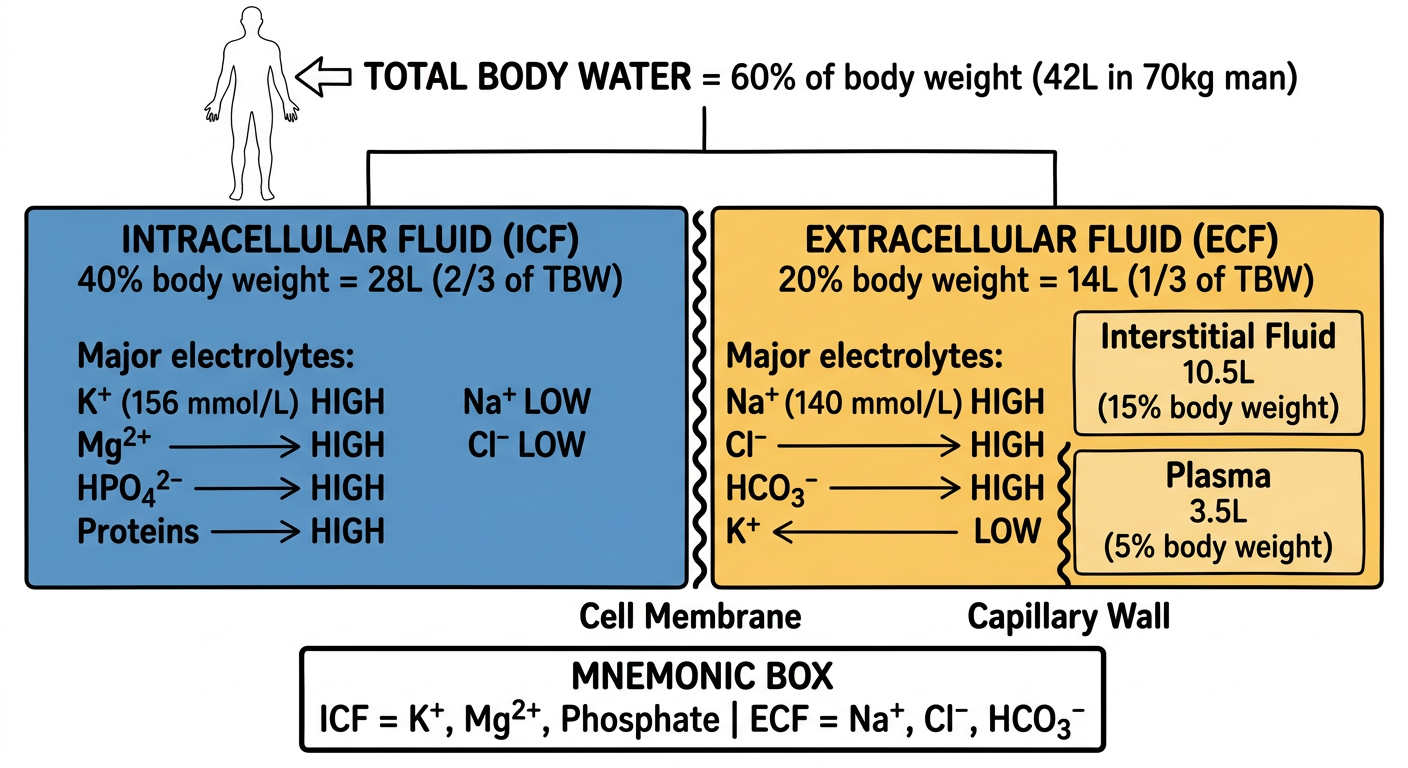
Simple Compartment Layout
TOTAL BODY WATER (TBW) = 42 L (60% body weight)
              │
    ┌─────────┴──────────┐
    │                    │
  ICF (28L)           ECF (14L)
 40% BW - 2/3 TBW    20% BW - 1/3 TBW
                          │
              ┌───────────┴────────────┐
              │                        │
    Interstitial Fluid (10.5L)    Plasma (3.5L)
        ~15% BW                    ~5% BW
    (separated by                (separated from ISF
     cell membrane)               by capillary wall)
- 60% = Total Body Water
- 40% = Intracellular Fluid
- 20% = Extracellular Fluid
Electrolyte Distribution (Key Table)
| Electrolyte | Plasma | Interstitial Fluid | ICF |
|---|---|---|---|
| Na⁺ | 140 mmol/L ↑↑ | 145 mmol/L ↑↑ | 12 mmol/L ↓ |
| K⁺ | 4 mmol/L ↓ | 4 mmol/L ↓ | 156 mmol/L ↑↑ |
| Ca²⁺ | 2.4 | 2-3 | 0.3 ↓ |
| Mg²⁺ | 1 | 0.5-1 | 13 ↑ |
| Cl⁻ | 103 ↑↑ | 114 ↑↑ | 4 ↓ |
| HCO₃⁻ | 27 ↑ | 31 ↑ | 12 |
| HPO₄²⁻ | 1 ↓ | - | High ↑↑ |
| Protein⁻ | 16 ↑ | Trace | 55 ↑↑ |
Key Features of Each Compartment
Intracellular Fluid (ICF)
- Major cation: K⁺ (Potassium) - 156 mmol/L
- Major anion: HPO₄²⁻ (Phosphate) + Proteins
- Also high: Mg²⁺, SO₄²⁻
- Low Na⁺, Low Cl⁻, Low Ca²⁺
- Separated from ECF by cell membrane (Na⁺/K⁺ ATPase maintains gradient)
Extracellular Fluid (ECF)
- Major cation: Na⁺ (Sodium) - 140 mmol/L
- Major anions: Cl⁻ and HCO₃⁻
- Low K⁺, Low Mg²⁺, Low Phosphate
- Plasma has extra proteins (albumin, globulin) vs. interstitial fluid
Separation Barriers
| Barrier | Separates | Permeable to |
|---|---|---|
| Cell membrane | ICF from ECF | Water freely; NOT to most electrolytes |
| Capillary wall | Plasma from Interstitial fluid | Almost all solutes; NOT to proteins |
The Donnan effect: Plasma proteins (negatively charged) hold extra Na⁺/K⁺ in plasma, so cation concentration in plasma is slightly higher (~2%) than interstitial fluid.
Mnemonic
"K⁺ inside, Na⁺ outside"
- Kitchen is Inside (K⁺ = ICF)
- Nation is Outside (Na⁺ = ECF)
Water Balance (Daily)
| Input | Output |
|---|---|
| Oral intake: ~1500 mL | Urine: 1200-1500 mL |
| Food water: ~700 mL | Insensible loss (skin + lungs): 400-700 mL |
| Metabolic water: ~300 mL | Stool: ~100-200 mL |
| Total: ~2500 mL | Total: ~2500 mL |
Regulation of Gene Expression Principle of Electrophoresis Northern Blotting Chromatography — Principle & Types Products derived from Glycine Salient Features of Genetic Code Quality Control
Simple MBBS biochemistry exam diagram: Regulation of Gene Expression showing Prokaryote (Lac Operon) vs Eukaryote comparison. Left side: Prokaryote - show Operator, Promoter, Structural genes (lacZ, lacY, lacA) with Repressor protein binding operator (negative control) and Inducer (allolactose) removing repressor. Label: "Operon model". Right side: Eukaryote - show DNA wrapped on Histones, Chromatin remodeling by Acetylation, Transcription Factors binding Enhancer and TATA box, RNA Polymerase II assembling. Label levels: DNA level, Transcription level, Post-transcriptional level, Translation level. Clean white background, color-coded, bold exam-style labels, simple arrows.
Simple MBBS biochemistry exam diagram for Electrophoresis principle and Northern Blotting. TOP HALF - Electrophoresis: Show a gel slab with wells at top (negative electrode - cathode) and bottom (positive electrode - anode). Show DNA/RNA fragments migrating from negative to positive pole. Smaller fragments migrate farther (bottom), larger fragments stay near top. Label: "Separation based on size and charge", "Smaller = faster", "Migration in electric field". Show agarose gel with band pattern. BOTTOM HALF - Northern Blotting steps in numbered sequence: 1. RNA isolation from cells 2. Gel electrophoresis (RNA separation by size) 3. Transfer to nitrocellulose/nylon membrane (blotting) 4. Hybridization with labeled DNA/RNA probe 5. Autoradiography - bands detected Label: "Northern Blot = detects mRNA size and quantity" Clean white background, numbered steps, bold text, simple arrows, exam-ready.
Simple MBBS biochemistry exam diagram: Products derived from Glycine. Central box labeled GLYCINE. Show arrows going outward to all products: 1. PORPHYRINS (Heme synthesis) - arrow to hemoglobin icon 2. PURINES (AMP, GMP) - arrow to DNA/RNA icon 3. CREATINE → Creatinine - arrow to muscle icon 4. GLUTATHIONE (with Cys, Glu) - arrow to antioxidant label 5. BILE SALTS (glycocholic acid) - arrow to liver icon 6. HIPPURIC ACID (with benzoic acid) - arrow to kidney/urine 7. SERINE (via hydroxymethyl transfer) - arrow 8. ONE CARBON POOL (via THF) - arrow 9. GLYOXYLATE → Oxalate - arrow Also label: Glycine is simplest amino acid, non-essential, glucogenic. Clean white background, color-coded arrows, bold text, exam-style radial diagram.
Simple MBBS biochemistry exam diagram showing Chromatography Principle and Types. TOP: Principle box - "Separation of molecules based on differential distribution between stationary phase and mobile phase". Show two phases labeled. MAIN DIAGRAM - Tree/classification: CHROMATOGRAPHY ├── Paper Chromatography - show paper strip, solvent front, Rf value formula (distance of spot / distance of solvent) ├── Column Chromatography │ ├── Ion Exchange (separates by charge - cation/anion exchange resin) │ ├── Gel Filtration/Size Exclusion (separates by size - small molecules enter pores, large elute first) │ └── Affinity Chromatography (separates by specific binding - ligand attached to matrix) ├── HPLC (High Performance Liquid Chromatography - high pressure, very precise) └── Gas Chromatography (volatile compounds, gas mobile phase) Bottom box: Rf value = Distance moved by solute / Distance moved by solvent. Used in paper & TLC. Clean white background, bold text, color-coded boxes, exam-ready style.
429 Too Many Requests. {'message': '{\n "error": {\n "code": 429,\n "message": "Resource exhausted. Please try again later. Please refer to https://cloud.google.com/vertex-ai/generative-ai/docs/error-code-429 for more details.",\n "status": "RESOURCE_EXHAUSTED"\n }\n}\n', 'status': 'Too Many Requests'}
We encountered an error while processing your request. Please try again or reach out to us for help.