
Hopi for 3 years female having fever for 2 months and abdo.inal pain for 2 months
systemic juvenile idiopathic arthritis salmon pink rash child fever
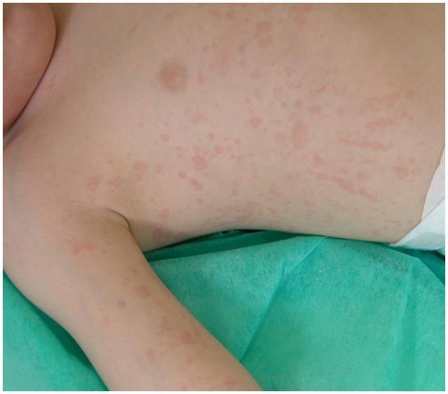
This clinical photograph displays a widespread cutaneous manifestation on a pediatric patient's torso and upper extremity. The presentation is characterized as a salmon-macular rash, featuring numerous discrete and confluent salmon-pink erythematous lesions. The lesions are primarily macular, appearing flat and flush with the skin surface, with irregular margins and varying sizes. Some areas exhibit linear or streaky patterns among the scattered spots. This specific visual finding is a hallmark clinical feature of systemic juvenile idiopathic arthritis (sJIA), often presenting during fever spikes. The image demonstrates the classic morphology and distinctive hue essential for differentiating sJIA from other pediatric exanthems or inflammatory conditions. The distribution includes the chest, abdomen, and the inner aspect of the arm, highlighting the diffuse and migratory nature of the rash in rheumatological contexts.
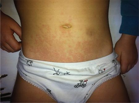
Clinical photograph of a pediatric patient's torso demonstrating the classic cutaneous manifestations of systemic juvenile idiopathic arthritis (sJIA). The image displays a salmon-pink colored, evanescent, maculopapular rash distributed across the abdomen and extending toward the proximal extremities. The individual lesions consist of small, circumscribed, pink macules and slightly elevated papules that appear non-pruritic and lack significant scaling. The arrangement is scattered and confluent in areas, creating a mottled or reticular pattern that fades gradually into the surrounding healthy skin. This specific dermatological finding is a hallmark diagnostic feature of sJIA, typically occurring during daily febrile spikes. The clinical relevance of this visual is to aid in the differential diagnosis of pediatric inflammatory conditions, distinguishing the characteristic sJIA rash from other childhood exanthems based on its specific color, distribution, and association with systemic symptoms.
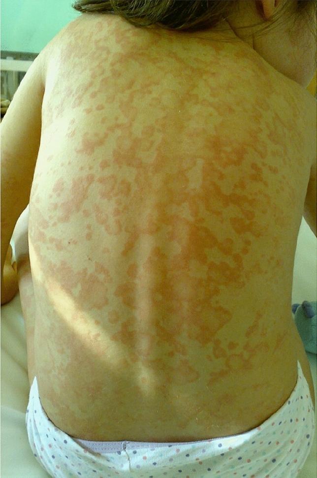
This clinical photograph shows the back of a pediatric patient exhibiting a widespread, salmon-pink erythematous rash. The lesions are distributed extensively across the upper and lower back and shoulders. Morphologically, the rash is composed of discrete macules and slightly raised papules that frequently coalesce into larger, irregularly shaped, nonfixed patches. The borders of the lesions are indistinct and poorly demarcated, blending into the surrounding fair skin. There is a notable absence of scaling, crusting, vesiculation, or blistering. Clinically, this type of evanescent rash is a hallmark cutaneous manifestation of systemic juvenile idiopathic arthritis (sJIA), typically characterized by its transient nature, often appearing during peaks of spiking fever and subsiding as the temperature normalizes. The image serves as a key educational example for diagnosing autoinflammatory disorders through cutaneous physical examination.
History of Presenting Illness (HOPI) - Structured Analysis
Characterizing the Fever (Key History Points to Elicit)
| Feature | What to Ask | Clinical Significance |
|---|---|---|
| Pattern | Continuous / intermittent / spiking? | Spiking (quotidian) once or twice daily suggests sJIA; remittent suggests TB/infection |
| Height | Measured temperature? | >39°C daily spikes classic for sJIA |
| Response to antipyretics | Settles with paracetamol? | Broad, not discriminatory |
| Associated chills/rigors | Present? | Suggests malaria, bacteremia |
| Night sweats | Present? | TB, lymphoma |
| Child's appearance during fever | Well vs. unwell | In sJIA, child appears ill during fever but recovers when afebrile |
Characterizing the Abdominal Pain
| Feature | What to Ask |
|---|---|
| Location | Diffuse / RIF / epigastric / periumbilical |
| Character | Dull ache / colicky / constant |
| Associated symptoms | Vomiting, diarrhea, constipation, distension, jaundice |
| Relation to fever | Worsens during fever spikes (sJIA serositis) |
| Weight loss | Present? Suggests TB, malignancy, VL |
Differential Diagnosis - Ranked by Likelihood
1. Systemic Juvenile Idiopathic Arthritis (sJIA) - TOP DIFFERENTIAL
- Quotidian spiking fevers - peak ≥39°C once or twice daily, child appears ill during fever but recovers when afebrile
- Evanescent salmon-pink rash in 80% - migratory, macular, worsens with fever, can be elicited by Koebner phenomenon
- Hepatosplenomegaly + lymphadenopathy
- Serositis - pericarditis, pleural effusion, abdominal pain/peritonitis
- Arthritis may be absent for weeks to months at onset (arthralgias/myalgias nearly universal)
- Markedly elevated inflammatory markers (ESR, CRP, ferritin)
- Life-threatening complication: Macrophage Activation Syndrome (MAS)
2. Visceral Leishmaniasis (Kala-Azar) - HIGH PRIORITY IN ENDEMIC REGION
- Caused by Leishmania donovani complex; endemic in Indian subcontinent, East Africa, Brazil
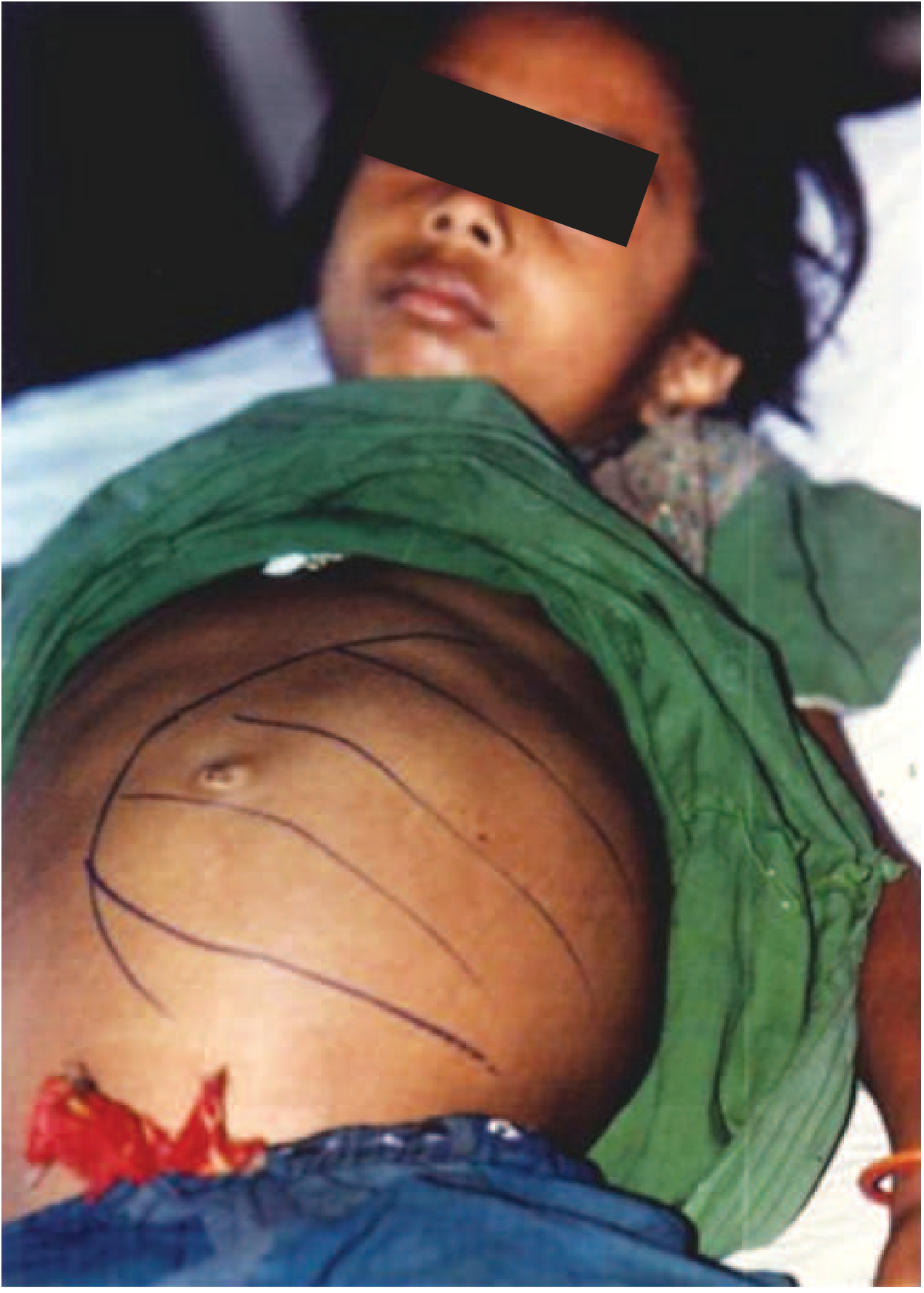
- Massive splenomegaly is the most important feature - producing abdominal distension and pain
- Prolonged fever (weeks to months) - the hallmark
- Pancytopenia, weight loss, weakness, anorexia
- Hyperpigmentation of skin in some patients (hence "black fever")
- Differential diagnosis: easily mistaken for malaria, typhoid, tuberculosis, brucellosis
- "Fever with neutropenia or pancytopenia in patients from an endemic region strongly suggests VL; hypergammaglobulinemia in long-standing illness strengthens the diagnosis"
3. Abdominal Tuberculosis
- Chronic abdominal pain with peritonitis and intermittent partial intestinal obstruction (particularly M. bovis)
- Gastrointestinal TB can mimic inflammatory bowel disease
- Fever, night sweats, weight loss/poor weight gain are characteristic
- Associated pulmonary TB in most cases - check CXR
- Important in children in endemic regions
4. Lymphoma (Abdominal)
- Prolonged fever + abdominal pain + abdominal mass/lymphadenopathy
- Burkitt lymphoma common in children in endemic malaria regions; presents with abdominal mass
- B symptoms: fever, night sweats, weight loss
- Must be excluded with imaging and LDH
5. Other Differentials
| Condition | Clues |
|---|---|
| Malaria | Episodic fever, travel/endemic area, hepatosplenomegaly |
| Typhoid fever | Stepladder fever, rose spots, relative bradycardia, diarrhea/constipation |
| Brucellosis | Animal exposure (unpasteurized milk, livestock), undulant fever |
| Systemic lupus (SLE) | Rare at 3 years, but serositis + fever possible; more common in older girls |
| Inflammatory bowel disease | Chronic abdominal pain, diarrhea, weight loss |
| Intra-abdominal abscess | Localized pain, signs of sepsis |
Recommended Workup
First-Line Investigations
- CBC with differential - Pancytopenia (VL), leukocytosis + anemia (sJIA, infection), lymphocytosis (TB)
- ESR, CRP - Markedly elevated in sJIA, TB, lymphoma
- Serum ferritin - Very high (often >500 ng/mL, can be >10,000) in sJIA; also elevated in MAS
- LFTs - Hepatitis in VL, sJIA, TB
- Blood culture - Typhoid, bacteremia
- Peripheral blood smear - Malaria parasites
- Mantoux/TST or IGRA - TB screening
- Chest X-ray - Hilar adenopathy (TB), cardiomegaly (pericarditis in sJIA)
Second-Line / Targeted
- Abdominal ultrasound - Splenomegaly (VL), lymphadenopathy, ascites, mass
- rK39 rapid antigen test - Point-of-care for VL (high sensitivity in Indian subcontinent)
- Bone marrow aspirate - Gold standard for VL; also rules out leukemia/lymphoma
- ANA, anti-dsDNA - If SLE suspected
- Serum LDH, uric acid - Lymphoma workup
- Echocardiogram - If pericarditis suspected (sJIA)
Key Red Flags Requiring Urgent Action
- Rapidly falling platelets + rising ferritin → MAS (complication of sJIA, life-threatening)
- Pancytopenia + splenomegaly in endemic region → VL until proven otherwise
- Abdominal mass → Lymphoma/Wilms tumor must be excluded urgently
In paragraph