
Treatment of osteoporosis in postmenopausal women
"osteoporosis" AND "postmenopausal women" AND "treatment"
osteoporosis bone density treatment postmenopausal
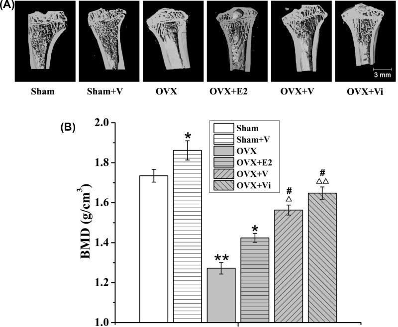
This composite image evaluates the impact of Low-Magnitude Vibration (LMV) and estrogen (E2) on bone mineral density (BMD) in a rat model of postmenopausal osteoporosis. Panel (A) presents micro-CT sectional views of the left proximal tibiae across six experimental groups: Sham, Sham+V (vibration), OVX (ovariectomized), OVX+E2, OVX+V, and OVX+Vi (immediate vibration). Visually, the OVX group shows significant trabecular bone loss and reduced density compared to Sham controls. The OVX+V and OVX+Vi groups demonstrate partial restoration of trabecular architecture, appearing superior to the OVX+E2 group. Panel (B) provides a corresponding bar graph measuring BMD in g/cm³. Statistical significance is noted relative to the Sham group (*, **), the OVX group (Δ, ΔΔ), and the OVX+E2 group (#). The data indicates that while ovariectomy significantly reduces BMD, LMV—particularly when initiated early (Vi)—effectively mitigates bone loss, showing higher efficacy in preserving bone mass and structural integrity than traditional estrogen treatment in this preclinical model.
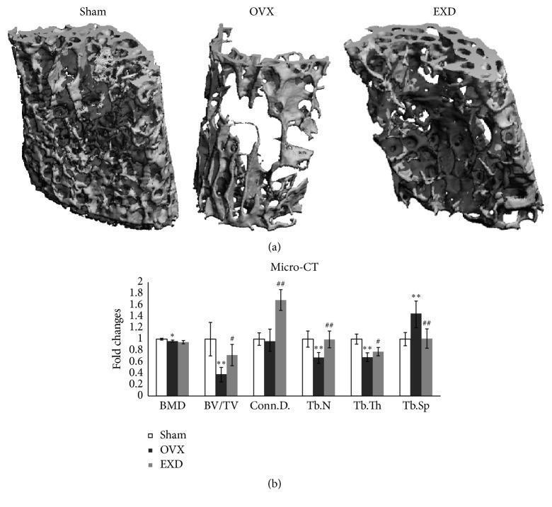
This medical figure presents micro-computed tomography (μCT) analysis of bone density and architecture in an osteoporotic mouse model. Section (a) displays three-dimensional reconstructed images of the L4 vertebra across three experimental groups: Sham (control), OVX (ovariectomized), and EXD (treated). The Sham group illustrates normal, dense trabecular connectivity. The OVX group demonstrates severe bone loss characterized by sparse, fragmented trabeculae and increased void spaces, typical of postmenopausal osteoporosis pathophysiology. The EXD group shows a significant recovery of bone volume and structural integrity compared to the OVX group. Section (b) provides a bar graph with morphometric data quantified as fold changes. Key parameters evaluated include Bone Mineral Density (BMD), Bone Volume/Total Volume (BV/TV), Connectivity Density (Conn.D.), Trabecular Number (Tb.N), Trabecular Thickness (Tb.Th), and Trabecular Separation (Tb.Sp). The data confirms that ovariectomy induces significant architectural deterioration, while EXD treatment provides a protective effect, specifically improving connectivity and trabecular density, highlighting its potential therapeutic role in bone resorption prevention.
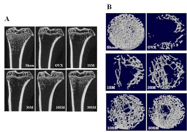
This diagnostic image set displays micro-computed tomography (μ-CT) analysis of rat medial-proximal tibiae, used as a preclinical model for postmenopausal osteoporosis. Panel A presents coronal cross-sections across six experimental groups: Sham control, Ovariectomized (OVX), and four dose-dependent treatment groups (1SM, 3SM, 10SM, 30SM). The Sham group shows robust trabecular bone density and thick cortical bone. The OVX group demonstrates significant osteopenia with a marked loss of trabecular architecture and cortical thinning. Treatment groups show a dose-dependent increase in trabecular volume, with the 30SM group appearing most similar to the Sham control. Panel B provides corresponding 3D reconstructions of the trabecular bone network. The images illustrate variations in trabecular connectivity and microarchitecture, highlighting the transition from a dense, interconnected honeycomb lattice in the Sham group to a fragmented, sparse structure in the OVX and low-dose groups, followed by gradual restoration of bone volume fraction (BV/TV) and connectivity density in the high-dose SM treatment groups. This visual demonstrates the efficacy of a therapeutic agent in preventing bone loss in a hormone-deficient model.
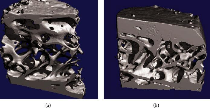
This diagnostic image displays 3D micro-computed tomography (micro-CT) reconstructions of transiliac bone biopsies from postmenopausal women, illustrating the microstructural effects of osteoporosis treatment. The side-by-side comparison focuses on the trabecular and cortical bone architecture. Panel (a) shows the bone microstructure after 36 months of strontium ranelate therapy, characterized by a more open trabecular network with visible connectivity and thickness of individual trabeculae. Panel (b) represents the placebo group, demonstrating a contrasting bone density and architecture. The educational focus is on identifying changes in bone mineral density (BMD) and the preservation of trabecular microarchitecture, which are critical markers in assessing the efficacy of anti-osteoporotic pharmacological interventions. Key visible features include the porosity of the cancellous bone, the thickness of the cortical shell, and the overall structural integrity of the bone matrix. This material is highly relevant for medical education in rheumatology, endocrinology, and orthopedics, specifically regarding the diagnosis and management of postmenopausal osteoporosis.
Treatment of Osteoporosis in Postmenopausal Women
1. Diagnosis and Indications for Treatment
- Prior hip or vertebral fragility fracture
- BMD T-score ≤ -2.5 at spine or proximal femur
- BMD T-score between -1.0 and -2.5 (osteopenia) plus either:
- 10-year risk of major osteoporotic fracture ≥ 20%, or
- 10-year risk of hip fracture ≥ 3% (calculated via FRAX tool)
Box: Indications for Therapy - Rheumatology, 2-Volume Set (2022, Elsevier), p. 1837
2. Non-Pharmacologic Measures (Foundation of All Treatment)
| Measure | Details |
|---|---|
| Calcium | 1000-1200 mg/day total (diet + supplement); combined Ca+Vit D reduces fracture risk |
| Vitamin D | 800-2000 IU/day; target serum 25-OHD > 30 ng/mL (ensure before starting pharmacotherapy) |
| Exercise | Weight-bearing aerobic + resistance training; balance training (e.g., Tai Chi) reduces falls |
| Fall prevention | Multidisciplinary assessment; Timed Up-and-Go test; remove home hazards |
| Lifestyle | Smoking cessation; limit alcohol (>3 units/day increases fracture risk); adequate BMI |
Rheumatology, 2-Volume Set (Elsevier 2022), p. 1837-1838
3. Evaluate for Secondary Causes
Endocrine: hyperparathyroidism, hyperthyroidism, Cushing's syndrome, hypogonadism, diabetes
GI/Nutritional: celiac disease, malabsorption, inflammatory bowel disease
Medications: glucocorticoids, aromatase inhibitors, GnRH agonists, PPIs, anticonvulsants, SSRIs, heparin
Other: chronic kidney disease, RA, multiple myeloma, organ transplant
4. Pharmacotherapy
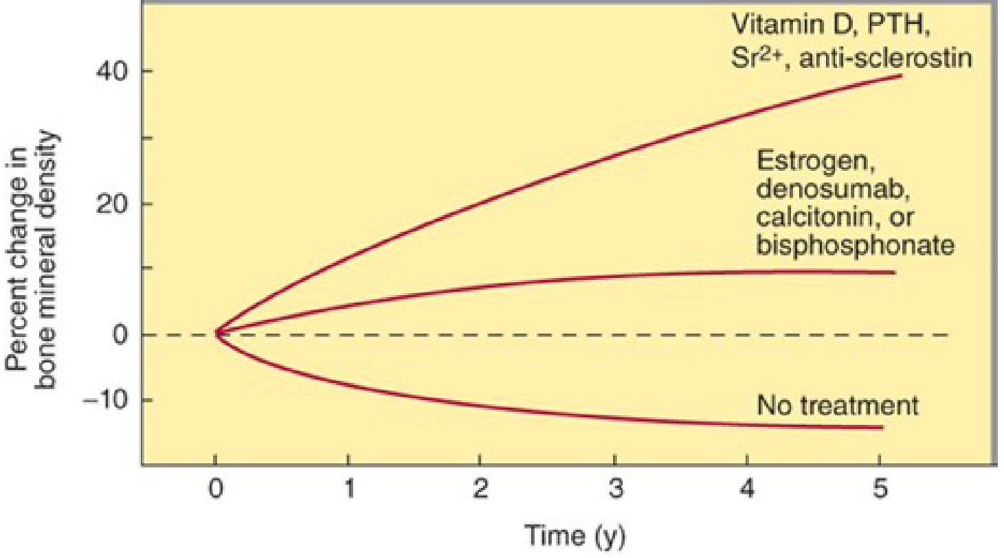
A. Bisphosphonates (First-Line Antiresorptives)
| Drug | Oral dose | IV dose |
|---|---|---|
| Alendronate | 10 mg/day or 70 mg/week | - |
| Risedronate | 5 mg/day, 35 mg/week, or 150 mg/month | - |
| Ibandronate | 2.5 mg/day or 150 mg/month | 3 mg IV every 3 months |
| Zoledronic acid | - | 5 mg IV once yearly |
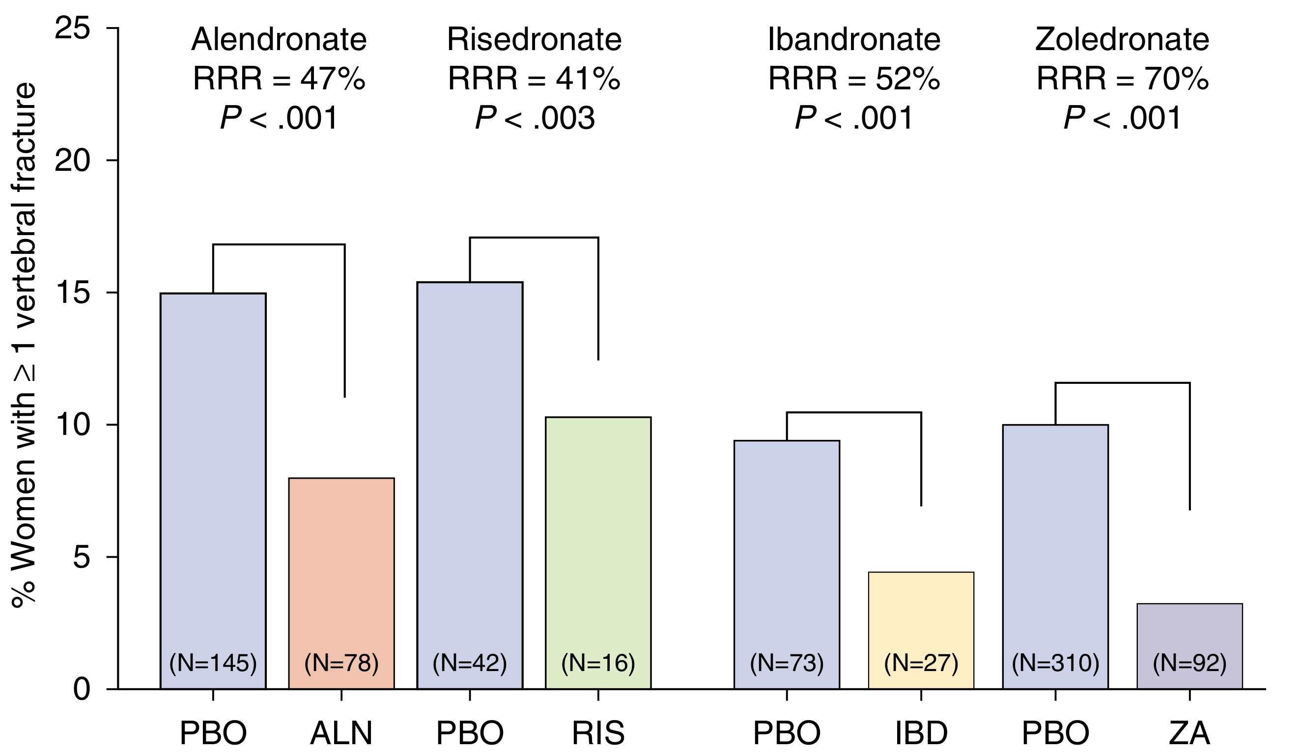
Firestein & Kelley's Textbook of Rheumatology, p. 1379
- Upper GI effects: nausea, esophagitis, esophageal ulceration (oral forms) - take with full glass of water, remain upright ≥30 min
- Osteonecrosis of the jaw (ONJ): rare, mostly in cancer patients on high-dose IV bisphosphonates
- Atypical femoral fractures (subtrochanteric): rare, associated with >5 years of use
- Acute phase reaction (fever, myalgia): especially with first IV zoledronate infusion
- Renal toxicity: IV formulations require eGFR monitoring (avoid if eGFR <30-35 mL/min)
B. Denosumab (Antiresorptive, Biologic)
- Vertebral fractures reduced by 68% (p < 0.001)
- Hip fractures reduced by 40% (p = 0.04)
- Nonvertebral fractures reduced by 20% (p = 0.01)
- At 10 years of continuous therapy: lumbar spine BMD +21.6%, total hip BMD +9.1% - significantly greater than bisphosphonates
Rheumatology, 2-Volume Set (Elsevier), p. 1843-1844
C. Raloxifene (Selective Estrogen Receptor Modulator - SERM)
D. Hormone Therapy (Estrogen ± Progestin)
Katzung's Basic and Clinical Pharmacology, 16th Edition, p. 1226
E. Teriparatide and Abaloparatide (PTH/PTHrP Analogs - Anabolics)
- Mechanism: Intermittent PTH stimulates osteoblast differentiation and decreases osteoblast apoptosis → net bone formation (when given as daily injection, in contrast to continuous PTH elevation in hyperparathyroidism which causes resorption)
- Dose: 20 mcg subcutaneous daily
- Efficacy: Reduces vertebral fractures by ~65% and nonvertebral fractures by ~53%
- Duration: Maximum 24 months lifetime (concern over osteosarcoma risk from rodent studies; no osteosarcoma signal seen in humans)
- Contraindications: Prior radiation therapy to bone, Paget's disease, hypercalcemia, bone metastases, pediatric patients with open epiphyses
- Must be followed by an antiresorptive (bisphosphonate or denosumab) to consolidate BMD gains
- Similar mechanism and efficacy to teriparatide
- Lower rate of hypercalcemia (3.4% vs 6.4% with teriparatide)
- More vasoactive: more palpitations, dizziness, nausea
- 80 mcg subcutaneous daily
F. Romosozumab (Sclerostin Antibody - Dual Mechanism)
- FRAME trial (n=7,180 postmenopausal women): Romosozumab for 12 months followed by denosumab for 12 months
- Reduced new vertebral fractures by 73% (0.5% vs 1.8%, p < 0.001)
- Clinical fractures reduced by 36% at 12 months (p = 0.008)
- ARCH trial: Romosozumab vs alendronate in high-risk women - romosozumab superior at fracture reduction
Rheumatology, 2-Volume Set (Elsevier), p. 1845-1846
5. Sequential (Treat-to-Target) Strategy
Severe osteoporosis / very high risk
    ↓
Start with anabolic agent (teriparatide, abaloparatide, or romosozumab)
    ↓
Transition to antiresorptive (bisphosphonate or denosumab) to consolidate gains

Moderate-high risk / typical postmenopausal osteoporosis
    ↓
Start with bisphosphonate (alendronate or zoledronic acid preferred)
    ↓
Drug holiday after 5 years (oral) / 3 years (IV) if low-moderate ongoing risk
    ↓
Resume if fracture, significant BMD loss, or high ongoing risk
- Bisphosphonate after teriparatide/abaloparatide/romosozumab - consolidates BMD
- Bisphosphonate after denosumab - prevents rebound fractures
- Switching from denosumab to teriparatide causes transient bone loss (avoid this sequence)
- Do NOT use teriparatide and bisphosphonate concurrently (bisphosphonate blunts the anabolic effect)
Firestein & Kelley's Textbook of Rheumatology, p. 1379
6. Summary Table: Drug Classes at a Glance
| Drug | Class | Route | Key Benefit | Key Risk |
|---|---|---|---|---|
| Alendronate | Bisphosphonate | PO weekly | First-line; hip + vertebral fractures | GI intolerance, ONJ, atypical fracture |
| Risedronate | Bisphosphonate | PO weekly | First-line; better GI tolerability | Same as alendronate |
| Zoledronic acid | Bisphosphonate | IV yearly | Best adherence; strongest BP | Flu-like symptoms, renal toxicity |
| Denosumab | Anti-RANKL mAb | SC q6mo | Greatest BMD gain; no renal limit | Rebound fractures if stopped |
| Raloxifene | SERM | PO daily | Breast cancer prevention bonus | No hip fracture benefit, VTE risk |
| Teriparatide | PTH analog | SC daily | Anabolic; severe/failed cases | 24-mo limit, cost |
| Abaloparatide | PTHrP analog | SC daily | Less hypercalcemia than TPTD | Vasomotor SE |
| Romosozumab | Anti-sclerostin | SC monthly x12 | Largest 12-mo BMD gain | CV risk (avoid post-MI/stroke) |
| Estrogen/HRT | Hormone | PO/patch | Symptom + fracture benefit | Breast CA, VTE, stroke |
7. Monitoring
- Repeat DEXA every 1-2 years during treatment (or every 2 years once stable)
- Bone turnover markers (serum CTX for resorption, P1NP for formation) can be measured at 3-6 months to confirm treatment response
- Check serum calcium, phosphate, and 25-OHD before starting pharmacotherapy (especially IV bisphosphonate and denosumab)
- Monitor renal function (eGFR) before IV bisphosphonate
- Dental evaluation before IV bisphosphonate or denosumab therapy (ONJ risk)
Recent Evidence Update
- A network meta-analysis on romosozumab cardiovascular safety (PMID: 39227560, Drug Safety 2025) confirms that when compared to commonly used anti-osteoporosis agents, romosozumab carries a cardiovascular signal warranting caution in at-risk patients.
- A systematic review of calcium+vitamin D supplementation (PMID: 40087804, Eur J Med Res 2025) confirms combined calcium and vitamin D are essential adjuncts to pharmacological management.
- An updated meta-analysis of romosozumab efficacy (PMID: 40323656, J Clin Rheumatol 2025) confirms its superior BMD and fracture reduction vs bisphosphonates in the first 12 months.
- Rheumatology, 2-Volume Set (Elsevier, 2022), Ch. 202 - Management of Osteoporosis
- Firestein & Kelley's Textbook of Rheumatology - Ch. 67 (Bisphosphonates) and Ch. 103
- Katzung's Basic and Clinical Pharmacology, 16th Edition, Ch. 42
- Swanson's Family Medicine Review, Ch. 4