
flow chart of the steps of phagocytes in acute inflammation form harsh mohan pathology and mao depends and independent killing
A detailed medical flowchart titled "Steps of Phagocytosis in Acute Inflammation" showing: STEP 1: RECOGNITION & ATTACHMENT - Phagocyte detects microbe via pattern recognition receptors (TLRs, lectins), Fc receptors (IgG), complement receptors (C3b), mannose receptors. Opsonization enhances attachment (IgG, C3b = opsonins). STEP 2: ENGULFMENT - Pseudopods extend around microbe, plasma membrane zips around particle, phagosome forms, phagosome fuses with lysosome forming PHAGOLYSOSOME. STEP 3: KILLING & DEGRADATION - splits into two branches: LEFT BRANCH: O2-DEPENDENT (MAO-DEPENDENT) KILLING - NADPH oxidase activated, Respiratory burst occurs, Superoxide (O2-) generated, converted to H2O2 by superoxide dismutase, Myeloperoxidase (MPO) + H2O2 + Cl- = HOCl (hypochlorous acid = most potent), Hydroxyl radicals (OH-) formed, NO produced by iNOS (macrophages). RIGHT BRANCH: O2-INDEPENDENT (MAO-INDEPENDENT) KILLING - Lysosomal enzymes (acid hydrolases), Lysozyme (degrades cell wall), Lactoferrin (chelates iron), Cationic proteins/Defensins (disrupt membrane), Cathepsin G, BPI (bactericidal permeability-increasing protein), Major basic protein (eosinophils). FINAL STEP: DEGRADATION - Proteases digest microbial remnants, Antigenic peptides presented on MHC II, Phagolysosome breakdown products released. Use color coding: blue for attachment, green for engulfment, orange for O2-dependent, purple for O2-independent. Clean, professional medical textbook style.
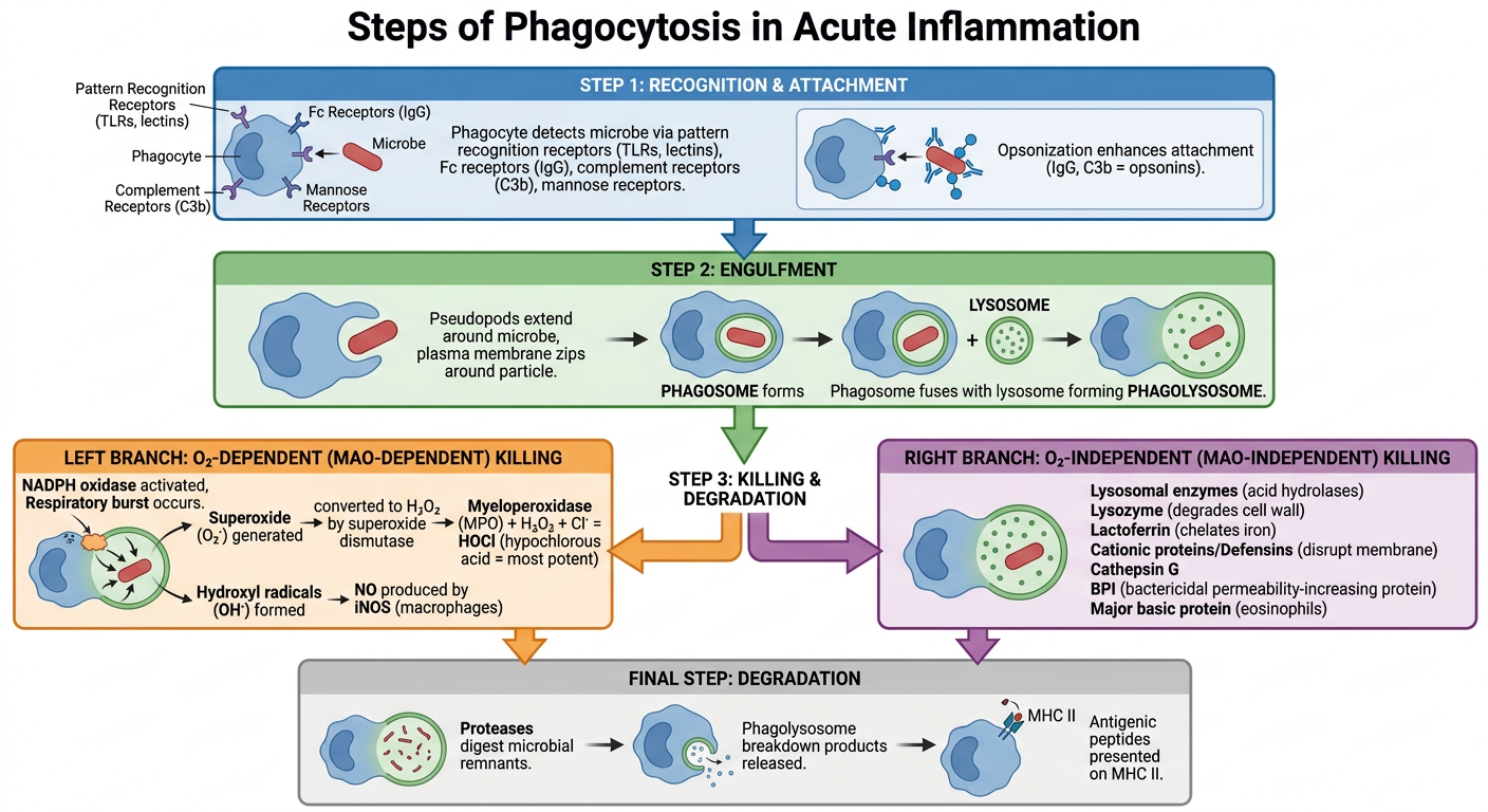
Steps of Phagocytosis in Acute Inflammation
Note: Harsh Mohan's Textbook of Pathology is not in the digital library, but the content below is drawn directly from Robbins Cotran & Kumar (the primary reference Harsh Mohan is based on) and cross-referenced with Medical Microbiology 9e and Histology (Gartner), which cover the same topic identically.
FLOWCHART: Steps of Phagocytosis
MICROBE / FOREIGN PARTICLE AT SITE OF INFECTION
                    │
                    ▼
═══════════════════════════════════════════════
STEP 1: RECOGNITION & ATTACHMENT (Opsonization)
═══════════════════════════════════════════════
    Phagocyte surface receptors bind microbe:
    ├── Mannose receptors (binds mannose on microbe surface)
    ├── Scavenger receptors (binds LPS, lipoteichoic acid)
    ├── Toll-like receptors (TLRs) - Pattern recognition
    ├── Fc receptors (binds IgG-coated microbes)
    └── Complement receptors CR1/CR3 (binds C3b-coated microbes)

    OPSONINS enhance attachment:
    → IgG antibody (Fc region recognized)
    → C3b (complement fragment)
    → Mannose-binding lectin
                    │
                    ▼
═══════════════════════════════════════════════
STEP 2: ENGULFMENT (Internalization)
═══════════════════════════════════════════════
    Phagocyte membrane extends pseudopods
                    │
                    ▼
    Pseudopods surround & engulf microbe
    (actin cytoskeleton rearrangement)
                    │
                    ▼
    PHAGOSOME formed (membrane-bound vacuole)
                    │
                    ▼
    Phagosome fuses with lysosomes/granules
                    │
                    ▼
    PHAGOLYSOSOME formed
                    │
                    ▼
═══════════════════════════════════════════════════════════════
STEP 3: INTRACELLULAR KILLING & DEGRADATION
═══════════════════════════════════════════════════════════════
              ┌────────────┴────────────┐
              ▼                         ▼
  ┌─────────────────────┐   ┌──────────────────────────┐
  │  O₂-DEPENDENT       │   │  O₂-INDEPENDENT           │
  │  (MAO-DEPENDENT)    │   │  (MAO-INDEPENDENT)         │
  │  KILLING            │   │  KILLING                   │
  └────────┬────────────┘   └──────────┬───────────────-┘
           │                           │
           ▼                           ▼
  [See detailed branches below]  [See detailed branches below]
O₂-DEPENDENT (MAO-DEPENDENT) KILLING - Detailed
PHAGOLYSOSOME FORMED
        │
        ▼
NADPH OXIDASE ACTIVATED (assembled on phagosomal membrane)
        │
        ▼
RESPIRATORY BURST (increased O₂ consumption)
        │
        ▼
O₂ + NADPH → SUPEROXIDE (O₂⁻) + NADP⁺
        │
        ├──► Superoxide Dismutase ──► H₂O₂ + ¹O₂ (singlet oxygen)
        │           │
        │           │    H₂O₂ + O₂⁻ (Haber-Weiss/Fenton rxn)
        │           ▼
        │    HYDROXYL RADICAL (OH•) — most reactive ROS
        │
        └──► MYELOPEROXIDASE (MPO) pathway:
                    │
                    ▼
             MPO + H₂O₂ + Cl⁻
                    │
                    ▼
             HYPOCHLOROUS ACID (HOCl) ← MOST POTENT BACTERICIDAL
             (1000× more effective than H₂O₂)
                    │
                    ▼
             OCl⁻ (hypochlorite/bleach) + Cl₂
                    │
                    ▼
             MICROBIAL KILLING

+ NO (Nitric Oxide) — produced by iNOS in MACROPHAGES
  (IFN-γ activates iNOS → L-arginine → NO → antimicrobial)
  [Note: NO plays lesser role in neutrophils in humans]
O₂-INDEPENDENT (MAO-INDEPENDENT) KILLING - Detailed
PHAGOLYSOSOME FORMED
        │
        ▼
GRANULE FUSION with phagosome
        │
    ┌───┴────────────────────────────┐
    │                                │
    ▼                                ▼
SPECIFIC GRANULES              AZUROPHILIC (PRIMARY) GRANULES
(secondary/tertiary)
    │                                │
    ▼                                ▼
• Lysozyme                    • Myeloperoxidase (MPO)
  (cleaves peptidoglycan)     • Defensins (cationic peptides)
• Lactoferrin                 • Cathepsin G (serine protease)
  (chelates iron → starves    • Elastase
  bacteria)                   • Azurocidin/CAP37
• Collagenase                 • BPI (Bactericidal Permeability-
• Gelatinase                    Increasing protein)
                              • Acid hydrolases
                              • Lysozyme
        │                            │
        └───────────┬────────────────┘
                    │
                    ▼
        MECHANISMS OF KILLING:
        • Enzyme digestion of cell wall (lysozyme)
        • Iron deprivation (lactoferrin)
        • Membrane disruption (defensins, BPI)
        • Proteolytic degradation (cathepsins)
        • Low pH in phagolysosome (pH ~3.5-4)
        │
        ▼
        MICROBIAL KILLING
        (less efficient than O₂-dependent; gram-negative
         bacteria more susceptible; gram-positive and fungi
         killed principally by O₂-dependent pathway)
STEP 4: DEGRADATION & ANTIGEN PRESENTATION
KILLED MICROBE in phagolysosome
        │
        ▼
Proteases & hydrolases digest microbial proteins
        │
        ├──► MACROPHAGES: Peptides loaded onto MHC Class II
        │    → Antigen presentation to CD4+ T cells
        │
        └──► Debris exocytosed or retained
Summary Table: MAO-Dependent vs MAO-Independent Killing
| Feature | O₂-Dependent (MAO-dependent) | O₂-Independent (MAO-independent) |
|---|---|---|
| Key enzyme | NADPH oxidase, Myeloperoxidase | Lysozyme, Cathepsin G, Elastase |
| Products | O₂⁻, H₂O₂, HOCl, OH•, NO | Cationic proteins, enzymes |
| Most potent | HOCl (hypochlorous acid) | Defensins, BPI |
| Requires O₂? | Yes | No |
| Defect disease | Chronic Granulomatous Disease (CGD) | Less clinically significant |
| Effective against | Gram-positive bacteria, fungi | Gram-negative bacteria (mainly) |
| Neutrophils | Primary pathway | Secondary pathway |
| Macrophages | Less prominent; iNOS produces NO | Major pathway (with activation) |
Key Points to Remember (Harsh Mohan Style)
- Opsonization is the coating of microbes with IgG or C3b to enhance phagocytosis - this is the critical "recognition" step.
- Respiratory burst = sudden increase in O₂ consumption when NADPH oxidase is activated. Measured by NBT (nitroblue tetrazolium) test.
- MPO-H₂O₂-Cl⁻ system is the most potent bactericidal mechanism. MPO is found in azurophilic granules of neutrophils.
- Chronic Granulomatous Disease (CGD) = NADPH oxidase deficiency → absent respiratory burst → recurrent infections with catalase-positive organisms (S. aureus, Aspergillus).
- Macrophage activation by IFN-γ is required for efficient killing via iNOS-generated NO.
- The term "MAO" in Harsh Mohan refers to Myeloperoxidase-Antibody-Oxygen or oxygen-dependent killing; this is equivalent to the ROS/MPO-dependent system in Robbins.
- Robbins Cotran & Kumar Pathologic Basis of Disease (Phagocytosis and Clearance, pp. 89-92)
- Robbins & Kumar Basic Pathology (Phagocytosis section, block 1)
- Medical Microbiology 9e (Phagocytic Responses, p. 78)
- Histology: A Text and Atlas - Gartner (Neutrophil killing mechanisms, p. 701)