
There are 3 ways a person can be deficient of growth hormone hypothalamus, pituitary and liver state each of its different causes. And identification symptoms
Reading File
Now let me search the medical library for growth hormone deficiency causes and symptoms.
Finding Sources
Finding Sources
Reading File
Reading File
Finding Sources
Reading File
Finding Sources
Reading File
Reading File
Finding Sources
Finding Sources
Reading File
Reading File
I now have comprehensive information from multiple authoritative textbooks to provide a thorough answer. Let me fetch the GH-IGF axis diagram to include visually.
Loading Image
Here is a thorough breakdown of the three anatomical levels at which growth hormone (GH) deficiency can arise, with the causes at each level and the resulting clinical features.
The GH-IGF Axis
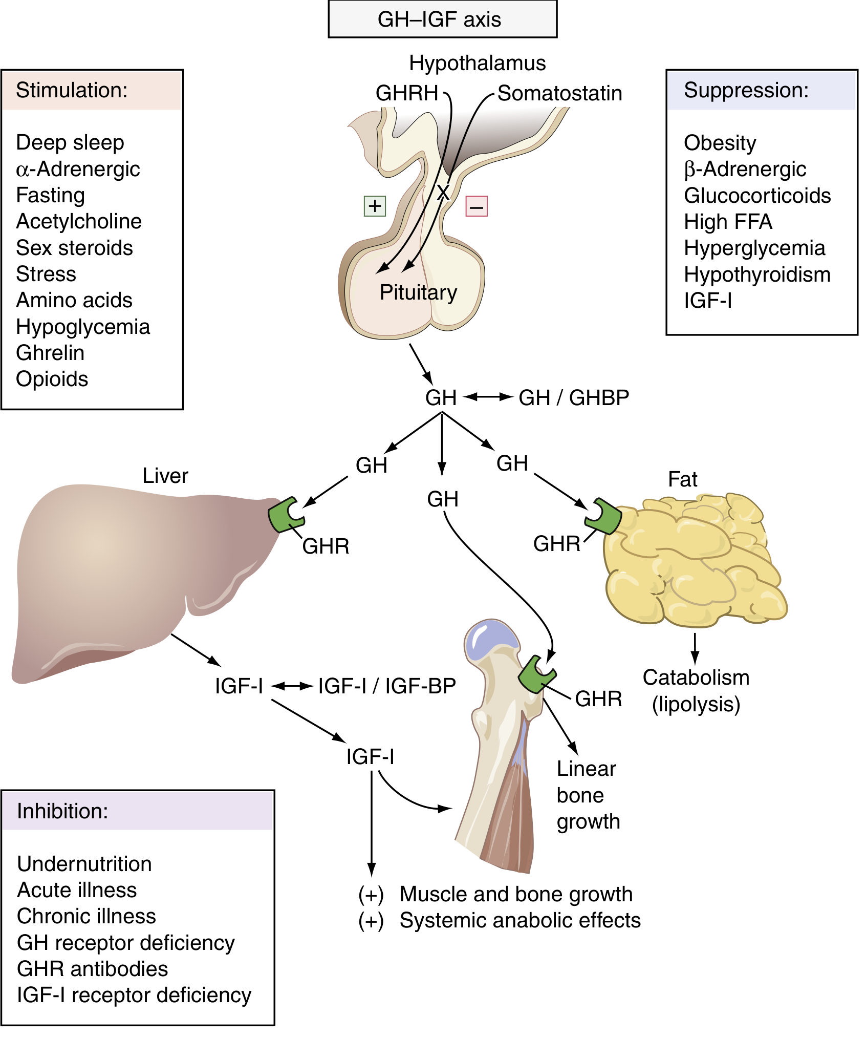
The full axis works like this: the hypothalamus secretes GHRH → stimulates the anterior pituitary to release GH → GH acts on the liver (primarily) to produce IGF-1 (somatomedin C) → IGF-1 drives tissue growth. A defect at any of these three levels produces the same end result - growth failure - but through different mechanisms.
Level 1: Hypothalamic Causes (Tertiary Deficiency)
This is a failure to produce, release, or respond to GHRH, so the pituitary never gets the signal to secrete GH.
Causes:
- Congenital/Idiopathic GHRH deficiency - the most common cause in children; reduced GHRH secretion from the hypothalamic nuclei (arcuate and paraventricular) with no identifiable structural lesion
- Hypothalamic tumors - craniopharyngioma is the classic culprit; also germinomas, gliomas, and ependymomas compressing the hypothalamus
- Head trauma / traumatic brain injury (TBI) - disrupts hypothalamic-pituitary connections; estimated 10-30% of TBI patients develop GHD
- Cranial irradiation - radiation damage to the hypothalamus; GHD is dose-dependent
- Infiltrative diseases - histiocytosis X (Langerhans cell histiocytosis), sarcoidosis, hemochromatosis, lymphocytic hypophysitis
- Infections - encephalitis, meningitis, tuberculosis
- Pituitary stalk disruption - any lesion compressing the pituitary stalk severs GHRH delivery ("stalk effect"); paradoxically prolactin rises while all other anterior pituitary hormones fall
Key point: Repetitive GHRH administration can restore GH secretion in purely hypothalamic-type deficiency, helping distinguish it from true pituitary deficiency.
(Sources: Goldman-Cecil Medicine; Costanzo Physiology 7th Ed.; Scott-Brown's Otorhinolaryngology)
Level 2: Pituitary Causes (Secondary Deficiency)
Here the hypothalamus is working but the pituitary somatotroph cells cannot produce or release adequate GH.
Causes:
- Idiopathic GH deficiency (isolated GHD) - the most common form of nonfamilial deficiency; somatotroph dysfunction without identifiable cause; accounts for the majority of hypopituitarism cases in children
- Pituitary adenoma - the most common etiology in adult-onset GHD; destroys or compresses normal somatotrophs
- Craniopharyngioma - also directly compresses pituitary tissue (>70% of children with craniopharyngioma present with GHD)
- Congenital pituitary aplasia or hypoplasia - complete or partial failure of pituitary gland development (including septo-optic dysplasia)
- Pituitary apoplexy - sudden hemorrhage into or infarction of the pituitary gland; can arise spontaneously or in a pre-existing adenoma; causes acute hypopituitarism
- Sheehan syndrome - postpartum pituitary ischemic necrosis from obstetric hemorrhage and shock; presents as failure to lactate, then progressive panhypopituitarism
- Surgery / radiotherapy - postsurgical disruption of the hypothalamic-pituitary axis
- Genetic mutations - mutations in transcription factors (PIT1, PROP1) that control pituitary development result in combined pituitary hormone deficiency (CPHD)
- Panhypopituitarism (Simmonds syndrome) - loss of all anterior pituitary hormones including GH; patients are pale, lethargic, with dry skin and low BP
(Sources: Goldman-Cecil Medicine; Bradley & Daroff's Neurology; Katzung's Pharmacology; Textbook of Family Medicine 9e)
Level 3: Liver/Peripheral Causes (Primary IGF-1 Deficiency / GH Resistance)
In this scenario, GH is secreted normally (levels are normal or even elevated), but the liver cannot produce IGF-1 in response. This is sometimes called GH insensitivity or GH resistance.
Causes:
- Laron syndrome (Laron dwarfism) - mutations in the GH receptor (GHR) gene → the receptor is defective or absent → GH cannot bind → no IGF-1 is produced by the liver despite high GH levels. Treatment with exogenous GH is ineffective; these patients require recombinant IGF-1 (mecasermin)
- Post-receptor signaling defects - mutations in the GH receptor signaling pathway (e.g., JAK2/STAT5 pathway) downstream of the receptor
- Neutralizing antibodies to GH - immune-mediated blockade of GH before it can act on the liver
- IGF-1 gene defects - rare mutations in the IGF-1 gene itself prevent production
- Severe liver disease - the liver is the primary source of circulating IGF-1; hepatic failure reduces IGF-1 synthesis even with normal GH levels
- Malnutrition / chronic illness / undernutrition - these states suppress hepatic IGF-1 production independently of GH levels
- IGF-1 receptor deficiency - the most distal defect; IGF-1 is produced but cannot signal in target tissues (e.g., African pygmy phenotype - normal/elevated GH and IGF-1 but receptor unresponsive)
(Sources: Costanzo Physiology 7th Ed.; Guyton & Hall Medical Physiology; Katzung's Pharmacology; Bradley & Daroff's Neurology)
Clinical Identification - Signs and Symptoms of GH Deficiency
The symptoms differ between children and adults:
In Children:
| Feature | Detail |
|---|---|
| Short stature / dwarfism | Most prominent sign; failure to grow, growth velocity below the 25th percentile |
| Delayed bone age | Epiphyseal plates close late; body proportions remain normal |
| Mild obesity | Increased body fat, especially truncal; child appears younger than stated age |
| Delayed puberty | Puberty depends partly on achieving critical body mass; GHD delays this |
| Small genitalia | Hypogonadism or micropenis may be variably present |
| Delayed dental eruption | Secondary tooth eruption is delayed (primary teeth are normal) |
| Fasting hypoglycemia | Occurs in up to 10% of children with GHD; GH is a counter-regulatory hormone opposing insulin |
| Normal intelligence | Cognitive development is typically unaffected |
In Adults:
| Feature | Detail |
|---|---|
| Decreased muscle mass | Reduced lean body mass and exercise capacity |
| Increased adiposity | Especially visceral fat; worsens cardiovascular risk |
| Reduced bone density | Osteoporosis risk increases |
| Dyslipidemia | Elevated LDL cholesterol; reduced HDL cholesterol |
| Decreased cardiovascular function | Increased risk of cardiovascular events; reduced cardiac output |
| Diminished well-being | Fatigue, low energy, depression, reduced sense of well-being |
| Impaired wound healing | |
| Insulin sensitivity changes | Reduced insulin requirements in diabetics; fasting hypoglycemia possible |
Key diagnostic clue: In hypothalamic and pituitary causes, serum GH is low. In liver/GH-resistance causes (e.g., Laron syndrome), serum GH is high but IGF-1 is low - this is the distinguishing laboratory finding.
Sources:
- Costanzo Physiology, 7th Ed. - Pathophysiology of Growth Hormone section
- Goldman-Cecil Medicine - Growth Hormone Deficiency (Pathobiology, Clinical Manifestations)
- Bradley & Daroff's Neurology in Clinical Practice - Abnormalities of Anterior Pituitary Function
- Textbook of Family Medicine, 9e - GH Deficiency manifestations
- Guyton & Hall Medical Physiology - IGF-1 and Laron Syndrome
- Katzung's Basic & Clinical Pharmacology, 16th Ed. - GH Deficiency and Mecasermin
Tell how to identify its which case
The key is a stepwise approach using hormone levels, stimulation tests, and imaging to pinpoint exactly where in the axis the problem lies.
Searching sources...
Finding Sources
Finding Sources
Reading File
Reading File
Reading File
Finding Sources
Reading File
Reading File
I now have everything needed for a clear, diagnostic differentiation guide. Here it is:
How to Identify Which Level is Causing GH Deficiency
The approach is a stepwise ladder: measure baseline hormones first, then use provocative tests to stress the axis, then image the anatomy.
Step 1 - Baseline Blood Tests (First Draw)
Measure these simultaneously in a fasting morning sample:
| Test | What it tells you |
|---|---|
| Serum GH | Usually low in hypothalamic + pituitary causes; high in liver/GH-resistance causes |
| Serum IGF-1 | Low in ALL three causes - it's the universal marker of GH deficiency effect |
| IGFBP-3 | Low in GHD; useful especially in children under 3 years |
| GH-binding protein (GHBP) | Low in Laron syndrome (reflects GH receptor density on liver cells) |
Key pattern already emerges here:
- Low GH + Low IGF-1 → problem is at hypothalamus OR pituitary (can't yet distinguish)
- High/Normal GH + Low IGF-1 → problem is at the liver/receptor level (GH resistance)
Step 2 - The GHRH Stimulation Test (Separates Hypothalamus from Pituitary)
This is the critical differentiating test between Level 1 and Level 2.
How it works: Give exogenous GHRH intravenously → measure GH at 0, 15, 30, 45, 60 minutes
| Result | Interpretation |
|---|---|
| GH rises normally (>7 µg/L) | The pituitary somatotrophs are intact - problem is hypothalamic (they just needed GHRH to be given externally) |
| GH does NOT rise | The pituitary somatotrophs themselves are defective - problem is pituitary (they cannot respond even when given GHRH) |
This single test cleanly separates hypothalamic from pituitary GH deficiency. Repetitive GHRH administration can even therapeutically restore GH secretion in purely hypothalamic cases.
Step 3 - Gold Standard Provocative Tests (Confirm GH Deficiency Itself)
Since GH secretion is pulsatile and undetectable for most of the day even in healthy people, a single low GH level means nothing. Provocative tests force a maximum GH secretion response:
| Test | Protocol | Normal Response |
|---|---|---|
| Insulin Tolerance Test (ITT) - gold standard | Regular insulin 0.05-0.15 U/kg IV; glucose must drop below 40 mg/dL | GH should rise >3-5 µg/L |
| Arginine stimulation | 0.5 g/kg IV arginine HCl over 30 min; sample at 0, 30, 60, 90 min | GH should rise >5-7 µg/L |
| Glucagon test | 1 mg IM; sample at 0, 30, 60, 90, 120, 150, 180 min | GH should rise >3 µg/L (BMI-adjusted) |
| GHRH + Arginine combined | GHRH 1 µg/kg + arginine 30 g IV | Normal is BMI-dependent (>4-11 µg/L) |
| L-Dopa / Clonidine | Used primarily in children | GH should rise >7 µg/L |
A subnormal response on TWO separate provocative tests is required to diagnose GH deficiency (one test alone is insufficient).
Step 4 - The IGF-1 Generation Test (Confirms Liver/GH Resistance)
Used when Step 1 already shows high GH + low IGF-1. This test proves the liver is not responding to GH.
How it works: Give daily injections of exogenous recombinant GH for 4-7 days → measure IGF-1 before and after
| Result | Interpretation |
|---|---|
| IGF-1 rises | GH receptor is functional - the problem was lack of endogenous GH (hypothalamic or pituitary) |
| IGF-1 does NOT rise | GH receptor or post-receptor signaling is broken - confirms Laron syndrome or liver GH resistance |
Additional confirmation for Laron: measure GH-binding protein (GHBP) - it is markedly low or absent because GHBP is the extracellular fragment of the GH receptor.
Step 5 - Imaging (Find the Structural Cause)
Once you know the biochemical level of the defect, image accordingly:
| Finding | What to look for |
|---|---|
| MRI of hypothalamus and pituitary | Hypothalamic tumors (craniopharyngioma - often calcified), pituitary adenomas, pituitary aplasia/hypoplasia, Rathke's cleft cyst, empty sella, stalk interruption |
| Bone age X-ray (left hand/wrist) | Delayed bone age confirms long-standing GH deficiency |
| Liver function tests | If liver disease suspected as cause of low IGF-1 production |
The Complete Decision Algorithm
Child with short stature + low growth velocity
↓
Measure GH + IGF-1 + IGFBP-3
↓
┌────────────────────────────────────┐
│ │
Low GH + Low IGF-1 High/Normal GH + Low IGF-1
↓ ↓
Do GHRH stimulation test IGF-1 Generation Test + GHBP
↓ ↓
GH rises? → HYPOTHALAMIC No rise + Low GHBP → LARON
GH no rise? → PITUITARY No rise + Normal GHBP → post-receptor defect
↓
Confirm with ITT or Arginine test (×2)
↓
MRI brain/pituitary for structural cause
Summary Table - Distinguishing the 3 Levels
| Marker | Hypothalamic | Pituitary | Liver/GH Resistance |
|---|---|---|---|
| Basal GH | Low | Low | High/Normal |
| IGF-1 | Low | Low | Low |
| GHBP | Normal | Normal | Low (Laron) |
| Response to GHRH | GH rises (pituitary intact) | GH does NOT rise | GH already high |
| Response to IGF-1 gen. test | IGF-1 rises | IGF-1 rises | IGF-1 does NOT rise |
| MRI | Hypothalamic lesion | Pituitary lesion/aplasia | Normal pituitary |
| GH treatment | Works | Works | Does NOT work (Laron) |
Sources: Harrison's Principles of Internal Medicine 22E (2025); Tietz Textbook of Laboratory Medicine 7th Ed.; Goldman-Cecil Medicine; Henry's Clinical Diagnosis and Management by Laboratory Methods
Redo
Before I redo this, let me make sure I give you exactly what you need.
Asking for Preferences
What would you like me to change when I redo this?
I want to know how to differentiate between each primary or secondary or territary
Great clarification. You want to know how to tell apart:
- Primary = Liver/target tissue failure (GH resistance)
- Secondary = Pituitary failure
- Tertiary = Hypothalamic failure
Searching sources...
Finding Sources
Finding Sources
Finding Sources
Finding Sources
Reading File
Finding Sources
Reading File
Reading File
I have everything needed. Here is the clean, focused differentiation guide:
Differentiating Primary, Secondary, and Tertiary GH Deficiency
First, let's lock in the terminology:
| Term | Site of Problem |
|---|---|
| Tertiary | Hypothalamus - not producing GHRH |
| Secondary | Pituitary - not producing GH |
| Primary | Liver/target tissue - not responding to GH (GH resistance) |
The Logic: Work Down the Axis
The key insight is that each level upstream still produces its hormone normally - only the step below is broken.
Hypothalamus → GHRH → Pituitary → GH → Liver → IGF-1 → Growth
Tertiary defect = broken here ↑
Secondary defect = broken here ↑
Primary defect = broken here ↑
Step 1 - Two Blood Tests Tell You Almost Everything
Draw GH and IGF-1 together:
| GH level | IGF-1 level | Conclusion |
|---|---|---|
| LOW | LOW | Hypothalamus or Pituitary problem (tertiary or secondary) - need Step 2 to separate them |
| HIGH or NORMAL | LOW | Liver/receptor problem (primary) - GH is being made but the body can't use it |
This single pattern already identifies primary (GH resistance) vs the other two.
Step 2 - The GHRH Stimulation Test (Separates Tertiary from Secondary)
Once you know GH is low, inject exogenous GHRH and watch what happens:
Give: GHRH 1 µg/kg IV
Measure: GH at 0, 15, 30, 45, 60 minutes
| GH response after GHRH | Meaning |
|---|---|
| GH rises normally (>7 µg/L) | The pituitary works fine - it just wasn't getting the signal. Problem is TERTIARY (hypothalamic) |
| GH does NOT rise | The pituitary itself is broken and can't respond even when given GHRH directly. Problem is SECONDARY (pituitary) |
Why this works: In tertiary deficiency the pituitary is starved of GHRH but fully functional. Give it GHRH from outside and it responds. In secondary deficiency the pituitary cells are damaged, so no matter how much GHRH you give, nothing comes out.
Step 3 - Confirm Primary with the IGF-1 Generation Test
When GH is high but IGF-1 is low, give exogenous recombinant GH for 4-7 days and remeasure IGF-1:
| IGF-1 after GH injections | Meaning |
|---|---|
| IGF-1 rises | Liver receptor is intact - this was not primary. Review the diagnosis |
| IGF-1 stays LOW | The liver cannot respond to GH at all - confirms PRIMARY deficiency (Laron syndrome / GH resistance) |
Add-on test: Measure GH-binding protein (GHBP) - it is simply the shed extracellular portion of the GH receptor. If GHBP is very low or absent, the GH receptor itself is missing/defective, sealing the diagnosis of Laron syndrome.
Step 4 - MRI to Find the Structural Cause
| Finding on MRI | Points to |
|---|---|
| Hypothalamic tumor (craniopharyngioma), infiltration, trauma scar | Tertiary |
| Pituitary adenoma, pituitary aplasia/hypoplasia, empty sella, Sheehan necrosis | Secondary |
| Normal pituitary and hypothalamus | Primary (GH resistance - the problem is at the receptor) |
Full Summary in One Table
| Feature | Tertiary (Hypothalamic) | Secondary (Pituitary) | Primary (Liver/Receptor) |
|---|---|---|---|
| GHRH | LOW | Normal/High | Normal/High |
| GH | Low | Low | High or Normal |
| IGF-1 | Low | Low | Low |
| GHBP | Normal | Normal | Low (Laron) |
| GHRH stimulation test | GH rises ✓ | GH stays low ✗ | GH already high |
| IGF-1 generation test | IGF-1 rises | IGF-1 rises | IGF-1 stays low ✗ |
| GH replacement therapy | Works ✓ | Works ✓ | Does NOT work ✗ (need IGF-1 directly) |
| MRI | Hypothalamic lesion | Pituitary lesion | Normal |
Quick Mental Shortcut
- High GH + Low IGF-1 = Primary (GH is screaming but the liver is deaf)
- Low GH + GHRH test fixes it = Tertiary (pituitary was just waiting for the signal)
- Low GH + GHRH test does nothing = Secondary (pituitary is broken)
Sources: Harrison's Principles of Internal Medicine 22E; Goldman-Cecil Medicine; Scott-Brown's Otorhinolaryngology; Tietz Textbook of Laboratory Medicine 7th Ed.
Tell me step 3
You're asking about Step 3 - the IGF-1 Generation Test, which confirms Primary (liver/GH resistance) deficiency.
Searching sources...
Finding Sources
Finding Sources
Reading File
Finding Sources
Reading File
Reading File
Here is a detailed breakdown of Step 3:
Step 3 - The IGF-1 Generation Test (Confirms Primary / GH Resistance)
When Do You Use This Test?
You reach Step 3 when your Step 1 blood results show:
GH is HIGH or NORMAL + IGF-1 is LOW
This pattern tells you GH is being secreted fine - the body just isn't responding to it. The question now is: where exactly is the resistance?
The Problem It Detects
Normally the chain looks like this:
GH secreted → binds GH receptor on liver → liver produces IGF-1 → growth occurs
In primary deficiency, this chain breaks at the liver receptor level:
GH secreted ✓ → GH receptor BROKEN on liver ✗ → IGF-1 NOT produced → no growth
The liver is "deaf" to GH. No matter how much GH is present, IGF-1 never rises.
How the Test is Done
Give: Daily injections of recombinant GH (exogenous, at a supraphysiological dose) for 4-7 consecutive days
Measure: IGF-1 blood level before the injections start and after the course ends
The logic: You are flooding the body with very high doses of GH from outside. If the GH receptor on the liver is working - even partially - IGF-1 must rise. If it does not rise at all, the receptor is confirmed broken.
Reading the Result
| IGF-1 after GH injections | What it means |
|---|---|
| IGF-1 rises significantly | Liver GH receptor is intact. The patient was just lacking GH from their own pituitary/hypothalamus. This rules out primary deficiency - go back and look at secondary or tertiary |
| IGF-1 stays low / does NOT rise | The liver cannot respond to GH at all. Confirms PRIMARY deficiency (GH resistance / Laron syndrome) |
Supporting Test - GH-Binding Protein (GHBP)
Run this blood test alongside the IGF-1 generation test. It locks in the diagnosis.
What is GHBP?
The GH receptor is a large protein sitting on liver cells. Its outer portion breaks off into the bloodstream and is called GH-binding protein (GHBP). So GHBP is essentially a mirror of how many functional GH receptors the liver has.
| GHBP level | Meaning |
|---|---|
| Very low or absent | GH receptors are absent or severely defective → confirms Laron syndrome (GH receptor mutation) |
| Normal GHBP but IGF-1 still doesn't rise | GH receptor is present but the signaling machinery inside the liver cell is broken (e.g., STAT5B mutation, post-receptor defect) |
Variants of Primary GH Resistance
Once you confirm primary deficiency, the sub-type matters for treatment:
| Sub-type | GH | IGF-1 | GHBP | What's broken |
|---|---|---|---|---|
| Laron syndrome | High | Low | Very LOW | GH receptor gene mutation |
| STAT5B defect | High | Low | Normal | JAK-STAT signaling pathway inside liver cell |
| IGF-1 gene mutation | High | Low | Normal | IGF-1 itself cannot be made |
| GH receptor antibodies | High | Low | Normal | Antibodies block the receptor externally |
| Severe liver disease | High/Normal | Low | Normal | Liver tissue destroyed, receptors lost |
| African pygmy phenotype | Normal/High | Low | Normal | IGF-1 receptor insensitivity in tissues |
Why This Matters for Treatment
This is the most critical clinical point:
If primary GH resistance is confirmed - giving GH injections will do absolutely nothing. The liver cannot respond to GH, so IGF-1 will never be produced.
Treatment instead: Recombinant human IGF-1 directly - called Mecasermin (bypasses the broken GH receptor entirely and delivers IGF-1 straight to the tissues). It is FDA-approved specifically for GH resistance syndromes.
- Secondary and tertiary deficiencies respond to GH replacement
- Primary deficiency responds only to direct IGF-1 replacement
Summary of Step 3 in One Line
Inject high-dose GH for 4-7 days → if IGF-1 still doesn't rise + GHBP is low → the GH receptor on the liver is broken → Primary deficiency confirmed → treat with Mecasermin, NOT GH.
Sources: Harrison's Principles of Internal Medicine 22E (2025); Medical Physiology (Boron & Boulpaep); Katzung's Basic & Clinical Pharmacology 16th Ed.; Goodman & Gilman's Pharmacological Basis of Therapeutics
What is Mecasermin and how is it dosed?
Mecasermin is recombinant human IGF-1, used when the GH receptor is broken and GH replacement won't work. It bypasses the whole GH-liver axis entirely.
Searching sources...
Finding Sources
Finding Sources
Reading File
Reading File
Here is everything you need to know about Mecasermin:
Mecasermin (Increlex)
What It Is
Mecasermin is recombinant human IGF-1 (rhIGF-1) - a lab-manufactured copy of the exact same IGF-1 the liver normally produces in response to GH. It is injected directly into the body, completely bypassing the broken GH → GH receptor → liver → IGF-1 chain.
Brand name: Increlex
FDA approved: 2005
There is also a second form called mecasermin rinfabate - this is rhIGF-1 combined with its binding protein IGFBP-3, which extends the half-life of IGF-1 in the bloodstream. However, this combined form was discontinued for short stature indications due to patent issues (still available for other conditions like severe insulin resistance, muscular dystrophy, and HIV-related fat redistribution).
Who It Is For (Indications)
Mecasermin is specifically used when IGF-1 deficiency cannot be fixed by giving GH, meaning the defect is downstream of GH itself:
| Indication | Why GH won't work |
|---|---|
| GH receptor mutations (Laron syndrome) | Receptor is absent or non-functional |
| Post-receptor signaling defects (e.g. STAT5B mutation) | Signal can't pass from receptor into the cell |
| Neutralizing antibodies against GH | GH is blocked before it reaches the receptor |
| IGF-1 gene defects | IGF-1 cannot be synthesized even if signaling is intact |
It can also stimulate linear growth in GH deficiency and idiopathic short stature, but it is less effective than GH replacement for those conditions, so GH is preferred when available.
How It Is Dosed
Route: Subcutaneous (SC) injection - just under the skin, like insulin
Frequency: Twice daily (BID)
| Phase | Dose |
|---|---|
| Starting dose | 40-80 µg/kg per injection, twice daily |
| Escalation | Increased gradually each week as tolerated |
| Maximum dose | 120 µg/kg per injection, twice daily |
Key rule: The dose is increased weekly based on tolerance and response, not immediately started at the maximum.
The Most Important Rule - Timing with Food
Must eat a carbohydrate-containing meal or snack within 20 minutes of each injection.
This rule exists because IGF-1, like insulin, lowers blood glucose. Injecting on an empty stomach causes significant hypoglycemia. If a child cannot eat (vomiting, illness), the dose should be skipped that time.
Expected Effect
- First year: Height velocity increases by approximately 4-5 cm/year above baseline
- After 2 years: Height SDS (standard deviation score) increases by about 0.5
- This is less than what GH replacement achieves in GH-deficient children (9-14 cm/year in year one), because IGF-1 alone does not trigger all the same anabolic pathways GH does
Side Effects
| Side Effect | Detail |
|---|---|
| Hypoglycemia | Most important and most common - reason for the mandatory food rule |
| Lipohypertrophy | Fat tissue buildup at injection sites - rotate injection sites |
| Adenotonsillar hypertrophy | Enlarged tonsils from lymphoid tissue growth - may need surgical removal |
| Intracranial hypertension | Raised pressure inside the skull - headaches, visual changes |
| Elevated liver enzymes | Usually asymptomatic; monitor LFTs |
Contraindications
- Closed epiphyses - once growth plates are fused, there is no linear growth to stimulate, so using it for growth promotion is pointless
- Active or suspected cancer/neoplasia - IGF-1 is a potent growth factor and could accelerate tumor growth; stop if cancer develops during treatment
Pharmacokinetics (How the Body Handles It)
- Absorption: Virtually complete after subcutaneous injection
- Binding: More than 80% of circulating IGF-1 binds to a ternary complex with IGFBP-3 and the acid-labile subunit - this prolongs the half-life
- Half-life: Approximately 6 hours
- Metabolism: Both liver and kidney metabolize IGF-1
Quick Summary
| Feature | Detail |
|---|---|
| Drug | Recombinant human IGF-1 |
| Brand | Increlex |
| Route | SC injection |
| Frequency | Twice daily |
| Starting dose | 40-80 µg/kg twice daily |
| Maximum dose | 120 µg/kg twice daily |
| Must eat within | 20 minutes of injection |
| Main side effect | Hypoglycemia |
| Do NOT use when | Epiphyses closed, active cancer |
Sources: Katzung's Basic & Clinical Pharmacology 16th Ed.; Goodman & Gilman's Pharmacological Basis of Therapeutics
Hormones produced by anterior pituitary and posterior pituitary
Quick answer: Anterior pituitary produces GH, TSH, ACTH, FSH, LH, and Prolactin. Posterior pituitary releases ADH (vasopressin) and Oxytocin - though these are actually made in the hypothalamus and just stored/released from the posterior pituitary.
Searching sources...
Finding Sources
Finding Sources
Reading File
Reading File
Reading File
Loading Image
Loading Image
Here is a complete breakdown of all pituitary hormones from both lobes:
Anterior Pituitary Hormones
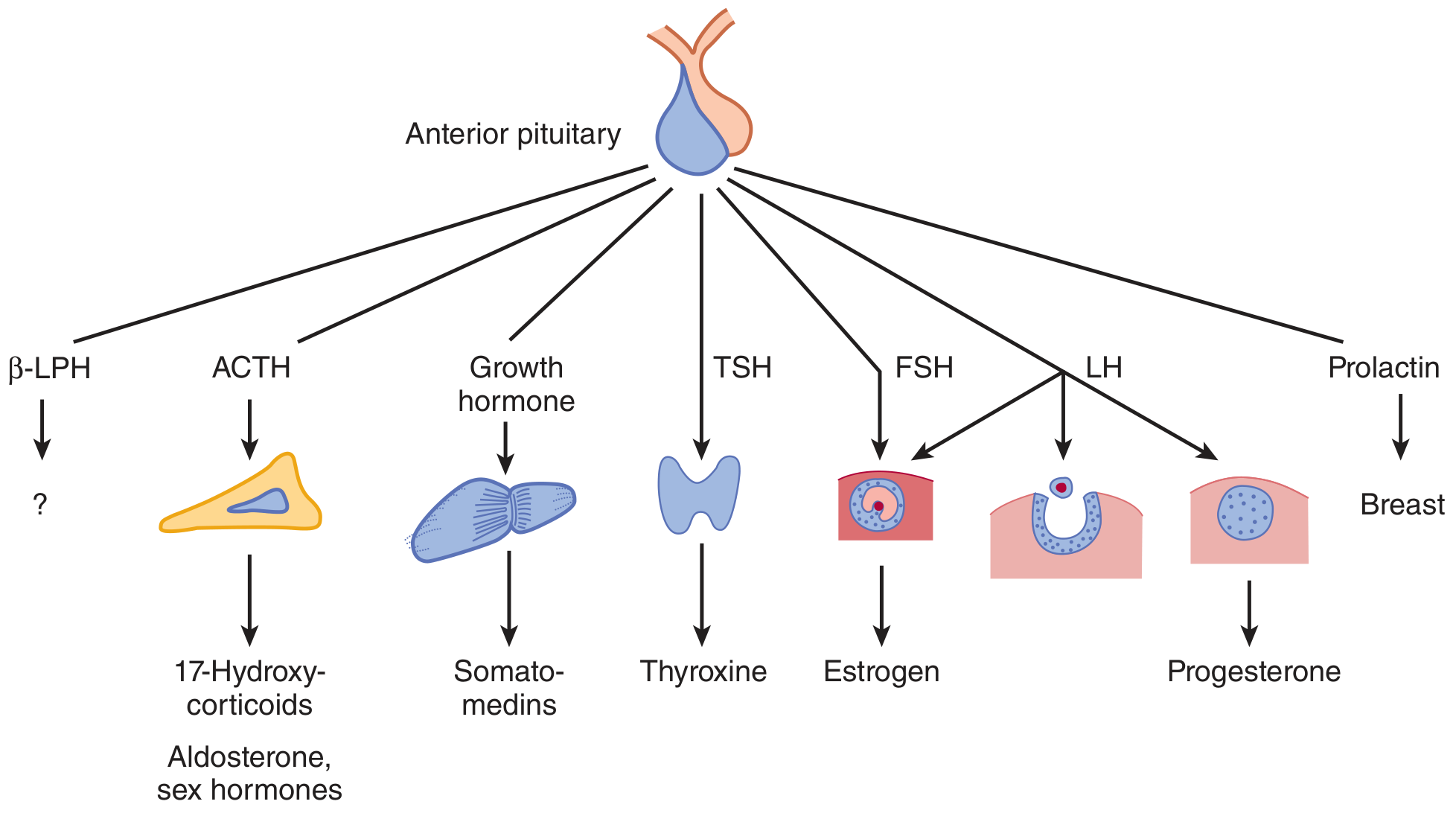
The anterior pituitary (also called the adenohypophysis) produces 6 main hormones, each secreted by a specific cell type:
| # | Hormone | Abbreviation | Cell Type | Hypothalamic Control | Target | Effect |
|---|---|---|---|---|---|---|
| 1 | Growth Hormone | GH | Somatotrope | GHRH (+) / Somatostatin (-) | Liver, bone, muscle | Growth, IGF-1 production, protein synthesis, lipolysis |
| 2 | Prolactin | PRL | Lactotrope | Dopamine (-) / TRH, estrogen (+) | Breast | Milk production (lactogenesis); breast development |
| 3 | Adrenocorticotropic Hormone | ACTH | Corticotrope | CRH (+) / Glucocorticoids (-) | Adrenal cortex | Stimulates cortisol, aldosterone, sex hormones |
| 4 | Thyroid-Stimulating Hormone | TSH | Thyrotrope | TRH (+) / T3, T4 (-) | Thyroid gland | Stimulates T3 and T4 production |
| 5 | Follicle-Stimulating Hormone | FSH | Gonadotrope | GnRH (+) / Sex steroids, inhibin (-) | Ovaries / Testes | Follicle development (F); sperm production (M) |
| 6 | Luteinizing Hormone | LH | Gonadotrope | GnRH (+) / Sex steroids (-) | Ovaries / Testes | Ovulation, progesterone (F); testosterone (M) |
Bonus hormone also from anterior pituitary:
- β-Lipotropin (β-LPH) - secreted alongside ACTH from the same POMC precursor; exact physiologic role is still not fully established
Important note about Prolactin: It is the only anterior pituitary hormone under predominant inhibitory control from the hypothalamus (via dopamine). All others are primarily stimulated by their hypothalamic releasing hormones. This is why a pituitary stalk cut causes all anterior hormones to fall except prolactin, which rises.
Posterior Pituitary Hormones
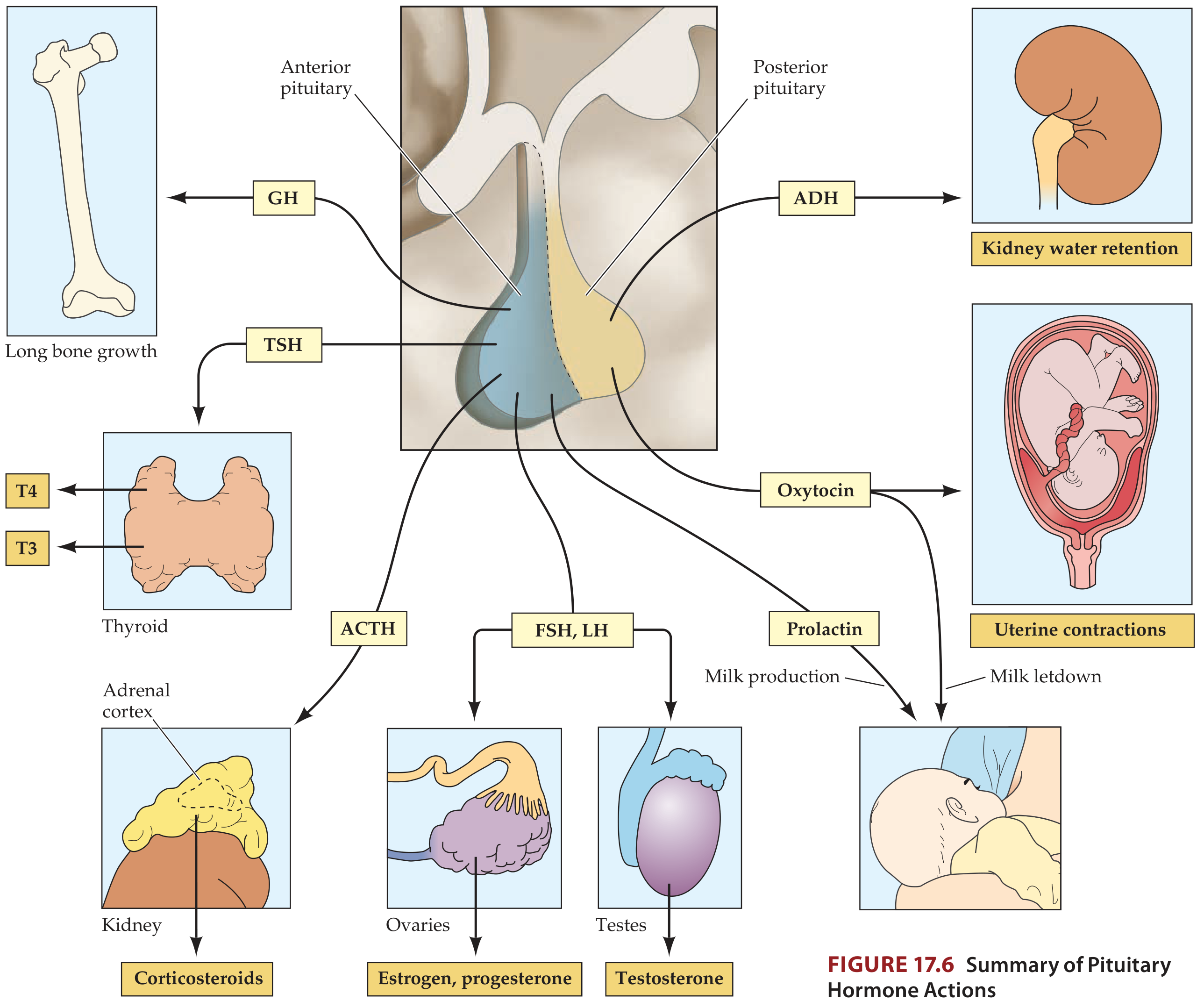
The posterior pituitary (also called the neurohypophysis) releases only 2 hormones:
| # | Hormone | Abbreviation | Where MADE | Where RELEASED | Target | Effect |
|---|---|---|---|---|---|---|
| 1 | Antidiuretic Hormone (Vasopressin) | ADH / AVP | Supraoptic nucleus of hypothalamus | Posterior pituitary | Kidney collecting ducts | Water retention; increases urine concentration; raises blood pressure at high doses |
| 2 | Oxytocin | OT | Paraventricular nucleus of hypothalamus | Posterior pituitary | Uterus; breast | Uterine contractions during labor; milk letdown (ejection) during breastfeeding |
Critical concept: The posterior pituitary does NOT manufacture its own hormones. ADH and oxytocin are produced in hypothalamic neurons (supraoptic and paraventricular nuclei), travel down axons into the posterior pituitary, and are stored there until released into the bloodstream. The posterior pituitary is essentially a storage and release depot, not a secretory gland.
The Complete Picture Side by Side
| Feature | Anterior Pituitary | Posterior Pituitary |
|---|---|---|
| Other name | Adenohypophysis | Neurohypophysis |
| Tissue type | Glandular epithelial cells | Nerve terminals (axon endings) |
| Hormones made here? | Yes - makes its own hormones | No - only stores and releases |
| Number of hormones | 6 (+ β-LPH) | 2 |
| Hormones | GH, PRL, ACTH, TSH, FSH, LH | ADH, Oxytocin |
| Hypothalamic control | Via portal blood vessels (releasing/inhibiting hormones) | Via direct axon projections from hypothalamic nuclei |
Easy Memory Mnemonics
Anterior pituitary (6 hormones) - "FLAT PiG"
- FSH
- LH
- ACTH
- TSH
- Prolactin
- iGF (via GH)
- GH
Posterior pituitary (2 hormones) - "ADO"
- ADH (antidiuretic hormone)
- Oxytocin
Sources: Ganong's Review of Medical Physiology 26th Ed.; Harrison's Principles of Internal Medicine 22E; Neuroanatomy through Clinical Cases 3rd Ed.
Tonsil surfaces
The palatine tonsil has two surfaces - a medial (free) surface covered in crypts, and a lateral (deep) surface covered by a fibrous capsule. Let me get the full detail from the textbooks.
Searching sources...
Finding Sources
Finding Sources
Finding Sources
Finding Sources
Reading File
Reading File
Reading File
Loading Image
Finding Sources
Reading File
Reading File
Here is a complete guide to the surfaces of the palatine tonsil:
Surfaces of the Palatine Tonsil
The palatine tonsil is a mass of lymphoid tissue sitting in the tonsillar fossa - the recess between the palatoglossal arch (anterior pillar) and the palatopharyngeal arch (posterior pillar) on the lateral wall of the oropharynx. It forms the anteroinferior part of Waldeyer's ring.
It has two main surfaces:
1. Medial Surface (Free Surface)
- This is the surface that faces into the oropharynx - it is visible when you open your mouth
- It is covered by stratified squamous epithelium (the same mucosa lining the oral cavity)
- It presents a pitted appearance - the pits are the openings of the tonsillar crypts
- These pits (usually 10-15 in number) lead into a system of blind-ending, often highly branching crypts that extend through the whole thickness of the tonsil
- The walls of the crypts are lined by epithelium that is heavily infiltrated by lymphocytes - so densely that the epithelial lining may be difficult to see microscopically
- The crypts trap debris, bacteria, and food particles - this is why the tonsil acts as a first-line immunological defense
The vascular supply enters primarily via the medial surface, with tonsillar branches of the ascending pharyngeal and facial arteries running through the superior constrictor muscle.
2. Deep (Lateral) Surface
- This surface faces outward toward the pharyngeal wall and is not exposed to the pharyngeal lumen
- It is covered by a fibrous tissue hemicapsule (the tonsillar capsule)
- The capsule can be easily separated from the underlying superior constrictor muscle during tonsillectomy - this plane is the surgical dissection plane
- An important surgical landmark here is the external palatine vein (also called the paratonsillar vein) - it descends from the soft palate, runs lateral to the tonsillar hemicapsule, then pierces the pharyngeal wall. Hemorrhage from this vessel is a recognised complication of tonsillectomy.
Histological View
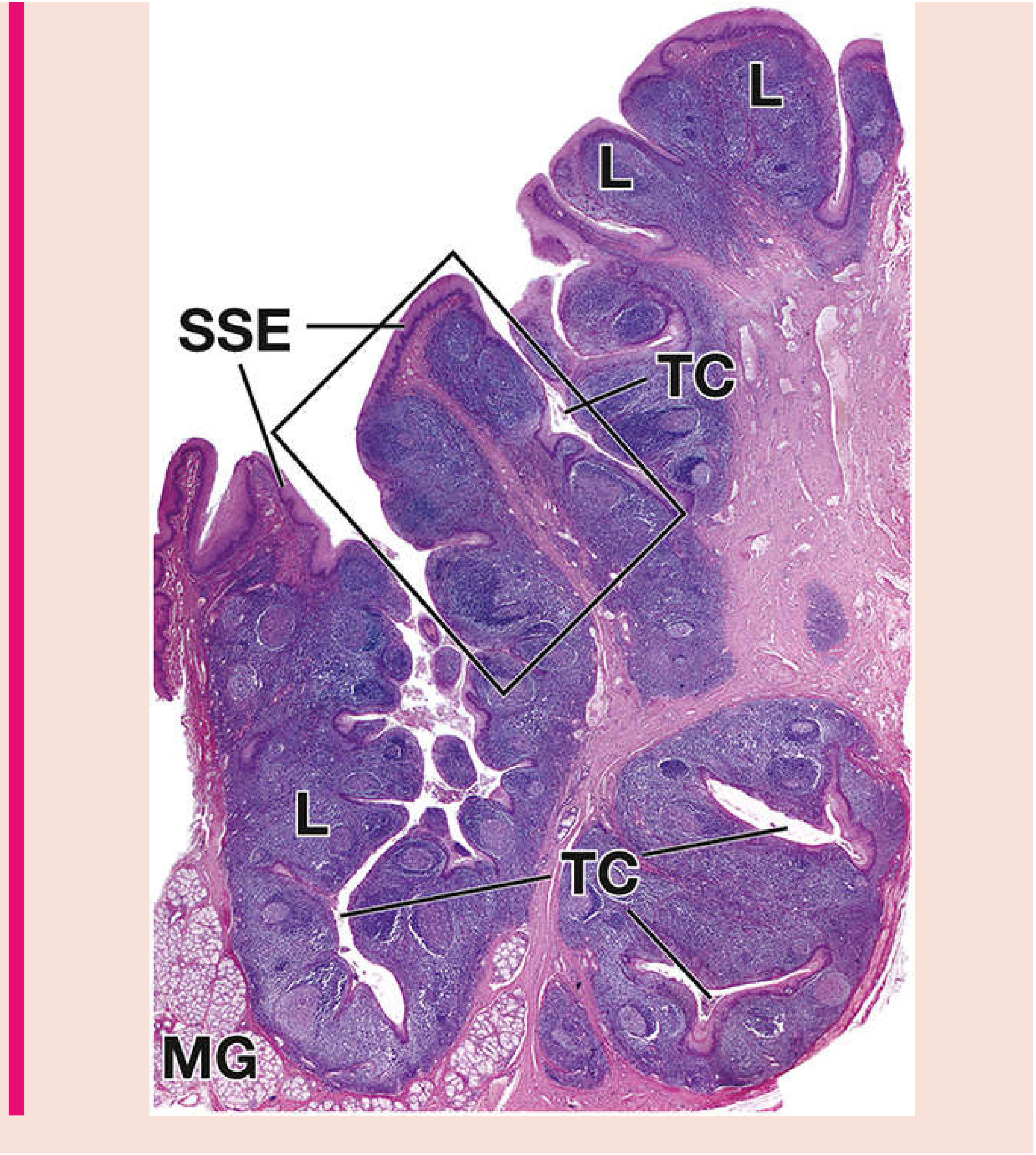
Under the microscope you can see:
- SSE = Stratified Squamous Epithelium covering the free (medial) surface
- TC = Tonsillar Crypts - deep invaginations of the surface epithelium
- L = Lymphoid tissue (nodules with germinal centers)
- MG = Mucous glands at the base of the crypts
- Beneath all of this lies the submucosa of dense connective tissue, continuous with the capsule on the deep surface
Tonsil Bed Borders (Surgical Anatomy)
When the tonsil is removed, the surrounding boundaries are:
| Direction | Structure |
|---|---|
| Superior | Soft palate |
| Inferior | Lingual tonsil |
| Anterior | Palatoglossus muscle (anterior pillar) |
| Posterior | Palatopharyngeus muscle (posterior pillar) |
| Deep | Superior constrictor muscle |
Size and Lifespan
- At puberty: approximately 10-15 mm transverse diameter, 20-25 mm vertical
- The tonsil begins to atrophy at puberty
- By old age, only a small remnant of lymphoid tissue remains
Summary: Two Surfaces at a Glance
| Feature | Medial (Free) Surface | Deep (Lateral) Surface |
|---|---|---|
| Faces | Oropharyngeal lumen | Pharyngeal wall / superior constrictor |
| Covering | Stratified squamous epithelium | Fibrous hemicapsule |
| Key feature | 10-15 pitted crypts | Surgical dissection plane |
| Clinical relevance | Traps bacteria → tonsillitis, tonsil stones | Paratonsillar vein → tonsillectomy bleeding |
Sources: Scott-Brown's Otorhinolaryngology Head & Neck Surgery; Histology: A Text and Atlas (Pawlina) 9th Ed.; Pfenninger & Fowler's Procedures for Primary Care
Wheel and flare formation
The wheal and flare reaction is the classic triple response of Lewis - a local skin reaction to injury or allergen involving a wheal (raised edematous bump) and flare (surrounding redness). Let me get the full detail.
Searching sources...
Finding Sources
Finding Sources
Reading File
Reading File
Reading File
Loading Image
Finding Sources
Reading File
Loading Image
Here is a complete explanation of the wheal and flare reaction:
Wheal and Flare - The Triple Response of Lewis
The triple response of Lewis is the classic skin reaction to injury or allergen. It has three sequential components - the red reaction, the flare, and the wheal - and involves three separate mechanisms working together.
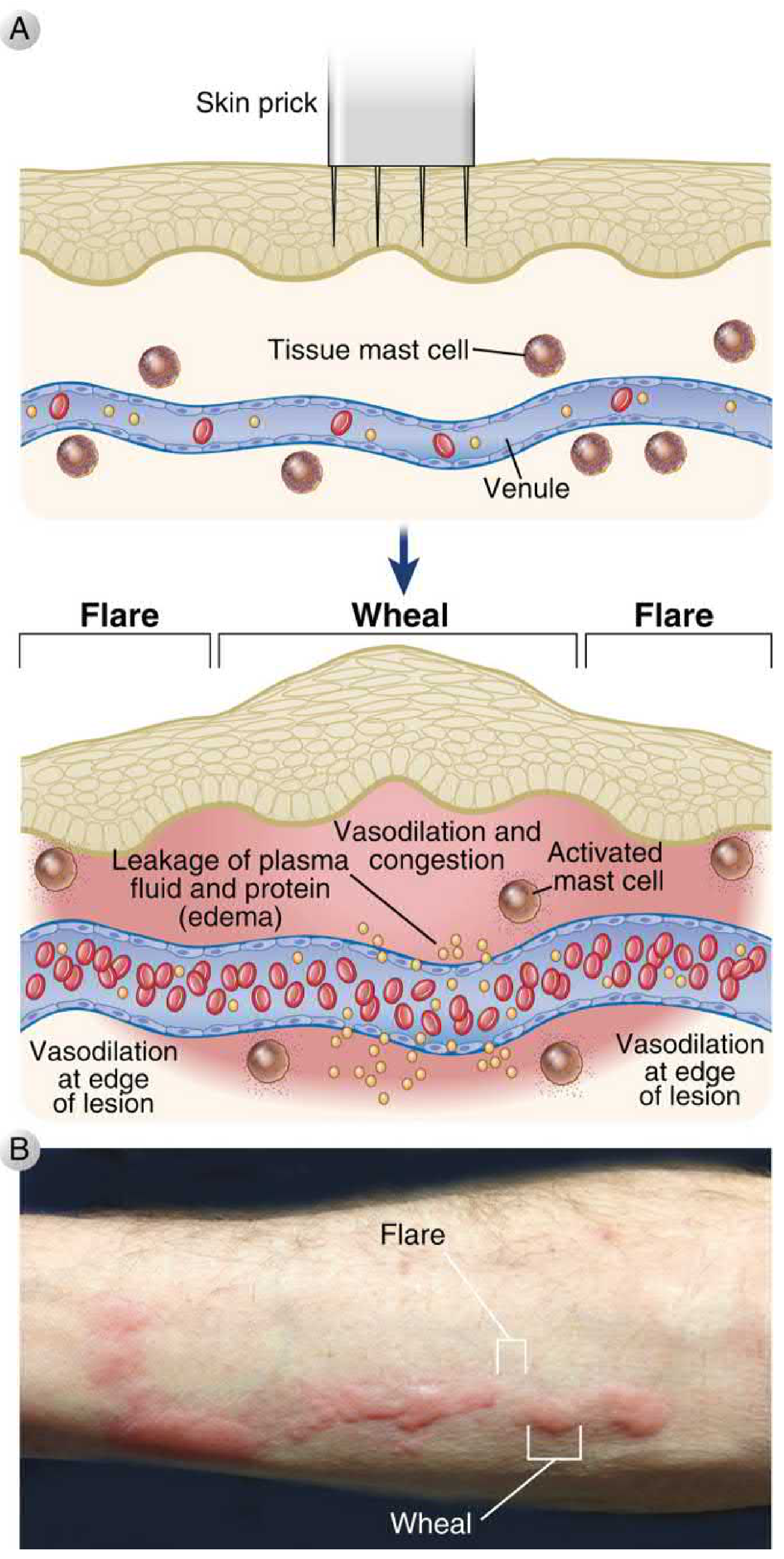
How It Is Triggered
Context 1 - Physical Injury (Dermatographism)
When a pointed instrument is drawn firmly across the skin, the mechanical force directly stimulates local cells and nerve endings, releasing vasoactive mediators (primarily histamine) and triggering a nerve reflex.
Context 2 - Allergic/Immunological (Skin Prick Test)
When an allergen contacts skin containing sensitized mast cells:
- The patient already has allergen-specific IgE bound to FcεRI receptors on dermal mast cells
- The allergen cross-links two adjacent IgE molecules on the mast cell surface
- This triggers mast cell degranulation - explosive release of preformed granule contents
- Histamine is the primary mediator of the wheal and flare response
- Other mediators released: tryptase, chymase, PGI₂, nitric oxide, leukotrienes
The Three Components
Component 1 - Red Reaction (within seconds)
- A small spot of reddening appears within a few seconds directly at the site of injury or injection
- Maximal at about 1 minute
- Caused by direct vasodilation of local capillaries and venules - a direct effect of histamine on capillary walls
- Mediated by H1 receptor activation → Ca²⁺-dependent activation of eNOS in endothelial cells → NO production → rapid vasodilation
- Independent of innervation - persists even after complete denervation
Component 2 - Flare (within 1-2 minutes)
- A bright red flush spreading ~1 cm beyond the original red spot
- Caused by arteriolar dilation in the surrounding skin
- The mechanism is the axon reflex - this is unique to the flare:
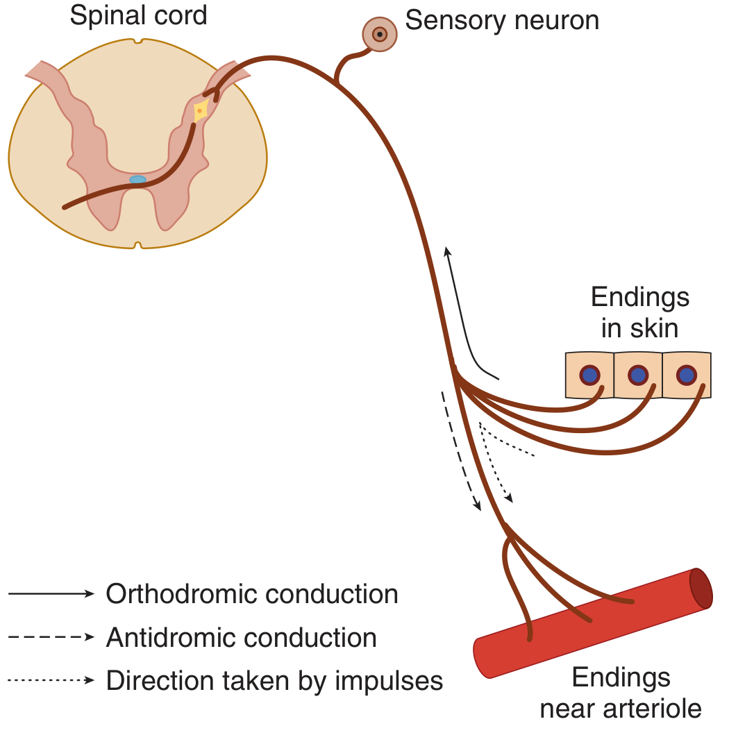
How the axon reflex works:
- Injury stimulates a sensory C-fiber nerve ending in the skin
- An action potential travels orthodromically toward the spinal cord
- At the branch point of the nerve fiber, the signal also travels antidromically (backward) down collateral branches to nearby arterioles
- These collateral branches release Substance P and CGRP (calcitonin gene-related peptide) from their terminals
- Substance P and CGRP cause arteriolar dilation → the flare
- This is why the flare is absent in:
- Locally anesthetized skin (nerve blocked)
- Denervated skin after sensory nerves have degenerated
- BUT it IS present immediately after nerve block or section above the injury site (nerve still intact distally)
Component 3 - Wheal (within 1-5 minutes)
- A raised, pale, soft swelling at the exact injection/injury site
- This is edema - fluid and plasma proteins leaking out of the circulation into the skin
- Caused by increased capillary and venular permeability:
- Histamine binds H1 receptors on venular endothelial cells
- Activates RhoA/ROCK pathway → contracts endothelial cells → gaps open between them
- Plasma proteins and fluid leak through these gaps into the tissue → edema = wheal
- Substance P also contributes to the wheal by increasing vascular permeability
- The wheal replaces the initial red reaction as it develops
Summary of All Three Components
| Component | Appearance | Timing | Cause | Mediator | Mechanism |
|---|---|---|---|---|---|
| Red Reaction | Small red spot at site | Within seconds | Capillary dilation | Histamine (H1) | Direct vasodilation via NO |
| Flare | Spreading redness ~1 cm around | 1-2 minutes | Arteriolar dilation | Substance P, CGRP | Axon reflex (antidromic conduction) |
| Wheal | Raised pale soft swelling at site | 1-5 minutes | Plasma leakage/edema | Histamine (H1) | Increased vascular permeability |
Key Clinical Points
In allergy testing (skin prick test):
- A wheal >3 mm greater than negative control = positive test
- It confirms allergen-specific IgE is present on dermal mast cells
- Antihistamines must be stopped before testing because they suppress the wheal and flare response:
- First-generation antihistamines (e.g., diphenhydramine): suppress for 48-72 hours
- Second-generation antihistamines (e.g., cetirizine, loratadine): suppress for 3-10 days
- Tricyclic antidepressants (e.g., amitriptyline): suppress for up to 7 days
The late-phase reaction follows 2-4 hours after the wheal and flare:
- Mast cell cytokines (TNF) upregulate endothelial adhesion molecules (E-selectin, ICAM-1)
- Influx of eosinophils, basophils, neutrophils, Th2 helper T cells
- Peaks at ~24 hours then subsides
- Responsible for the prolonged inflammation in conditions like chronic asthma and atopic dermatitis
Clinical manifestations of widespread wheal and flare:
- Urticaria (hives) - disseminated wheal-and-flare across the skin from systemic allergen exposure
- Dermographism - exaggerated physical wheal and flare on stroking the skin
Sources: Goodman & Gilman's Pharmacological Basis of Therapeutics; Ganong's Review of Medical Physiology 26th Ed.; Medical Physiology (Boron & Boulpaep); Cellular and Molecular Immunology (Abbas et al.)
How does inflammation happens
Inflammation is a protective vascular and cellular response to tissue injury or infection. The classic signs are redness, heat, swelling, pain, and loss of function - all driven by a coordinated sequence of events.
Searching sources...
Finding Sources
Finding Sources
Reading File
Finding Sources
Reading File
Reading File
Reading File
Reading File
Finding Sources
Finding Sources
Loading Image
Reading File
I now have all the detail needed for a thorough answer. Here it is:
How Inflammation Happens
Inflammation is the body's protective response to any harmful stimulus - infection, physical injury, chemical damage, or dead cells. It aims to eliminate the cause, clear damaged tissue, and begin repair. The whole process can be remembered as the Five R's:
Recognition → Recruitment → Removal → Regulation → Repair
Step 1 - Recognition of the Injurious Agent
When tissue is injured or invaded, sentinel cells (tissue macrophages, dendritic cells, mast cells, epithelial cells) immediately detect the threat through pattern recognition receptors:
- Toll-like receptors (TLRs) - detect bacterial cell wall components (LPS, peptidoglycan), viral RNA/DNA
- NOD-like receptors (NLRs) - detect intracellular pathogens
- Scavenger receptors - detect dead/necrotic cells
- Circulating plasma proteins - recognize microbes that enter the bloodstream
Once triggered, these cells release a flood of chemical mediators that kick off the next steps.
Step 2 - Vascular Changes (The Visible Signs of Inflammation)
The mediators act on local blood vessels, producing the classic cardinal signs:
2a. Vasodilation → Heat and Redness
- Histamine (from mast cells) and nitric oxide act on postcapillary venules
- Vessels dilate → blood flow increases dramatically
- This produces rubor (redness) and calor (heat)
2b. Increased Vascular Permeability → Swelling
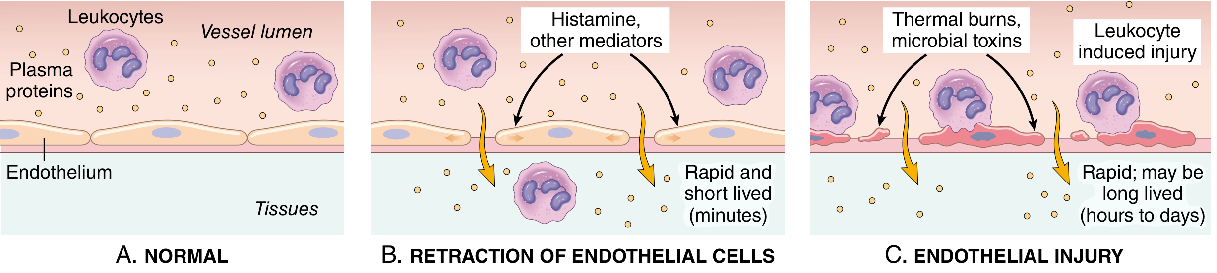
Two main mechanisms open the vessel wall:
1. Endothelial cell retraction (most common):
- Histamine, bradykinin, leukotrienes bind endothelial H1 receptors
- Activates RhoA/ROCK pathway → endothelial cells contract and pull apart
- Gaps form between adjacent cells → plasma proteins and fluid pour out
- Rapid and short-lived (15-30 minutes)
2. Direct endothelial injury (severe cases):
- Burns, microbial toxins, activated neutrophils directly damage the endothelium
- Endothelial cells die and detach, creating large leaks
- Sustained for hours to days
The protein-rich fluid that escapes is called an exudate. Its accumulation in tissue causes tumor (swelling / edema). The inflammatory pain - dolor - comes from bradykinin and prostaglandins stimulating nociceptors. The fifth sign, functio laesa (loss of function), follows from the combined effect of pain and swelling.
Step 3 - Leukocyte Recruitment (The Cellular Response)
As blood flow slows (stasis) from vessel dilation and fluid loss, leukocytes (mainly neutrophils first, then monocytes) begin a choreographed journey from blood to tissue in four phases:
Phase 1 - Margination
- Normal blood flow keeps cells in the central axial column
- As blood slows, leukocytes drift to the vessel wall and start rolling along the endothelium
Phase 2 - Rolling (Selectin-mediated)
- Activated endothelium expresses E-selectin and P-selectin (induced by TNF, IL-1, histamine)
- Leukocytes carry sialylated carbohydrate ligands (e.g., sialyl-Lewis X) that bind selectins
- This creates a loose, tethering and rolling interaction - like velcro at low strength
Phase 3 - Firm Adhesion (Integrin-mediated)
- Chemokines (e.g., IL-8/CXCL8) are displayed on the endothelial surface
- They activate leukocyte integrins (LFA-1, MAC-1) → conformational change → high-affinity binding
- Integrins bind their endothelial ligands ICAM-1 and VCAM-1 (upregulated by TNF and IL-1)
- Leukocyte now stops firmly on the endothelial surface
Phase 4 - Transmigration / Diapedesis
- Leukocyte squeezes between endothelial cells (paracellular) at intercellular junctions
- Guided by CD31 (PECAM-1) homotypic interactions between the leukocyte and endothelial cell
- Once through the vessel wall, it crosses the basement membrane using collagenases
Phase 5 - Chemotaxis
- Now in the tissue, the leukocyte follows a gradient of chemoattractants toward the source:
- Bacterial products (e.g., fMLP - formyl-met-leu-phe)
- C5a (complement fragment)
- Leukotriene B4 (LTB4)
- IL-8 / CXCL8 (cytokine chemokine)
- The leukocyte reorganizes its cytoskeleton, extends filopodia at the leading edge, and migrates toward the highest concentration
Step 4 - Elimination (Phagocytosis and Killing)
Once at the site, neutrophils (first 6-24 hrs) and macrophages (24-48 hrs onward) destroy the invader:
| Cell | Timing | Mechanism |
|---|---|---|
| Neutrophils | First 6-24 hours | Rapid degranulation, respiratory burst (ROS), myeloperoxidase (bleach-like HOCl), NETs |
| Macrophages | After 24-48 hours | Phagocytosis, nitric oxide via iNOS, prolonged cytokine production, antigen presentation |
How phagocytosis works:
- Opsonins (IgG antibodies and C3b complement) coat the target
- Leukocyte Fc receptors and complement receptors recognize the coated target
- Pseudopods engulf it into a phagosome
- Phagosome fuses with lysosome → phagolysosome
- Killing by:
- Reactive oxygen species (ROS) via NADPH oxidase (respiratory burst)
- Nitric oxide (NO) via iNOS
- Lysosomal enzymes (elastase, cathepsins, lysozyme)
- Defensins - antimicrobial peptides
Key Chemical Mediators
| Mediator | Source | Main Action |
|---|---|---|
| Histamine | Mast cells, basophils | Vasodilation, increased permeability (rapid, transient) |
| Prostaglandins (PGE₂, PGI₂) | All cells via COX pathway | Vasodilation, fever, pain sensitization |
| Leukotrienes (LTB₄, LTC₄, LTD₄) | Leukocytes via LOX pathway | Chemotaxis (LTB₄), bronchoconstriction, permeability |
| Bradykinin | Plasma kinin system | Pain, vasodilation, permeability |
| TNF and IL-1 | Macrophages | Endothelial activation, fever, acute phase response |
| IL-8 / CXCL8 | Macrophages, endothelium | Neutrophil chemotaxis |
| Complement (C3a, C5a) | Plasma | Mast cell degranulation, opsonization, chemotaxis |
| Nitric oxide (NO) | Endothelium, macrophages | Vasodilation, bactericidal |
Outcomes of Acute Inflammation
Injurious agent eliminated?
↓
YES → Resolution → Tissue repair → Full restoration
↓
NO → Persistent injury
↓
Chronic inflammation (macrophages, lymphocytes, fibrosis)
↓
OR Abscess formation (walled-off pus)
↓
OR Systemic spread → Sepsis
The Classic 5 Signs (Cardinal Signs)
| Latin | English | Mechanism |
|---|---|---|
| Rubor | Redness | Vasodilation → increased blood flow |
| Calor | Heat | Increased blood flow + metabolic activity |
| Tumor | Swelling | Exudate accumulation (edema) |
| Dolor | Pain | Bradykinin + PGE₂ stimulate nociceptors |
| Functio laesa | Loss of function | Consequence of pain and swelling |
Sources: Robbins & Cotran Pathologic Basis of Disease (10th Ed.) - Chapter 3: Inflammation and Repair
This is a shared conversation. Sign in to Orris to start your own chat.