
How to remember facial nerve course branches clinical significance
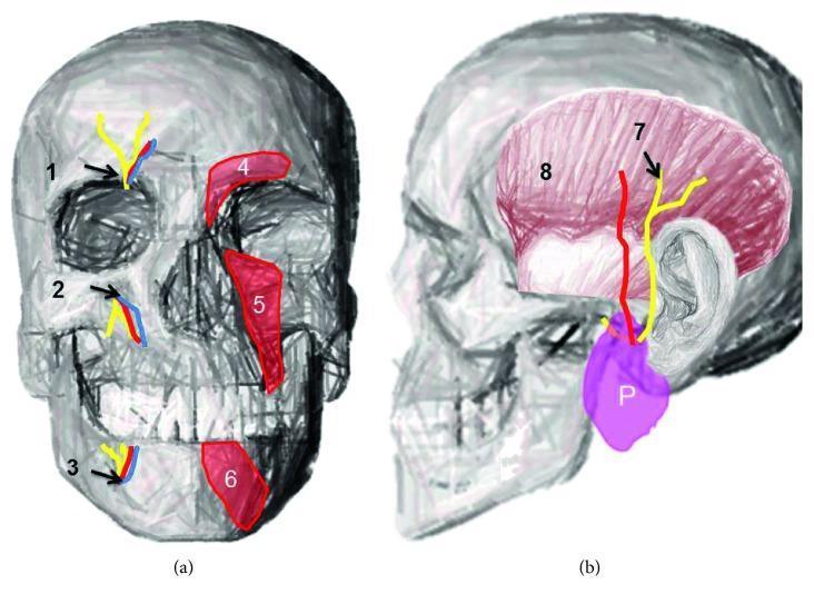
five branches of facial nerve temporal zygomatic buccal marginal mandibular cervical
Bell's palsy peripheral facial nerve palsy forehead involvement
Remembering the Facial Nerve (CN VII): Course, Branches, and Clinical Significance
1. The Course — 6 Segments ("I'M LT ME" walk)
| # | Segment | Location | Memory hook |
|---|---|---|---|
| 1 | Intracranial (cisternal) | Pons -> cerebellopontine angle (CPA) -> internal acoustic meatus | Travels with CN VIII here - acoustic neuromas compress both |
| 2 | Meatal (canalicular) | Inside internal acoustic meatus | Narrowest, most crowded segment - most vulnerable to compression edema in Bell's palsy |
| 3 | Labyrinthine | Meatal fundus to geniculate ganglion | Shortest segment; geniculate ganglion = first branch point (greater petrosal nerve) |
| 4 | Tympanic (horizontal) | Along medial wall of middle ear, below lateral semicircular canal | Runs just under the incus - vulnerable in ear/mastoid surgery |
| 5 | Mastoid (vertical) | Descends to stylomastoid foramen | Gives off nerve to stapedius and chorda tympani here |
| 6 | Extratemporal | Exits stylomastoid foramen -> through parotid gland | Splits into 5 terminal branches ("pes anserinus") |
2. The Three Intratemporal Branches — "Greater, Stapedius, Chorda" (in order given off)
- Greater petrosal nerve (at geniculate ganglion) - parasympathetic to lacrimal gland + mucous glands of nose/palate; taste doesn't travel here.
- Nerve to stapedius (mastoid segment) - dampens stapedius muscle.
- Chorda tympani (near end of mastoid segment) - taste anterior 2/3 tongue + parasympathetic to submandibular/sublingual glands.
- Lesion at/before geniculate ganglion (e.g., CPA tumor) -> everything affected: weakness + dry eye + hyperacusis + loss of taste + dry mouth.
- Lesion between geniculate ganglion and stapedius branch -> hyperacusis + taste loss + dry mouth, but tearing preserved.
- Lesion between stapedius and chorda tympani -> taste loss + dry mouth only, no hyperacusis.
- Lesion distal to stylomastoid foramen (parotid tumor, facial trauma, most Bell's palsy variants) -> pure motor weakness only - taste, tearing, and hearing all normal.
3. The Five Extratemporal (Terminal) Branches — Classic Mnemonic
- Temporal
- Zygomatic
- Buccal
- Marginal mandibular
- Cervical
Clinical significance of each - tie the muscle to the deficit:
| Branch | Muscle(s) | If injured | Where it matters clinically |
|---|---|---|---|
| Temporal | Frontalis, corrugator | Can't raise eyebrow/wrinkle forehead | Brow lifts, temporal craniotomy, forehead flaps |
| Zygomatic | Orbicularis oculi | Can't close eye -> exposure keratopathy | Superficial parotidectomy - protect the eye during surgery/anesthesia if injured |
| Buccal | Buccinator, orbicularis oris | Drooping cheek, food pocketing | Runs alongside the parotid duct - injured together in duct trauma/repair |
| Marginal mandibular | Depressor anguli oris | Asymmetric smile, drooling from corner of mouth | Most commonly injured branch - runs superficially below the mandibular angle; at risk in submandibular gland excision, neck dissection, facelifts |
| Cervical | Platysma | Minimal cosmetic deficit | Least clinically significant |
4. Localizing Upper vs Lower Motor Neuron Lesions - the "Forehead Rule"
- UMN lesion (stroke) -> forehead spared, only lower face droops.
- LMN lesion (Bell's palsy, nerve trunk injury) -> entire hemiface affected, including inability to raise the eyebrow or close the eye.
Quick recap mnemonic chain
- Course: Intracranial → Meatal → Labyrinthine → Tympanic → Mastoid → Extratemporal
- Intratemporal branches (order matters for localization): Greater petrosal → Stapedius nerve → Chorda tympani
- Extratemporal branches: To Zanzibar By Motor Car (Temporal, Zygomatic, Buccal, Marginal mandibular, Cervical)
- Forehead spared = UMN/stroke; forehead involved = LMN/Bell's palsy
Create a visual anatomy poster of facial nerve course and branches
A medical anatomy poster illustrating the course and branches of the facial nerve (cranial nerve VII). Show the nerve emerging from the brainstem at the pons, passing through the internal acoustic meatus, through the facial canal in the temporal bone including the geniculate ganglion, greater petrosal nerve, nerve to stapedius, and chorda tympani, then exiting the stylomastoid foramen. Show it entering the parotid gland and dividing at the pes anserinus into the five terminal branches: temporal, zygomatic, buccal, marginal mandibular, and cervical branches, spreading across the face to the muscles of facial expression. Label each segment and branch clearly with anatomical labels, include a small side panel showing the muscles innervated by each terminal branch (frontalis, orbicularis oculi, buccinator/orbicularis oris, depressor anguli oris, platysma). Educational medical illustration style, clean line diagram with labeled leader lines, suitable for a classroom or clinical teaching poster.
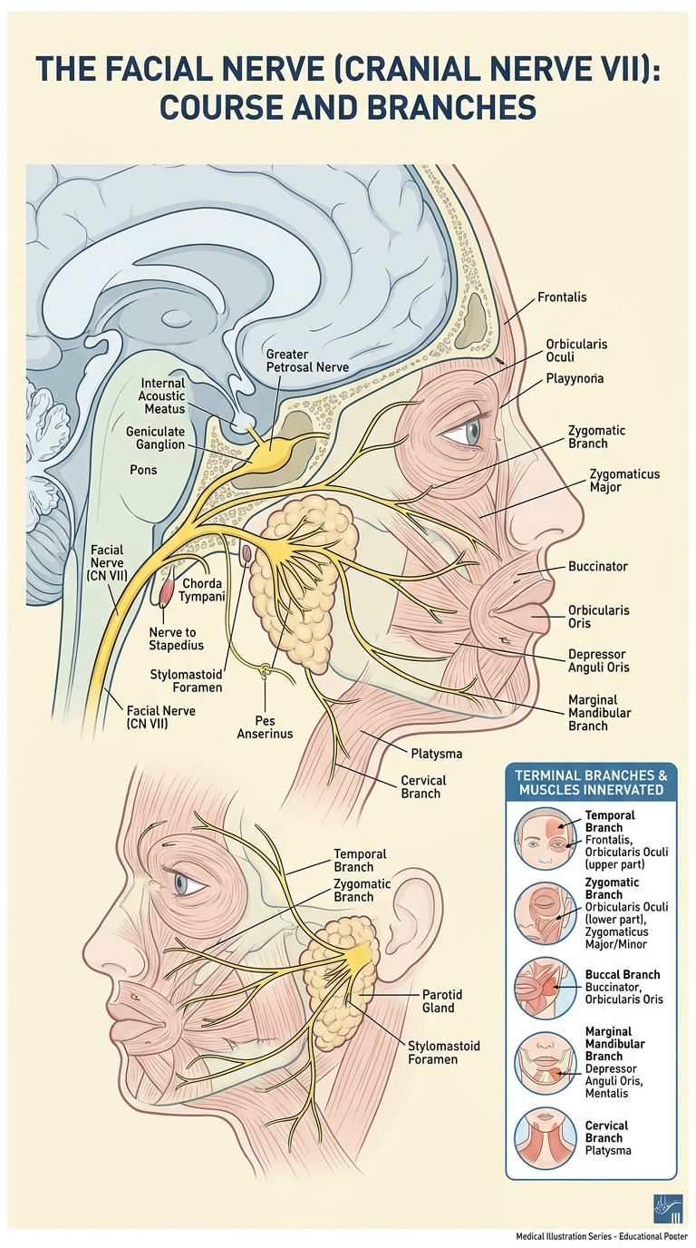
- Intracranial to intratemporal course: brainstem (pons) → internal acoustic meatus → facial canal (geniculate ganglion, greater petrosal nerve, nerve to stapedius, chorda tympani) → stylomastoid foramen
- Extratemporal course: entry into the parotid gland → pes anserinus bifurcation → five terminal branches (temporal, zygomatic, buccal, marginal mandibular, cervical)
- Muscle targets panel: frontalis, orbicularis oculi, buccinator/orbicularis oris, depressor anguli oris, and platysma, tied to each terminal branch