
Radiation enteritis
Reading File
Radiation enteritis is inflammation/injury of the bowel caused by radiation therapy, occurring as an acute or chronic complication. Let me search the medical library for comprehensive coverage.
Searching sources...
Finding Sources
Finding Sources
Searching PubMed
"radiation enteritis" AND management
Good results. Now let me read the most detailed sources in parallel.
Reading File
Reading File
Reading File
Excellent data. Let me read the Schwartz surgical management section and the Sleisenger GI treatment section for more depth.
Reading File
Finding Sources
Now let me read the Sleisenger section for more detailed medical management and the radiology findings section.
Reading File
Reading File
I now have very comprehensive data. Let me also fetch the two histopathology images for inclusion.
Reading File
Excellent - I have comprehensive data including the therapeutic options table. Now I have enough to write a detailed, well-structured answer.
Radiation Enteritis
Radiation enteritis refers to injury to the small intestine caused by ionizing radiation, most often as a complication of radiotherapy for abdominal and pelvic malignancies (cervix, endometrium, ovary, bladder, prostate, rectum). It presents as two clinically distinct syndromes.
Epidemiology
- Acute radiation enteritis occurs in approximately 75% of patients undergoing abdominal/pelvic radiation.
- Chronic radiation enteritis develops in 5-15% of irradiated patients.
- The risk rises sharply with doses exceeding 5000 cGy (50 Gy); serious late complications are unusual below 4000 cGy.
- At doses of 70 Gy or greater, toxicity rises precipitously.
(Schwartz's Principles of Surgery, 11th Ed.)
Pathophysiology
Acute Phase
Radiation acts directly by causing double-strand DNA breaks and indirectly through free radical generation. The primary mechanism of cell death is apoptosis.
- The small intestinal epithelium (with a turnover every 3-5 days) is acutely vulnerable.
- Radiation targets clonogenic stem cells in the crypts of Lieberkühn.
- Within 2-4 weeks: leukocyte infiltration, crypt microabscess formation, villus blunting, mucosal denudement, ulceration, and edema.
- Impaired absorption of fats, proteins, carbohydrates, bile salts, and vitamin B12.
- Impaired ileal bile salt absorption → conjugated bile salts reach the colon → deconjugation by bacteria → intraluminal salt/water accumulation → cholerheic diarrhea.
- Lactose maldigestion → bacterial fermentation → flatulence, distension, diarrhea.
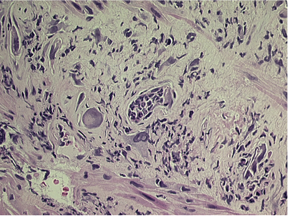
Fig. 41.1 - Histopathology of acute radiation enteritis: submucosal reaction with large, bizarre radiation fibroblasts and microabscesses (Sleisenger & Fordtran's)
Chronic Phase
The acute mucosal injury heals, but chronic changes are driven by vascular injury, not mucosal damage:
- Progressive occlusive vasculitis / obliterative endarteritis of submucosal blood vessels (foam cell formation, subintimal fibrosis).
- Leads to chronic ischemia, submucosal fibrosis, and fibrotic changes affecting all layers of the bowel wall.
- Results in strictures, abscesses, fistulas, and ultimately vascular insufficiency with potential necrosis/perforation.
- Endothelial cell apoptosis actually precedes epithelial cell apoptosis, confirming vascular injury as the primary event.
(Sleisenger & Fordtran's GI & Liver Disease, 7th Ed.; Goldman-Cecil Medicine)
Risk Factors
| Category | Factors |
|---|---|
| Radiation dose/technique | Total dose >4500-5000 cGy, large field size, high dose per fraction (>2 Gy/fraction) |
| Vascular comorbidities | Hypertension, diabetes mellitus, coronary artery disease (limit splanchnic perfusion) |
| Surgical history | Previous laparotomy → adhesions fixing bowel loops in radiation field |
| Patient factors | Female sex, older age, thin body habitus (more small bowel in pelvis) |
| Concurrent chemotherapy | 5-FU, capecitabine, doxorubicin, actinomycin D, methotrexate (radiation sensitizers) |
(Sabiston Textbook of Surgery, 21st Ed.; Sleisenger & Fordtran's)
Clinical Presentation
Acute Radiation Enteritis
- Onset: During the 3rd week of a fractionated radiation course (typically after ~18-22 Gy)
- Symptoms: Nausea, vomiting, diarrhea (reported in 20-70% of patients), crampy abdominal pain, anorexia, malaise
- Course: Resolves within 2-6 weeks after completion of radiotherapy
- Diagnosis is clinical; CT is indicated if peritonitis signs develop to rule out other acute abdominal pathology.
Chronic Radiation Enteritis
- Onset: Usually within 18 months to 6 years after radiation (can appear from several months to up to 30 years later)
- Most affected segment: Terminal ileum
- Common presentations:
- Diarrhea (from bile acid malabsorption, SIBO, dysmotility)
- Partial/complete small bowel obstruction (nausea, vomiting, distension, crampy pain, weight loss)
- Acute or chronic intestinal hemorrhage
- Abscess or fistula formation
- Malabsorption and nutritional deficiencies (B12, fat-soluble vitamins, steatorrhea)
- Lactose intolerance
- Protein-losing enteropathy
Pathophysiologic features of chronic radiation enteropathy and their manifestations:
| Pathophysiologic Feature | Clinical Manifestation |
|---|---|
| Mucosal dysfunction | Lactose intolerance, Vitamin B12 deficiency, Steatorrhea |
| Stricture/blind loop with SIBO | Diarrhea |
| Intestinal dysmotility | Bloating, constipation, diarrhea |
| Abnormal bile acid recirculation | Cholerheic diarrhea |
(Sleisenger & Fordtran's, Table 41.2)
Diagnosis
Acute: Clinical diagnosis based on context; no specific tests usually required.
Chronic:
- Review radiation treatment records: total dose, fractionation, treatment volume, high-dose areas.
- Enteroclysis (small bowel enema): Most accurate imaging for chronic radiation enteritis - sensitivity and specificity >90%. Shows widely separated loops, luminal narrowing, loss of mucosal folds, ulceration.
- CT scan: Less sensitive/specific than enteroclysis for mucosal detail, but best for assessing mural thickening.
- Acute/subacute phase: "Target sign" - submucosal edema + mucosal hyperenhancement
- Chronic fibrotic phase: Homogeneous mural thickening
- Radiological features (barium/CT): Thickening of valvulae conniventes, mural thickening, effacement of mucosal pattern, ulceration, fixation and angulation of loops. "Mucosal tacking" (spiking and distortion of antimesenteric mucosal folds from adhesions to inflamed mesentery) is characteristic. Luminal narrowing and sinuses/fistulae can be seen.
- Radiation-induced small bowel strictures are typically long and not amenable to endoscopic therapy.
(Schwartz's; Grainger & Allison's Diagnostic Radiology)
Treatment
Acute Radiation Enteritis
Most cases are self-limited and require only supportive care:
- Antiemetics
- Oral hydration; IV fluids if severe diarrhea-induced dehydration
- Rarely requires dose reduction or cessation of radiation therapy
Chronic Radiation Enteritis
Medical management (often unsatisfactory but attempted first):
| Pathophysiologic Feature | Therapeutic Options |
|---|---|
| Nutritional deficits | Correction of specific deficits (B12, fat-soluble vitamins), low-fat diet, lactose-free diet, elemental diet, TPN |
| Intestinal dysmotility | Loperamide (2 mg PO QID), loperamide-N-oxide (3 mg PO BID), octreotide, prokinetic agents |
| Bile acid malabsorption | Cholestyramine (PO three times daily) |
| Inflammation | Mesalamine, corticosteroids (limited success); NSAIDs (e.g., naproxen 250-500 mg PO BID) |
| SIBO from strictures/bypass | Antibiotics |
| Refractory disease | Hyperbaric oxygen therapy (promotes neoangiogenesis in hypoxic tissue) |
(Goldman-Cecil Medicine; Sleisenger & Fordtran's, Table 41.3)
A 2025 systematic review and meta-analysis (PMID: 41140687) confirmed hyperbaric oxygen therapy as a beneficial intervention for radiation enteritis with favorable clinical parameters.
Surgical Management
Surgery is difficult, high-morbidity, and should be reserved for specific indications:
- High-grade obstruction
- Perforation
- Hemorrhage
- Intra-abdominal abscess
- Fistula formation
Surgical principles:
- Goal: limited resection of diseased intestine with primary anastomosis between healthy (non-irradiated) bowel segments
- Distinguishing irradiated from normal bowel intraoperatively by gross inspection or frozen section is unreliable.
- Anastomosis between irradiated segments carries leak rates up to 50%.
- If limited resection is not achievable: intestinal bypass (except when hemorrhage is the indication, as bypass leaves the bleeding segment in situ).
- Extensive resections may lead to short bowel syndrome.
- Reported surgical mortality: ~10% (mean across series).
(Schwartz's Principles of Surgery, 11th Ed.)
Prevention
Radiation technique modifications:
- Keep total dose below 5000 cGy where clinically possible.
- Multibeam/multi-field radiation techniques to minimize peak exposure.
- Intensity-modulated radiotherapy (IMRT) and conformal radiotherapy.
- Tilt table positioning and bladder distention in prone position (moves bowel out of pelvis).
- Radiopaque clips (titanium) placed at original surgery for precise tumor targeting.
Surgical techniques (for patients undergoing pelvic surgery before planned RT):
- Reperitonealization
- Omental transposition
- Absorbable mesh slings to keep small bowel out of the pelvis/radiation field
Pharmacologic radioprotection:
- ACE inhibitors and statins - reduce acute GI symptoms during pelvic RT
- Sucralfate - may help bleeding from radiation proctitis (no evidence for small bowel prevention)
- Superoxide dismutase (free radical scavenger)
- Pentoxifylline + tocopherols (vitamin E compounds)
- Amifostine (WR-2721) - sulfhydryl compound that scavenges free radicals
- Antioxidants (vitamins A, E, beta-carotene)
- Oral sulfasalazine - may reduce acute radiation-induced enteritis
- Probiotics (under active investigation in randomized trials)
(Sabiston Textbook of Surgery, 21st Ed.; Schwartz's Principles of Surgery)
Key Summary Points:
- Acute enteritis (~75% incidence) is transient and self-limited; severity does NOT predict chronic disease.
- Chronic enteritis (5-15%) is driven by obliterative vasculitis and fibrosis - the vascular injury is the primary event.
- Terminal ileum is the most commonly affected site.
- Enteroclysis is the most accurate imaging test for chronic disease.
- Medical management is often unsatisfactory; surgery carries ~10% mortality.
- Anastomosis must be between healthy, non-irradiated bowel; irradiated-to-irradiated anastomoses leak in up to 50% of cases.
Management of crohn disease and ulcerative colitis
Your credits are exhausted. You can purchase a top-up or subscribe to a plan to continue using Orris. If you're already subscribed, your daily credits will reset tomorrow.
This is a shared conversation. Sign in to Orris to start your own chat.