Immune Response
Reading File
Asking for Preferences
Finding Sources
Finding Sources
Searching Images
innate vs adaptive immune response overview diagram
Finding Sources
Finding Sources
Reading File
Reading File
Reading File
Reading File
Reading File
Loading Image
Here is a comprehensive intermediate-level overview of the immune response, drawn from Robbins & Kumar Basic Pathology and Cellular and Molecular Immunology.
The Immune Response
The immune system defends against pathogens through two integrated arms that operate on different timescales and use different recognition strategies.
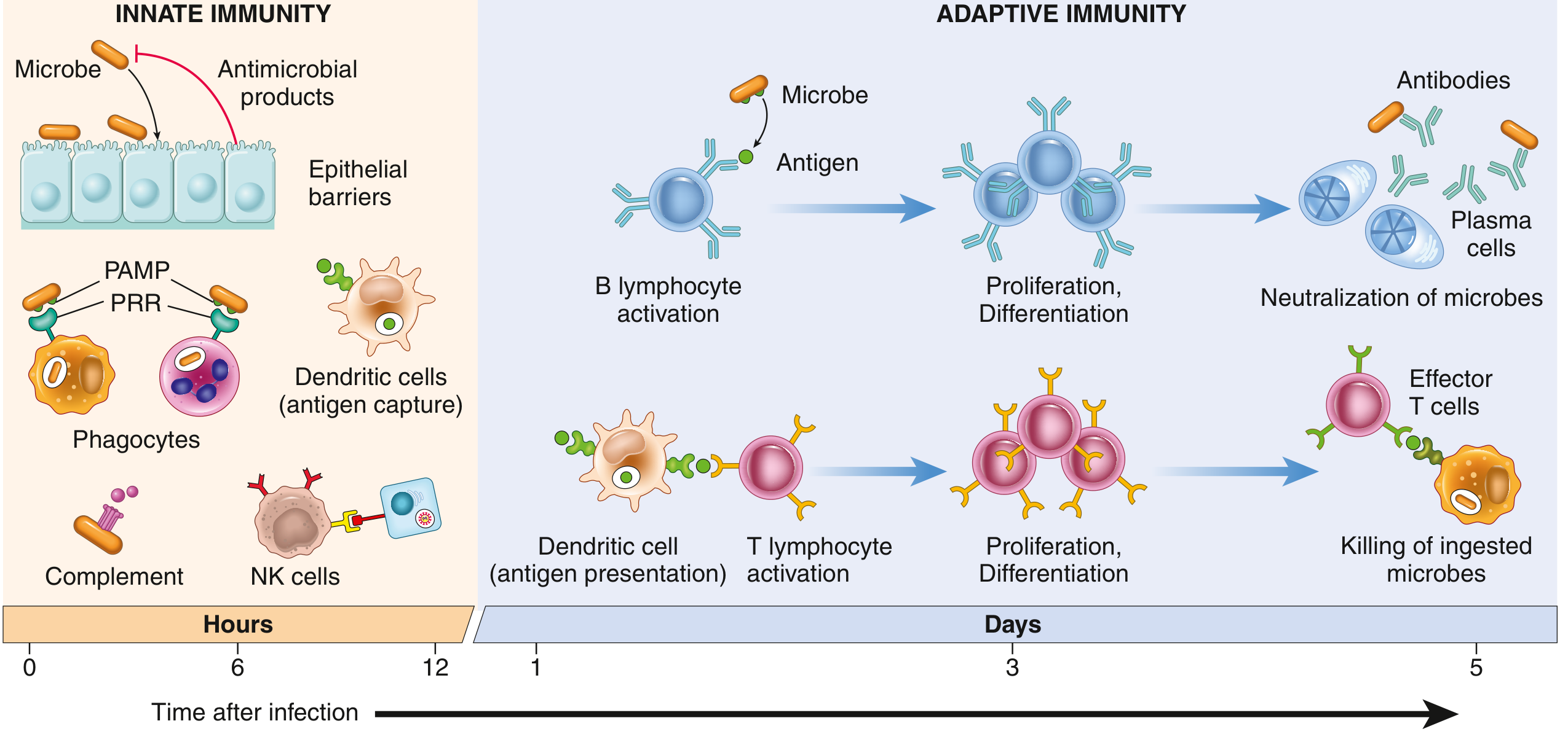
1. Innate Immunity (Hours 0–12)
Innate immunity is the first-line, rapid, non-specific response. It responds almost immediately to microbes and injured cells, and repeated exposures induce virtually identical responses — it does not improve with re-exposure.
Components
| Component | Role |
|---|---|
| Epithelial barriers | Physical + chemical block (skin, mucosa, antimicrobial peptides) |
| Phagocytes (neutrophils, macrophages) | Engulf and destroy pathogens |
| Dendritic cells (DCs) | Capture antigens; bridge innate → adaptive |
| NK cells | Kill virus-infected and tumor cells without prior sensitization |
| Complement system | Opsonization, lysis, recruitment of phagocytes |
| Mast cells & innate lymphoid cells (ILCs) | Cytokine release, barrier defense |
Pattern Recognition
Innate immune cells use pattern recognition receptors (PRRs) to detect conserved microbial structures:
- PAMPs (Pathogen-Associated Molecular Patterns) — structures shared by groups of microbes (e.g., LPS on gram-negative bacteria)
- DAMPs (Damage-Associated Molecular Patterns) — signals from injured/necrotic host cells (e.g., uric acid, ATP)
Major PRR families:
| Receptor | Location | What it detects |
|---|---|---|
| Toll-like receptors (TLRs) | Plasma membrane + endosomes | Bacterial LPS, viral RNA/DNA |
| NOD-like receptors (NLRs) | Cytosol | Necrotic cell products, ion disturbances, microbial fragments |
| RIG-like receptors | Cytosol | Cytoplasmic viral RNA |
| C-type lectin receptors | Plasma membrane (macrophages, DCs) | Bacterial/fungal polysaccharides |
| Cytosolic DNA sensors | Cytosol | Aberrant cytoplasmic DNA |
The Inflammasome: Several NLRs signal via this cytosolic multiprotein complex → activates caspase-1 → cleaves pro-IL-1β into active IL-1β. This drives fever and inflammation. Gain-of-function NLR mutations cause autoinflammatory syndromes; recognition of urate crystals by NLRs underlies gout inflammation.
Reactions of Innate Immunity
- Inflammation — cytokines + complement recruit leukocytes that destroy pathogens and clear dead cells
- Antiviral defense — Type I interferons (IFN-α/β) produced by virus-infected cells degrade viral nucleic acids and inhibit viral replication in neighboring cells
2. Adaptive Immunity (Days 1–5+)
Adaptive immunity is specific, diverse, and has memory. It can recognize an estimated 10⁷–10⁹ distinct antigenic determinants. The two major branches are humoral (antibody-mediated) and cell-mediated immunity.
Cardinal Features
| Feature | Description |
|---|---|
| Specificity | Each lymphocyte clone recognizes a unique epitope (antigen determinant) |
| Diversity | The lymphocyte repertoire can distinguish ~10⁷–10⁹ different antigens |
| Clonal selection | Antigen selects and activates pre-existing antigen-specific lymphocyte clones (Burnet, 1957) |
| Memory | Secondary responses are faster, larger, and qualitatively stronger |
| Contraction | Immune responses resolve after pathogen clearance, restoring homeostasis |
| Self-tolerance | The immune system normally does not attack self-tissues |
Cells of Adaptive Immunity
T Lymphocytes
Recognize peptide antigens presented on MHC molecules by antigen-presenting cells (APCs — mainly dendritic cells):
| T cell subset | MHC | Function | Key cytokines |
|---|---|---|---|
| CD4⁺ Helper T (Th1) | MHC II | Activates macrophages; helps CD8⁺ T cells; fights intracellular microbes | IFN-γ, TNF-α |
| CD4⁺ Helper T (Th2) | MHC II | Promotes IgE/IgG4 production; eosinophil activation; anti-parasite | IL-4, IL-5, IL-13 |
| CD4⁺ Helper T (Th17) | MHC II | Recruits neutrophils; mucosal defense vs. extracellular bacteria/fungi | IL-17A/F, IL-22 |
| CD4⁺ Regulatory T (Treg) | MHC II | Suppresses excessive immune responses; maintains tolerance | IL-10, TGF-β |
| CD8⁺ Cytotoxic T (CTL) | MHC I | Directly kills virus-infected cells and tumor cells | Perforin, granzymes |
Dendritic cells are the key bridge — they phagocytose antigens in peripheral tissues, process them, migrate to lymph nodes, and present peptides to naïve T cells to initiate the adaptive response.
B Lymphocytes & Antibodies
B cells recognize antigens directly via their B-cell receptors (BCRs), and with T-cell help undergo:
- Proliferation and differentiation → plasma cells (effector B cells)
- Antibody secretion (immunoglobulins: IgM, IgG, IgA, IgE, IgD)
- Class switching — driven by cytokines from T helper cells (e.g., IFN-γ → IgG; IL-4 → IgE)
- Memory B cell formation — enables rapid secondary responses
Antibody functions: neutralization, opsonization (marking for phagocytosis), complement activation, antibody-dependent cellular cytotoxicity (ADCC).
3. Innate–Adaptive Crosstalk
These two systems are mutually supportive, not separate:
- Innate cells (especially DCs) activate adaptive immunity by presenting antigens + providing co-stimulatory signals
- Cytokine crosstalk (e.g., IL-12 from DCs → Th1 differentiation; IL-4 → Th2; IL-6/IL-23 → Th17) matches the adaptive response to the type of threat
- Under normal mucosal conditions: tolerance is maintained (Tregs + controlled Th2 → IgA secretion without inflammation)
- Under pathogen threat: protective inflammatory responses are mounted with appropriate T-helper polarization
4. Summary Comparison
| Feature | Innate | Adaptive |
|---|---|---|
| Speed | Minutes–hours | Days–weeks |
| Specificity | Broad (PAMPs/DAMPs) | Highly specific (epitopes) |
| Memory | None | Yes |
| Key cells | Neutrophils, macrophages, NK cells, DCs | T cells, B cells |
| Key molecules | Cytokines, complement, interferons | Antibodies, TCR, BCR |
| Diversity | ~100 PRRs | 10⁷–10⁹ specificities |
Sources: Robbins & Kumar Basic Pathology (9780323790185); Cellular and Molecular Immunology (9780323757485); Cummings Otolaryngology Head and Neck Surgery (9780323612173)
Immune Response physiology
Finding Sources
Finding Sources
Finding Sources
Finding Sources
Finding Sources
Finding Sources
Finding Sources
Finding Sources
Reading File
Reading File
Reading File
Reading File
Reading File
Loading Image
Loading Image
Reading File
Here is a detailed physiological account of the immune response, drawn from Cellular and Molecular Immunology (Abbas et al.) and Robbins & Kumar Basic Pathology.
Immune Response — Physiology
The immune response operates as a highly coordinated physiological system involving cellular recognition, intracellular signaling, clonal expansion, effector function, and regulated resolution.
1. Physiological Sequence of Events
When a pathogen breaches host barriers, the immune response unfolds in overlapping phases:
Pathogen entry
     │
     ▼
Innate recognition (minutes–hours)
     │
     ▼
Antigen capture & presentation by DCs (hours–day 1)
     │
     ▼
Adaptive activation: T cell & B cell responses (days 3–5)
     │
     ▼
Effector phase: antibodies, cytotoxic killing, macrophage activation
     │
     ▼
Memory formation + response contraction
2. Innate Immune Physiology
Pattern Recognition & Signal Transduction
Innate immune cells (macrophages, DCs, neutrophils, epithelial cells) detect pathogens via pattern recognition receptors (PRRs):
| Receptor Class | Location | Triggers | Downstream Effect |
|---|---|---|---|
| TLRs (Toll-like receptors) | Plasma membrane / endosomes | Bacterial LPS, viral RNA/DNA | NF-κB activation → pro-inflammatory cytokines (TNF-α, IL-1, IL-6, IL-12), Type I IFNs |
| NLRs (NOD-like receptors) | Cytosol | Uric acid, ATP, microbial fragments | Inflammasome → Caspase-1 activation → IL-1β, IL-18 production |
| RIG-I-like receptors | Cytosol | Viral dsRNA | IRF3/IRF7 activation → Type I interferons (IFN-α/β) |
| C-type lectins | Plasma membrane | Fungal/bacterial polysaccharides | Phagocytosis, inflammatory signaling |
Inflammasome physiology: NLRs (e.g., NLRP3) sense cell stress → assemble a cytosolic platform → activate caspase-1 → cleave pro-IL-1β into IL-1β → fever, leukocyte recruitment, systemic acute-phase response. This pathway underlies gout (urate crystals), autoinflammatory syndromes, and contributes to atherosclerosis and metabolic syndrome.
Innate Effector Mechanisms
1. Inflammation:
- Cytokines (TNF-α, IL-1β, IL-6) and complement fragments (C3a, C5a) act on vascular endothelium
- Vasodilation + increased permeability → edema, heat, redness
- Leukocyte recruitment: selectins (rolling) → integrins (adhesion) → chemokines (migration to tissue)
- Recruited neutrophils and macrophages phagocytose and destroy pathogens
2. Antiviral defense:
- Virus-infected cells produce Type I IFNs (IFN-α, IFN-β)
- IFNs act on neighboring cells → upregulate antiviral enzymes (RNase L, PKR) → degrade viral RNA, halt translation
- NK cells recognize loss of MHC I on infected/tumor cells → release perforin + granzymes → target cell apoptosis
3. Complement System Physiology
The complement system amplifies innate defense via a cascade of >20 serum proteins. The critical step is cleavage of C3 by C3 convertase, generating C3a and C3b.
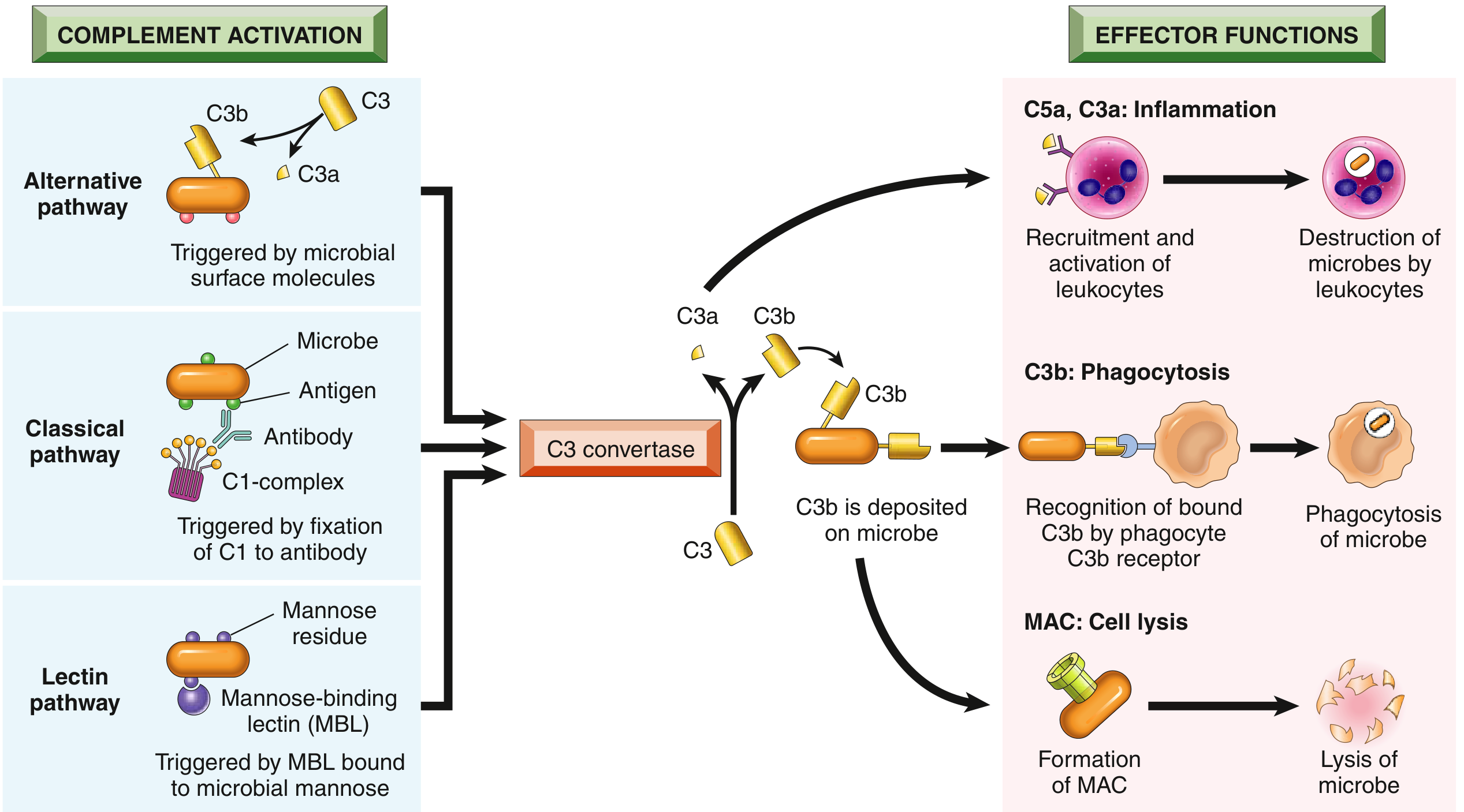
Three Activation Pathways
| Pathway | Trigger | Initiating Step |
|---|---|---|
| Classical | Antigen–antibody complexes (IgM or IgG bound to antigen) | C1q binds Fc region → C1r/s activated → cleaves C4 + C2 → C3 convertase (C4b2a) |
| Alternative | Microbial surfaces (LPS, polysaccharides) — no antibody needed | Spontaneous C3 hydrolysis → C3b deposits on surface → Factor B + D form C3 convertase (C3bBb), stabilized by properdin |
| Lectin | Mannose-binding lectin (MBL) binds microbial mannose | MBL–MASP complex activates C4 + C2 → same C3 convertase as classical pathway |
Effector Functions of Complement
| Product | Function |
|---|---|
| C3b, iC3b | Opsonization — coat microbes for phagocytosis via CR1/CR3 on phagocytes |
| C3a, C4a, C5a ("anaphylatoxins") | Mast cell degranulation (histamine), vasodilation, neutrophil recruitment |
| C5b-9 (MAC) | Membrane Attack Complex — polymerized C9 pores → osmotic lysis of thin-walled bacteria (especially Neisseria) |
Regulation of Complement
Normal host cells express inhibitory proteins to prevent self-damage:
- DAF (CD55) — displaces Bb from C3 convertase; prevents convertase assembly
- CD59 — blocks MAC formation on host cells
- Factor H — promotes iC3b formation (inactivates C3b); prefers sialic acid–rich host surfaces
- C1 INH — blocks classical pathway initiation; deficiency → hereditary angioedema
- Loss of GPI-anchored proteins (DAF + CD59) → paroxysmal nocturnal hemoglobinuria (PNH): uncontrolled complement lysis of RBCs
4. T Cell Activation Physiology
T cell activation requires three distinct signals:
| Signal | Mediator | Role |
|---|---|---|
| Signal 1 | TCR–MHC/peptide interaction | Antigen specificity; triggers TCR signaling cascade (ZAP-70, PLC-γ → IP3/DAG → Ca²⁺ → NFAT; PKC → NF-κB; RAS → MAPK/AP-1) |
| Signal 2 | CD28 (on T cell) + B7/CD80/CD86 (on APC) | Costimulation; activates PI3K → AKT (cell survival, metabolism), amplifies NF-κB, promotes IL-2 production |
| Signal 3 | Cytokines (IL-12, IL-4, IL-6, TGF-β) | Directs T-helper subset differentiation |
Without Signal 2: TCR engagement alone induces anergy (functional unresponsiveness) or apoptosis — a key mechanism of self-tolerance.
Physiological Sequence of T Cell Activation
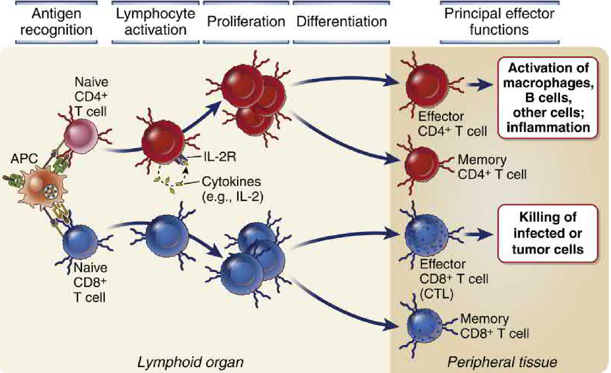
- Naïve T cells circulate through secondary lymphoid organs (lymph nodes, spleen), guided by CCR7 along fibroblastic reticular cell (FRC) conduits
- Mature DCs — loaded with antigen from peripheral tissues — migrate to T cell zones and display peptide–MHC complexes + B7 costimulators
- TCR scanning: T cells make transient contacts with DCs; antigen recognition causes T cell arrest → stable immunological synapse forms
- Signal cascade: TCR + CD28 → IL-2 secretion + upregulation of IL-2 receptor (CD25) → autocrine proliferation (clonal expansion)
- Differentiation into effector subsets (driven by cytokine milieu) and memory cells
- Effector T cells migrate to peripheral tissues; re-encounter antigen on local APCs/infected cells → execute effector functions
T Helper Subset Polarization (Signal 3)
| Cytokine environment | Subset | Key effector cytokines | Protection against |
|---|---|---|---|
| IL-12, IFN-γ | Th1 | IFN-γ, TNF-α | Intracellular pathogens (bacteria, viruses) |
| IL-4 | Th2 | IL-4, IL-5, IL-13 | Parasites; drives IgE, eosinophils |
| IL-6 + TGF-β (+ IL-23 for maintenance) | Th17 | IL-17A/F, IL-22 | Extracellular bacteria, fungi; neutrophil recruitment |
| TGF-β + IL-2 | Treg | IL-10, TGF-β | Suppression; self-tolerance |
CD8⁺ CTL Killing Mechanism
CTLs recognize peptide on MHC I (on all nucleated cells):
- Perforin: polymerizes in target cell membrane → forms pores
- Granzymes: serine proteases enter via pores → activate caspases → apoptosis
- Fas–FasL interaction: CTL expresses FasL → binds Fas on target → apoptosis
- CTLs are themselves protected from their own perforin by a lysosomal glycoprotein (LAMP-1)
5. B Cell Activation & Antibody Physiology
B cells are activated by T-cell help (for protein antigens — T-dependent) or by direct crosslinking (T-independent, e.g., polysaccharides):
T-dependent B cell activation (germinal center reaction):
- B cell receptor (BCR) binds antigen → internalizes and presents peptide on MHC II to helper T cell
- CD40L (on T cell) engages CD40 (on B cell) → key costimulatory signal
- Th2-derived IL-4/IL-13, Th1-derived IFN-γ, etc. → class switch recombination (CSR):
- IgM (primary, no T help needed) → IgG (opsonization, complement) → IgA (mucosal) → IgE (parasite/allergy)
- Somatic hypermutation in germinal centers → affinity maturation → selection of high-affinity B cell clones
- Differentiation into plasma cells (antibody factories) and memory B cells
Antibody Effector Mechanisms
| Mechanism | How it works |
|---|---|
| Neutralization | Antibodies block pathogen–receptor interactions; prevent cell entry |
| Opsonization | IgG Fc recognized by FcγRs on macrophages/neutrophils → enhanced phagocytosis |
| Complement activation | IgM/IgG–antigen complexes activate classical pathway → C3b deposition → MAC |
| ADCC | IgG-coated targets recognized by NK cell FcγRIII (CD16) → target cell killing |
| Mast cell degranulation | IgE crosslinked on mast cells → histamine, leukotrienes release (allergy/parasites) |
6. Physiological Resolution & Memory
- After pathogen clearance, 90–95% of effector cells undergo apoptosis (contraction) — mediated partly by withdrawal of survival signals (IL-2, costimulation)
- Surviving ~5% become long-lived memory cells (both T and B): lower activation threshold, faster kinetics, greater magnitude upon re-exposure
- Secondary immune response is faster (days not weeks), produces higher-affinity antibodies, and has qualitatively different effector mechanisms
Summary Flow
Pathogen → PRR signaling (TLRs/NLRs) → Cytokine release (IL-1β, TNF-α, IL-12)
                                              ↓
                                   Inflammation + DC maturation
                                              ↓
                           DC → lymph node → peptide–MHC presentation
                                              ↓
                   Signal 1 (TCR) + Signal 2 (CD28) + Signal 3 (cytokines)
                                              ↓
                     IL-2 → Clonal expansion → Th1/Th2/Th17/Treg / CTL
                                              ↓
              B cell help (CD40L, cytokines) → Germinal center → Antibodies
                                              ↓
                          Pathogen eliminated → Contraction → Memory
Sources: Cellular and Molecular Immunology — Abbas, Lichtman & Pillai (9780323757485); Robbins & Kumar Basic Pathology (9780323790185); Janeway's Immunobiology 10e (9780393884876)
Immune Response
Immune Response gyton
Finding Sources
Finding Sources
Finding Sources
Reading File
Finding Sources
Reading File
Reading File
Reading File
Reading File
Reading File
Your credits are exhausted. Your daily credits reset tomorrow, or you can purchase a top-up for more.
This is a shared conversation. Sign in to Orris to start your own chat.