
Thick white curdy discharge from vagina with itching?
vaginal candidiasis white curdy discharge vulvovaginitis
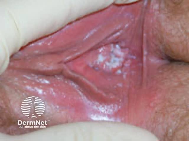
Clinical photography of the external female genitalia demonstrating vulvar mucosal inflammation with characteristic white curd-like plaques consistent with candidal vulvovaginitis. The view focuses on the labia majora and vestibular mucosa, highlighting diffuse erythema, edema, and superficial fissuring surrounding adherent, cottage-cheese-like plaques. The image captures the hallmark appearance of vulvovaginal candidiasis: intense itching, burning, and soreness often accompanying a thick, whitish discharge, though discharge quality may vary. From a diagnostic perspective, this photograph supports a clinical suspicion of Candida species infection, particularly Candida albicans, in an immunocompetent or mildly compromised host. Correlation with microscopy (KOH prep) and culture, or rapid antigen tests, can confirm budding yeast and pseudohyphae within mucosal epithelium. Normal vulvar variants are unlikely given erythema and plaque formation. Differential diagnoses include irritant contact dermatitis, bacterial vaginosis, and trichomoniasis, but the discrete white plaques favor fungal etiology. Timely antifungal therapy, such as azole antifungals, usually yields rapid improvement; recurrence prompts evaluation for diabetes, antibiotic exposure, or immunosuppression. This image is relevant for medical education, dermatology and gynecology training, clinical decision support, and research on vulvar infections, mucosal candidiasis pathophysiology, and patient counseling about symptom management and prevention. Educational value includes differential diagnosis, treatment guidelines, and patient education resources handouts.
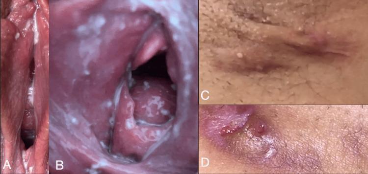
This composite of four clinical photographs illustrates gynecological and dermatological pathology. Images A and B are close-up clinical views of the vulva and vaginal mucosa demonstrating Candida vulvovaginitis. The images show significant mucosal erythema and a thick, white, adherent discharge with a curd-like or 'cottage cheese' texture localized within the vaginal folds and coating the vaginal walls. Images C and D display perianal or inguinal skin lesions consistent with hidradenitis suppurativa. Image C shows a linear, erythematous, and fibrotic-appearing scar or sinus tract. Image D demonstrates an inflammatory nodule with central suppuration and surrounding skin hyperpigmentation, characteristic of chronic follicular occlusion. This collection serves as an educational reference for distinguishing between fungal infections of the vaginal vault and chronic inflammatory skin diseases of the intertriginous regions, specifically highlighting the co-occurrence of vulvovaginitis and hidradenitis suppurativa.
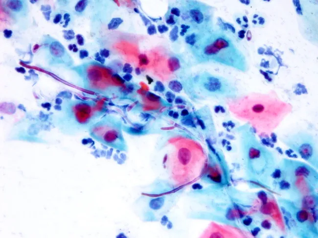
Conventional Pap smear cytology of a cervicovaginal sample stained with Papanicolaou stain. The smear demonstrates abundant squamous epithelial cells with clusters of intermediate and superficial cells. Within these cell clusters there are numerous oval budding yeast forms measuring 1–2 μm and filamentous pseudohyphae; the pseudohyphae are eosinophilic and stain unevenly, extending between cells and forming short branches. A prominent neutrophilic inflammatory component is present. The yeast elements and pseudohyphae are characteristic of Candida species (most commonly Candida albicans) causing vulvovaginitis or cervicitis in appropriate clinical settings. These features differentiate from non-filamentous yeasts and from malignant cells, and are best corroborated by clinical symptoms (pruritus, discharge) and culture or adjunct studies. Diagnostic significance lies in identifying fungal infection with inflammatory response; differential diagnoses include other Candida species, other yeasts lacking pseudohyphae, bacterial superinfection, or non-infectious causes of inflammation. The specimen shows no cytologic evidence of neoplasia. This image is useful for education on fungal cytology, diagnostic verification of candidiasis in cervical/vaginal cytology, and correlates with antifungal therapy planning. Clinicians should consider repeat smear or culture if symptoms persist despite therapy. Pathologic correlation: Candida infection; fungal morphology; budding yeast; pseudohyphae; neutrophils; cervicovaginal candidiasis.
Thick White Curdy Vaginal Discharge + Itching: Vulvovaginal Candidiasis
What Is It?
Classic Symptoms
| Feature | Description |
|---|---|
| Discharge | Thick, white, "cottage cheese" or curd-like; often adherent to vaginal walls |
| Itch (pruritus) | Intense vulvar itching - hallmark symptom |
| Burning / irritation | Common, especially on urination or intercourse (dyspareunia) |
| Odor | Usually absent (distinguishes it from bacterial vaginosis) |
| Vulvar signs | Erythema, edema, fissures, tenderness of the labia and vaginal walls |
Note: Although "curdy" discharge is classic, the discharge is frequently loose and may be hard to distinguish on appearance alone. Vaginal pH is usually normal (4.0-4.5), which is a key differentiating feature from bacterial vaginosis and trichomoniasis. - Goldman-Cecil Medicine
Predisposing / Risk Factors
- Antibiotic use (disrupts normal lactobacillus flora)
- Oral contraceptive pills
- Pregnancy
- Diabetes mellitus (uncontrolled)
- Immunosuppression (HIV, corticosteroids)
- Sexual activity
- Tight/synthetic clothing, poor perineal hygiene
Diagnosis
- Clinical - Classic symptoms + history of prior documented episodes is often sufficient
- KOH (10%) wet mount - Destroys other cells, reveals fungal elements (pseudohyphae/hyphae). Sensitivity ~50%, so a negative result does not rule out the diagnosis
- Saline wet mount / Gram stain - Shows pseudohyphae or hyphae
- Culture - Reserved for cases not responding to first-line treatment (to rule out non-albicans species like C. glabrata, which may be azole-resistant)

Treatment
Uncomplicated VVC (first episode or infrequent)
- Fluconazole 150 mg - single oral dose (highly effective)
- Clotrimazole 2% cream - 5g intravaginally daily x 3 days
- Miconazole 4% cream - 5g intravaginally daily x 3 days
- Miconazole 1,200 mg suppository - single dose
- Butoconazole 2% cream - 5g intravaginally once (Rx)
- Terconazole 0.8% cream - 5g intravaginally daily x 3 days (Rx)
- Terconazole 80 mg suppository - daily x 3 days (Rx)
"Short-course topical intravaginal azole drugs are effective for the treatment of uncomplicated vulvovaginal candidiasis. Single-dose oral treatment with fluconazole (150 mg) is also effective and is preferred by many patients." - Harrison's Principles of Internal Medicine, 22E (2025)
Complicated / Recurrent VVC (4+ episodes/year)
- Prolonged or periodic oral fluconazole therapy
- Oteseconazole (newer agent): 150 mg orally daily x 7 days, then once weekly x 11 weeks
- Investigate for underlying diabetes or HIV
- Note: C. glabrata is often azole-resistant - nystatin intravaginal suppositories may be needed
Differential Diagnosis
| Condition | Discharge | pH | Odor | Key Feature |
|---|---|---|---|---|
| Candidiasis | White, curdy/thick | Normal (4.0-4.5) | None | Intense itch, pseudohyphae on KOH |
| Bacterial vaginosis | Homogeneous, gray/white, thin | >4.5 | Fishy ("whiff" test +) | Clue cells; minimal itch |
| Trichomoniasis | Yellow-green, frothy | >4.5 | May be present | Motile trichomonads; cervical petechiae ("strawberry cervix") |
| Herpes | None/minimal | Normal | None | Ulcerative lesions; burning |
When to Suspect "Complicated" Candidiasis
- Recurs 4+ times per year
- Unusually severe episode
- Non-albicans species (especially C. glabrata)
- Pregnant patient
- Uncontrolled diabetes, immunosuppression, HIV
- Goldman-Cecil Medicine International Edition - Vaginitis section
- Harrison's Principles of Internal Medicine, 22E (2025) - Vulvovaginal Pruritus, Burning, or Irritation
- Henry's Clinical Diagnosis and Management by Laboratory Methods - Vaginal Candidiasis