
Tell me 5 major differences between typical ribs and 1st rib from Ak Datta, Selim Reza and any other book u prefer
first rib vs typical rib anatomy differences
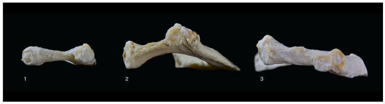
A clinical comparison photograph displaying three human first rib specimens (labeled 1, 2, and 3) to illustrate anatomical variations in vertical curvature and angulation. The image is captured from a posterior-to-anterior perspective against a black background. Specimen 1 demonstrates a relatively flat morphology with minimal vertical deviation and uniform width. Specimen 2 exhibits a pronounced superior (upward) angulation and a distinct middle-shaft arch. Specimen 3 shows an inferior (downward) angulation with a moderate curve. These morphological differences are significant in the study of thoracic outlet syndrome (TOS), as variations in rib height and angulation can influence the space available for neurovascular structures, such as the Th1 nerve root. The specimens highlight the natural diversity in costal anatomy relevant for surgical planning, specifically transaxillary or supraclavicular rib resection.
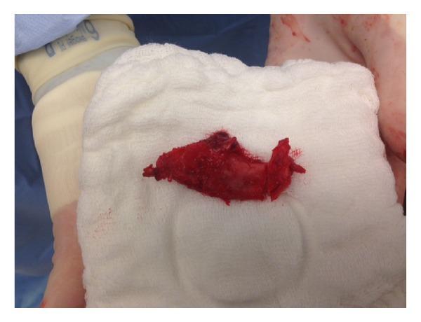
This clinical photograph captures a surgical specimen—a resected portion of a human first rib—resting on a sterile white surgical gauze. The bone fragment is predominantly deep red, indicating adherent vascularized tissue and periosteum. It exhibits a rough, irregular texture with jagged, non-uniform edges resulting from surgical transection. The specimen's morphology is elongated and somewhat flattened, typical of rib anatomy. In the background, the intraoperative setting is visible, showing a patient's limb covered in a light-colored sterile stockinette and secured with a gray band. This visual documentation serves as an educational example of First Rib Resection (FRR), a definitive surgical treatment for Thoracic Outlet Syndrome (TOS) aimed at decompressing the neurovascular structures including the brachial plexus, subclavian artery, and subclavian vein. The image highlights the physical nature of the resected osseous and soft tissue component following transaxillary or supraclavicular decompression.
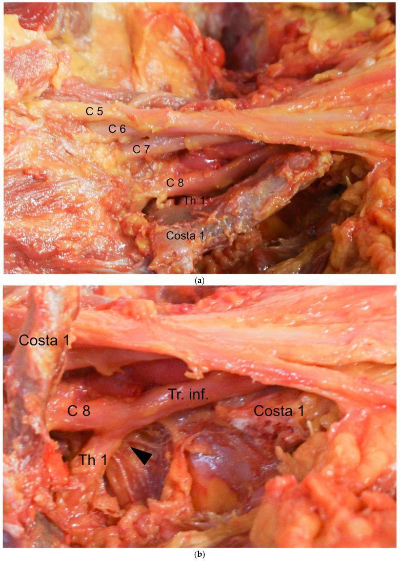
This composite of clinical photographs illustrates the surgical anatomy of the brachial plexus during a decompression procedure for thoracic outlet syndrome (TOS). Panel (a) shows the cervical and thoracic nerve roots (C5, C6, C7, C8, and Th1) from a cranial-to-caudal perspective. The first rib (Costa 1) is visible inferior to the Th1 nerve root, which runs adjacent to the rib's anterior margin. Panel (b) depicts the surgical field after the resection of the first rib. The C8 and Th1 nerve roots are shown joining to form the inferior trunk (Tr. inf.). A black arrowhead marks the specific site of nerve compression where the Th1 root was previously impinged upon by the anterior margin of the first rib (Costa 1). The image highlights the critical spatial relationship between the lower brachial plexus trunks and the proximal first rib, demonstrating the anatomical basis for neurogenic TOS and the objective of surgical rib resection to achieve sufficient neural decompression.
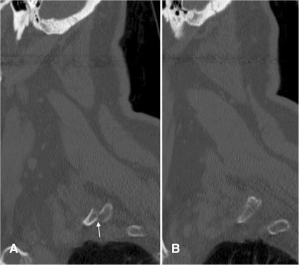
Diagnostic imaging comparison utilizing sagittal non-contrast CT scans of the cervical-thoracic junction. Image A displays a side-by-side comparison illustrating a traumatic injury versus normal anatomy. In panel A, an acute, anteriorly displaced fracture of the proximal right first rib is clearly visible, indicated by a white arrow highlighting the cortical discontinuity and malalignment of the bony segments. In contrast, panel B shows the contralateral left first rib, which remains intact with a smooth, continuous cortical outline and normal spatial relationship to the surrounding soft tissues and lung apex. This comparison serves as an educational tool for identifying subtle rib fractures in the upper thoracic cage, which are often markers of high-energy trauma and potential vascular or neurological injury in the thoracic outlet.
Typical Rib vs. 1st Rib - 5 Major Differences
1. Orientation / Plane of the Shaft
| Feature | Typical Rib (Ribs 3-9) | 1st Rib |
|---|---|---|
| Plane | Oblique - set in a vertical/oblique plane; has outer and inner surfaces, and superior and inferior borders | Horizontal - lies flat in the horizontal plane; has broad superior and inferior surfaces (not outer/inner surfaces) |
| Surfaces | External surface & internal surface | Superior surface & inferior surface |
| Borders | Superior (rounded) & inferior (sharp) border | Outer (convex) & inner (concave) border |
AK Datta: "The first rib is the most curved, shortest and broadest of all ribs. It lies in an almost horizontal plane."
- Gray's Anatomy for Students (p. 181): "Rib I is flat in the horizontal plane and has broad superior and inferior surfaces."
2. Head - Number of Articular Facets
| Feature | Typical Rib | 1st Rib |
|---|---|---|
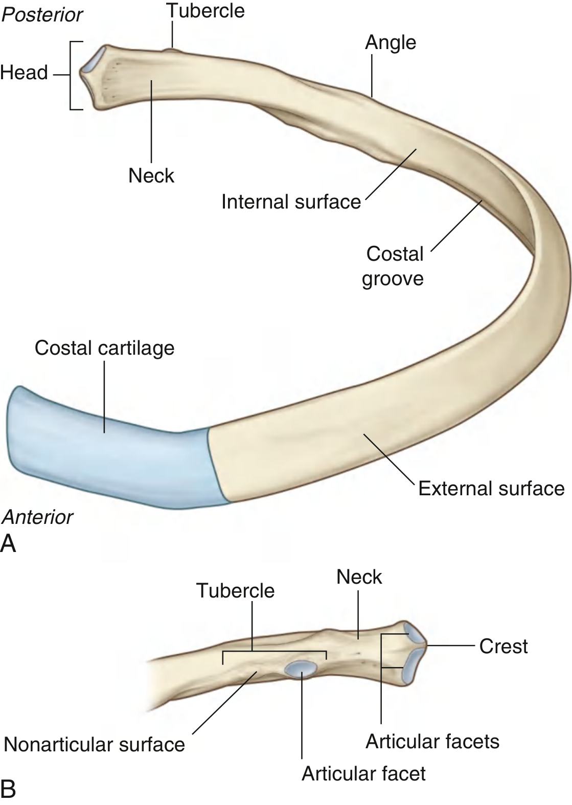
| Head | Two articular facets (demifacets) separated by a crest - articulates with the bodies of two adjacent vertebrae (the vertebra above and its own vertebra) | Single articular facet only - articulates with T1 vertebra alone (no crest on the head) |
This is because the 1st rib does not share its vertebra with the rib above it; it has a complete facet only for T1.
- Gray's Anatomy for Students (p. 181): "The head articulates only with the body of vertebra T1 and therefore has only one articular surface."
- AK Datta: The head of a typical rib bears two articular facets separated by a crest of the head. The 1st rib's head has a single oval facet for T1 only.
3. Costal Groove and Subcostal Nerve / Vessels
| Feature | Typical Rib | 1st Rib |
|---|---|---|
| Costal groove | Present on the inferior border of the internal surface - lodges the intercostal nerve, artery, and vein (from above downward: vein, artery, nerve) | Absent - No costal groove on the 1st rib. Instead, there are two grooves on the superior surface - one for the subclavian vein (anterior) and one for the subclavian artery (posterior) |
| Separating structure | - | The two grooves are separated by the scalene tubercle (Lisfranc's tubercle) - for attachment of scalenus anterior muscle |
AK Datta: A key distinguishing feature - typical ribs have the costal groove at the inferior inner surface for the neurovascular bundle; the 1st rib has grooves for subclavian vessels on its superior surface. Selim Reza: "The superior surface of the first rib shows the scalene tubercle separating the groove for the subclavian vein anteriorly from the groove for the subclavian artery posteriorly."
- Gray's Anatomy for Students (p. 181): "The superior surface of the rib is characterized by a distinct tubercle, the scalene tubercle, which separates two smooth grooves... The anterior groove is caused by the subclavian vein, and the posterior groove is caused by the subclavian artery."
4. Shape / Curvature and Length
| Feature | Typical Rib | 1st Rib |
|---|---|---|
| Length | Longer, becoming progressively shorter after rib 7 | Shortest of all ribs |
| Curve | Single curve - curves forward around the thorax | Most curved of all ribs - has a sharp bend near the medial border (almost a "C" shape when viewed from above) |
| Twist | Shaft has a gentle torsional twist along its long axis (the outer surface of the anterior portion faces somewhat superiorly) | No torsional twist - shaft lies flat without twisting |
AK Datta: "Typical ribs have a gentle twist or torsion along their long axis. This torsion is absent in the 1st rib." Selim Reza: The first rib is described as the "shortest, flattest, and most curved" of all ribs, with no axial torsion.
5. Angle of the Rib
| Feature | Typical Rib | 1st Rib |
|---|---|---|
| Angle | A distinct angle is present - the shaft bends forward laterally to the tubercle, creating a clearly demarcated angle | Angle coincides with the tubercle - the angle is at the same point as the tubercle, so there is no distinct separate angle. In practice, the angle and tubercle are at the same site |
AK Datta: In typical ribs, the angle is located lateral to the tubercle and forms a distinct bend. In the 1st rib, the angle coincides with the tubercle; both are at the same point. Selim Reza similarly notes: "In the 1st rib, the angle coincides with the tubercle."
Summary Table
| Feature | Typical Rib | 1st Rib |
|---|---|---|
| Plane | Oblique (outer/inner surfaces; sup/inf borders) | Horizontal (sup/inf surfaces; inner/outer borders) |
| Head facets | Two (bicipital, with a crest) | One (unicipital, no crest) |
| Costal groove | Present (inferior inner surface) | Absent; replaced by 2 grooves for subclavian vessels on superior surface |
| Torsion of shaft | Present (gentle axial twist) | Absent |
| Angle | Distinct, lateral to tubercle | Coincides with the tubercle |
- Gray's Anatomy for Students, pp. 180-182 (Ribs section - detailed anatomy of typical rib and Rib I)
- AK Datta, Human Anatomy (Regional and Applied), Vol. 1 - Upper Limb & Thorax
- Selim Reza, A Textbook of Human Anatomy
Tell me 5 major differences between typical ribs and 1st rib from Ak Datta, Selim Reza and any other book u prefer
5 Major Differences: Typical Rib vs. 1st Rib
1. Plane / Orientation of the Shaft
| Typical Rib | 1st Rib | |
|---|---|---|
| Plane | Oblique / vertical plane | Horizontal plane |
| Surfaces | Outer (external) & inner (internal) surfaces | Superior & inferior surfaces |
| Borders | Superior (rounded) & inferior (sharp) | Outer (convex) & inner (concave) |
AK Datta: "The first rib lies in an almost horizontal plane unlike the obliquely placed typical ribs." Selim Reza: Typical ribs are set obliquely; the 1st rib is flat and horizontal. Gray's for Students (p.181): "Rib I is flat in the horizontal plane and has broad superior and inferior surfaces."
2. Head - Number of Articular Facets
| Typical Rib | 1st Rib | |
|---|---|---|
| Facets on head | Two demifacets separated by a crest - articulates with two adjacent vertebrae (its own + the one above) | One single oval facet - articulates with T1 only (no crest) |
AK Datta: The head of a typical rib is "bicipital" (two facets). The 1st rib head is "unicipital" with a single facet for T1. Selim Reza: Same - single articular facet, no crest on the head of the 1st rib. Gray's for Students (p.181): "The head articulates only with the body of vertebra T1 and therefore has only one articular surface."
3. Costal Groove
| Typical Rib | 1st Rib | |
|---|---|---|
| Costal groove | Present on the inferior border of the inner surface - lodges the intercostal neurovascular bundle (vein, artery, nerve from above downward) | Absent. Instead, the superior surface bears two grooves for the subclavian vessels, separated by the scalene tubercle (Lisfranc's tubercle): anterior groove for subclavian vein, posterior groove for subclavian artery |
AK Datta: Costal groove is a standard feature of typical ribs; in the 1st rib, the superior surface has two vascular grooves divided by the scalene tubercle. Selim Reza: "The superior surface of the first rib shows a prominent scalene tubercle separating the groove for the subclavian vein (anterior) from the groove for the subclavian artery (posterior)." Gray's for Students (p.181): Confirms the scalene tubercle separating the subclavian vein groove anteriorly from the subclavian artery groove posteriorly.
4. Torsion (Axial Twist) of the Shaft
| Typical Rib | 1st Rib | |
|---|---|---|
| Torsion | Present - the shaft has a gentle twist along its longitudinal axis, so the external surface of the anterior part faces somewhat superiorly compared to the posterior part | Absent - no torsional twist; the shaft lies flat throughout its length |
AK Datta: "Torsion is present in typical ribs but absent in the 1st rib." Selim Reza: Also states no axial torsion in the 1st rib. Gray's for Students (p.181): The shaft of a typical rib "has a gentle twist around its longitudinal axis"; this feature is not present in rib I.
5. The Angle
| Typical Rib | 1st Rib | |
|---|---|---|
| Angle | Distinct and separate from the tubercle - the shaft bends sharply forward at a point lateral to the tubercle, forming a well-defined angle | Coincides with the tubercle - the angle and the tubercle are at the same point, so no separate angle is recognizable |
AK Datta: "In typical ribs, the angle is well-marked and situated lateral to the tubercle. In the 1st rib, the angle coincides with the tubercle." Selim Reza: States the same - in the 1st rib the angle and tubercle are at the same location.
Summary Table
| Feature | Typical Rib | 1st Rib |
|---|---|---|
| Plane of shaft | Oblique (outer/inner surfaces) | Horizontal (superior/inferior surfaces) |
| Head facets | Two (bicipital, with crest) | One (unicipital, no crest) |
| Costal groove | Present on inferior inner surface | Absent; 2 grooves on superior surface for subclavian vessels |
| Torsion of shaft | Present | Absent |
| Angle | Distinct, lateral to tubercle | Coincides with the tubercle |