
Abdominal Trauma And Death
Reading File
Searching sources...
Finding Sources
Finding Sources
Searching PubMed
"abdominal trauma" AND mortality
Now let me read the key textbook sections in parallel:
Reading File
Reading File
Reading File
Now let me read more from Tintinalli's for diagnosis/management and the Sabiston section:
Reading File
Reading File
I have rich material from multiple authoritative textbooks. Let me also fetch the CT/FAST images and read the specific organ injury sections:
Finding Sources
Finding Sources
Reading File
Loading Image
I now have comprehensive material from multiple authoritative sources. Let me compile the full answer.
Abdominal Trauma and Death
Overview and Mortality Burden
Abdominal trauma accounts for 15-20% of all trauma deaths. Death may occur as a direct consequence of massive hemorrhage, resulting in early demise shortly after injury. Patients who survive the initial insult face secondary risks from infection and sepsis. Firearm injuries - a major cause of penetrating abdominal trauma - are the third largest cause of trauma-related death in the United States, accounting for approximately 15% of all trauma-related deaths, and carry the highest case fatality rates across every age group.
- Tintinalli's Emergency Medicine, p. 1793
- Rosen's Emergency Medicine, p. 486
Mechanisms of Injury
Blunt Abdominal Trauma
The most common mechanism is motor vehicle collision (MVC). Falls are second in frequency. Pedestrians, motorcyclists, and bicyclists are at high risk due to absent abdominal protection. Three force types determine which organs are damaged:
- Compressive forces - direct crush of solid organs (spleen, liver, kidneys)
- Shearing/stretching forces - tears at fixed-mobile organ transitions (e.g., ligament of Treitz - a common site for mesenteric/small bowel injury)
- Acceleration/deceleration forces - retroperitoneal hemorrhage via axial skeleton transmission
The spleen is the most frequently injured intra-abdominal organ from blunt trauma (isolated in nearly two-thirds of cases). The liver is the most frequently injured overall.
Penetrating Abdominal Trauma
Stab wounds and gunshot wounds (GSWs) are the predominant mechanisms. GSWs account for approximately 90% of penetrating trauma mortality. Key facts:
-
Stab wounds most commonly injure the liver (large surface area, upper quadrants)
-
GSWs typically produce multiple organ injuries: small bowel > colon > liver
-
Simultaneous thoracic and abdominal penetration occurs in 20-40% of thoracoabdominal wounds
-
Any penetrating injury to the chest, pelvis, flank, or back must be assumed to have entered the abdominal cavity until proven otherwise
-
Rosen's Emergency Medicine, pp. 485-487
Pathophysiology of Death
1. Massive Hemorrhage (Early Death - "First Peak")
The dominant cause of early mortality. Solid organ injury (spleen, liver) and major vascular injury produce rapid exsanguination. Uncontrolled hemorrhage triggers the lethal triad:
| Component | Effect |
|---|---|
| Hypothermia | Impairs clotting enzyme function, decreases cardiac output |
| Coagulopathy | Loss of clotting factors, platelet dysfunction, trauma-induced coagulopathy |
| Acidosis | Lactic acidosis from tissue hypoperfusion, further depresses myocardial function |
Once the lethal triad is established, it becomes self-reinforcing and rapidly fatal without surgical intervention.
2. Infection/Sepsis (Late Death - "Third Peak")
Hollow viscus injury (bowel perforation) causes peritoneal contamination with gut flora. If missed or delayed in treatment, this progresses to peritonitis, sepsis, and multi-organ failure. Abdominal trauma kills via sepsis when:
- Hollow viscus injuries are occult on initial imaging
- Treatment is delayed beyond the window for source control
3. Abdominal Compartment Syndrome (ACS)
Post-damage control surgery, massive fluid resuscitation causes bowel edema and intra-abdominal hypertension. ACS compresses the inferior vena cava, reduces cardiac preload, impairs renal perfusion, and causes respiratory failure from diaphragm elevation. It is routinely anticipated by leaving the abdomen open ("open abdomen") after damage control laparotomy.
Diagnosis
Clinical Examination Limitations
Up to 45% of blunt trauma patients thought to have a benign abdomen on initial physical examination are later found to have significant intra-abdominal injury. Physical exam is unreliable when:
- Significant head injury or intoxication is present
- Distracting injuries exist
- The patient is elderly, on beta-blockers, or on anticoagulants
- Young, healthy patients physiologically compensate before decompensating suddenly
A "seatbelt sign" (abdominal contusion from lap belt) raises suspicion for hollow viscus and mesenteric injury, especially in children.
FAST Examination
Focused Assessment with Sonography for Trauma (FAST) is the primary rapid diagnostic tool. It detects free intraperitoneal fluid (surrogate for hemorrhage) in four windows: Morison's pouch, splenorenal interface, pelvic pouch, and pericardium.
FAST advantages: rapid, portable, non-invasive, no contrast, repeatable, detects pericardial/pleural fluid, identifies pneumothorax (eFAST).
FAST limitations: operator dependent; cannot identify the source of bleeding; limited retroperitoneal evaluation; cannot reliably distinguish blood from ascites.
In the unstable patient, a positive FAST is sufficient to take directly to the operating room.
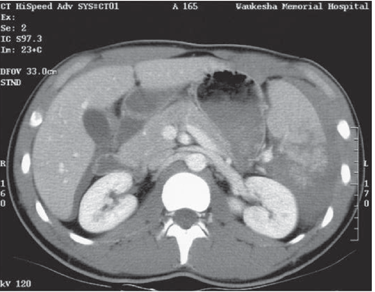
CT Abdominopelvic with IV Contrast
The gold standard for stable patients. Advantages:
- Precisely localizes and grades organ injuries
- Identifies retroperitoneal injuries (duodenum, pancreas, kidneys, major vessels)
- Detects active contrast extravasation (arterial blush) indicating ongoing hemorrhage
- Identifies free air (hollow viscus perforation)
- Guides nonoperative vs. operative decisions
Indications to avoid CT and go directly to OR: hemodynamic instability despite resuscitation, peritonitis, evisceration, positive FAST in unstable patient.
Indications for Emergency Laparotomy
| Setting | Absolute Indications |
|---|---|
| Blunt | Anterior abdominal injury with hypotension; abdominal wall disruption; peritonitis; free air under diaphragm; positive FAST/DPL in hemodynamically unstable patient; CT-diagnosed injury requiring surgery |
| Penetrating | Abdominal injury with hypotension; abdominal tenderness; GI evisceration; high suspicion for transabdominal GSW trajectory; CT-diagnosed injury requiring surgery |
- Tintinalli's Emergency Medicine, Table 263-2
Damage Control Surgery
Damage Control Laparotomy (DCL) is an abbreviated operative strategy designed to prevent/reverse the lethal triad. It is indicated when:
- Severe physiologic compromise with coagulopathy, hypothermia, or acidosis
- Bleeding from difficult-to-control injuries
- Large injury burden likely to cause physiologic decompensation if treated definitively at index operation
- Austere environments (rural, combat)
DCL sequence:
- Phase 1 - Stop hemorrhage (packing, ligation, vascular shunting; balloon tamponade for deep liver tracts) and control contamination (bowel stapling without anastomosis)
- Temporary abdominal closure - Fascia is left OPEN to prevent ACS; negative pressure wound therapy systems are used
- ICU resuscitation - Correct hypothermia, coagulopathy, acidosis; massive transfusion protocol (balanced resuscitation with packed RBCs:FFP:platelets in 1:1:1 ratio)
- Planned re-look - Definitive repair once physiology normalizes (typically 24-48 hours)
"The goal of this approach is to avoid the onset of the lethal triad: hypothermia, coagulopathy, and acidosis. The procedure is abbreviated and focused on maintaining the priorities of hemorrhage control." - Mulholland and Greenfield's Surgery, p. 1271
Organ-Specific Mortality Risks
| Organ | Notes |
|---|---|
| Spleen | Most commonly injured; high success with nonoperative management (NOM) in stable patients; angioembolization avoids splenectomy |
| Liver | Second most commonly injured; massive hemorrhage can be lethal; liver packing and balloon tamponade are DCL mainstays |
| Hollow viscus (bowel) | Occult injury common; missed perforation leads to delayed peritonitis and sepsis death |
| Pancreas | High morbidity; ductal injuries require delayed ERCP or surgical repair |
| Major vessels (aorta, IVC, mesenteric) | Rapidly fatal; may require REBOA (Resuscitative Endovascular Balloon Occlusion of the Aorta) as bridge to OR |
Nonoperative Management (NOM)
NOM is now the treatment of choice for hemodynamically stable patients with blunt hepatic and splenic injuries, irrespective of injury grade, age, or associated injuries (per Eastern Association for the Surgery of Trauma and Western Trauma Association guidelines). This is enabled by:
- High-resolution CT grading of solid organ injury
- Angiography/angioembolization for vascular injuries and pseudoaneurysms
- REBOA for unresponsive shock at Level I centers
- Serial clinical reassessment
Tranexamic acid (TXA) should be available within the ED for patients with hemorrhagic shock; use beyond 3 hours from injury is associated with increased risk of death.
- Tintinalli's Emergency Medicine, p. 1796
Special Populations
Pediatric Abdominal Trauma
- Approximately 1 in 10,000 children sustain abdominal trauma per year; blunt trauma causes 90% of cases
- Accounts for 10% of pediatric trauma fatalities (commonly in polytrauma with TBI)
- Non-accidental trauma (NAT) is the most common etiology in infants/toddlers; abdominal injury from child abuse is the second leading cause of death in abused children after abusive head trauma
- Children's less ossified ribs provide less protection to spleen and kidneys
- Seatbelts ridden above the ASIS in children transfer energy directly to abdomen, increasing hollow viscus injury risk
Pregnant Patients
- Penetrating trauma causes maternal death in fewer than 5% of cases
- Continuous fetal monitoring is indicated after more than minor abdominal trauma with a viable fetus
- Placental abruption is the most common cause of fetal death; when placental separation exceeds 50%, acute DIC and fetal death can occur
Causes of Preventable Death in Abdominal Trauma
- Delayed diagnosis - physical exam alone misses up to 45% of significant injuries
- Missed hollow viscus injury - CT has limited sensitivity for bowel/mesenteric injuries; serial exams essential
- Delayed hemorrhage control - every minute of uncontrolled bleeding worsens the lethal triad
- Failure to recognize ACS - post-DCL; bladder pressure monitoring is mandatory
- Inadequate damage control resuscitation - crystalloid-heavy resuscitation worsens dilutional coagulopathy; balanced blood product ratios are essential
- Delayed transfer - patients with significant abdominal trauma should be transferred to a trauma center without delay once the threatening injury is identified
Recent Evidence (2024-2026)
Three recent systematic reviews and meta-analyses are relevant:
- PMID 40785610 (2025) - Minimally invasive technologies in blunt abdominal trauma: mortality as primary endpoint
- PMID 39843466 (2025, Sci Rep) - Emergency room laparotomy in severe abdominal trauma: systematic review
- PMID 39671007 (2025, Pediatr Radiol) - Contrast-enhanced ultrasound for pediatric solid organ injuries: diagnostic performance meta-analysis
Sources:
- Tintinalli's Emergency Medicine, 9th ed., Ch. 263
- Rosen's Emergency Medicine, Ch. on Abdominal Trauma
- Mulholland and Greenfield's Surgery, 7th ed., Ch. 25
- Sabiston Textbook of Surgery, pp. 2730-2756
This is a shared conversation. Sign in to Orris to start your own chat.