
Rhumetaoid arthritis
Reading File
Now I'll search the medical library for comprehensive RA coverage, running parallel searches across key aspects.
Finding Sources
Searching PubMed
"rheumatoid arthritis"[MeSH Terms] AND treatment
Now let me search for the most dedicated RA sections in rheumatology/internal medicine textbooks and also do a paragraph-level search for key topics.
Finding Sources
Searching PubMed
"rheumatoid arthritis"[MeSH Terms] AND "biological therapy"[MeSH Terms]
Let me find dedicated rheumatology textbooks and search for the main RA sections there, reading the most relevant block files in parallel.
Finding Sources
Finding Sources
Excellent - I found Harrison's 22E (2025) and Firestein & Kelley's Rheumatology. Let me find and read the RA sections from these key textbooks.
Finding Sources
I found a rich section in Firestein & Kelley's Rheumatology (block6 line 530 has 24 paragraphs). Let me also find Harrison's RA chapter and read both simultaneously.
Finding Sources
Reading File
Good content from Firestein & Kelley's. Now let me read Harrison's RA chapter (block40) and also read Robbins for pathology, and get treatment info from Firestein block6 later lines.
Reading File
Reading File
Excellent content from Harrison's. I can see the treatment table and Harrison's chapter continues. Let me read more of Harrison's RA section (DMARDs table and key clinical features) and also Robbins' RA pathology section.
Reading File
Reading File
Excellent content. Let me fetch the RA histology image from Robbins and also read more of Harrison's chapter for clinical features, diagnosis criteria, and treatment table.
Reading File
Reading File
I have rich content. Let me fetch the pathogenesis diagram image and also read the Harrison's treatment table section and DMARDs section.
Loading Image
Reading File
I now have comprehensive content from Harrison's 22E, Firestein & Kelley's Rheumatology, and Robbins Pathology. I have enough to compile a thorough, well-organized answer on Rheumatoid Arthritis.
Rheumatoid Arthritis (RA)
A chronic, systemic autoimmune disease characterized by symmetric, erosive polyarthritis with extra-articular manifestations. It affects approximately 0.5-1% of adults worldwide, with a 2-3:1 female predominance, peak onset in the 4th-5th decade.
Pathogenesis
The diagram below (from Robbins & Kumar) illustrates the key steps:
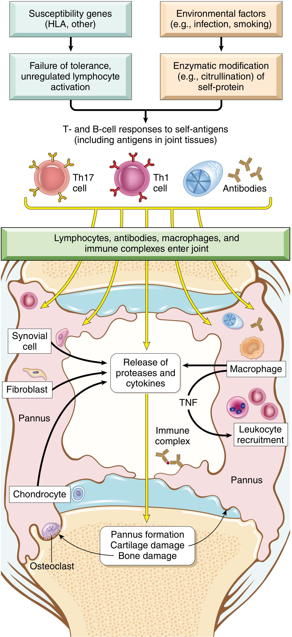
Genetic factors:
- ~50% of RA risk is inherited. The HLA-DR4 allele ("shared epitope") is strongly associated with ACPA-positive RA. HLA-DRB1 alleles that carry the shared epitope increase RA risk 4-6 fold; combined with smoking, this rises to 20-40 fold.
Environmental triggers:
- Smoking is the most reproducible risk factor: confers a relative risk of 1.5-3.5x. Risk is almost exclusively tied to RF/ACPA-positive disease and persists 15 years after cessation.
- Periodontal disease / oral microbiome: Porphyromonas gingivalis is uniquely capable of PAD (peptidyl arginine deiminase) enzyme production, catalyzing citrullination of host proteins - creating neo-antigens that trigger autoimmunity.
- Gut microbiome dysbiosis: Prevotella copri enrichment has been found in early untreated RA.
- EBV has been implicated but causation is unproven.
Autoantibodies:
- Anti-citrullinated protein antibodies (ACPA / anti-CCP): Present in ~70% of RA patients. Arginine residues in fibrinogen, type II collagen, vimentin, and α-enolase are citrullinated by PAD enzymes; these modified epitopes trigger autoimmunity. ACPAs may appear >10 years before clinical disease.
- Rheumatoid factor (RF): IgM/IgA autoantibodies against the Fc region of IgG, present in ~80% of patients. Not RA-specific.
Effector mechanisms in the joint:
- CD4+ Th1 cells secrete IFN-γ → activate macrophages and synovial cells
- Th17 cells secrete IL-17 → recruit neutrophils and monocytes
- TNF is a key mediator of inflammation and joint destruction (basis for biologic therapy)
- Macrophages and synovial fibroblasts release proteases (MMPs), prostaglandins, and cytokines (IL-1, IL-6, TNF)
- RANKL from activated T cells stimulates osteoclasts → bone resorption
- B cells and plasma cells form germinal centers in the synovium; secrete RF and ACPAs
- Robbins, Cotran & Kumar Pathologic Basis of Disease, p.1106; Harrison's Principles 22E, pp.2891-2892; Firestein & Kelley's Textbook of Rheumatology
Pathology / Morphology
The hallmarks are synovial inflammation, proliferation, focal bone erosions, and thinning of articular cartilage.
- Synovium becomes edematous, thickened, and hyperplastic - smooth contour replaced by delicate, bulbous villi
- Pannus formation: a mass of edematous synovium, inflammatory cells, granulation tissue, and fibroblasts that invades cartilage and bone. Composed of 6 cell types: T cells, B cells, plasma cells, dendritic cells, mast cells, granulocytes
- Histologic features: (1) Synovial cell hyperplasia/proliferation; (2) dense CD4+ T cell, B cell, plasma cell, macrophage infiltrates (often forming lymphoid follicles); (3) increased vascularity (angiogenesis); (4) fibrin-rich neutrophilic exudate on synovial surfaces; (5) osteoclastic subchondral bone erosion
- Bone damage: Osteoclasts at the pannus-bone interface form resorption lacunae, especially at radial sites of MCP joints
- Periarticular osteopenia: From bone marrow inflammation; visible as MRI signal alterations ("bone marrow lesions") - a forerunner of frank erosions
- End-stage: Fibrous ankylosis → ossification → bony ankylosis (bone fusion)
Rheumatoid nodules: Occur in ~20% of RF-positive patients. Firm, nontender subcutaneous masses at forearms, elbows, occiput, and lumbosacral area. Microscopically - central zone of fibrinoid necrosis surrounded by palisading activated macrophages and lymphocytes/plasma cells.
- Robbins, Cotran & Kumar, pp.1106-1107; Harrison's 22E, pp.2890-2891
Clinical Features
Onset:
- Often insidious - malaise, fatigue, generalized musculoskeletal pain in ~50% of patients, followed by joint involvement over weeks to months
- Can also have acute polyarticular onset
Joint involvement (symmetric):
- Small joints first: MCP and PIP joints of hands (sparing DIP - key distinction from OA), MTP joints of feet
- Then: wrists, ankles, elbows, knees
- Morning stiffness >1 hour (worse after inactivity - "gelling")
- Joints are swollen, warm, tender, and painful
Characteristic deformities (late disease):
- Ulnar deviation of fingers at MCPs
- Swan-neck deformity: PIP hyperextension + DIP flexion
- Boutonniere deformity: PIP flexion + DIP hyperextension
- Z-deformity of the thumb
- Subluxation of the carpus, atlantoaxial subluxation (C1-C2, causing neck pain/myelopathy risk)
Extra-articular manifestations:
| System | Manifestations |
|---|---|
| Skin | Rheumatoid nodules, vasculitis |
| Pulmonary | Interstitial lung disease, pleuritis, nodules, Caplan's syndrome (with pneumoconiosis) |
| Cardiac | Pericarditis, accelerated atherosclerosis (major cause of excess mortality) |
| Ocular | Keratoconjunctivitis sicca (secondary Sjogren's), scleritis, episcleritis |
| Hematologic | Normocytic normochromic anemia, Felty's syndrome (RA + splenomegaly + neutropenia) |
| Neurologic | Mononeuritis multiplex (from vasculitis), entrapment neuropathies, cervical myelopathy |
| Renal | Secondary amyloidosis (AA type) |
Diagnosis
2010 ACR/EULAR Classification Criteria (score ≥6/10 = definite RA):
| Domain | Score |
|---|---|
| Joint involvement: 1 large joint = 0; 2-10 large = 1; 1-3 small = 2; 4-10 small = 3; >10 joints (including ≥1 small) = 5 | 0-5 |
| Serology: Negative RF and ACPA = 0; Low-positive = 2; High-positive (>3x ULN) = 3 | 0-3 |
| Acute phase reactants: Normal CRP & ESR = 0; Abnormal CRP or ESR = 1 | 0-1 |
| Duration of symptoms: <6 weeks = 0; ≥6 weeks = 1 | 0-1 |
Serology:
- ACPA (anti-CCP): ~70% sensitivity, >95% specificity - the most specific test; predicts erosive disease
- RF: ~80% sensitivity but low specificity (also positive in SLE, Sjogren's, chronic infections, healthy elderly)
- ~20% of RA patients are seronegative (negative for both)
- ESR and CRP: elevated in active disease
- CBC: normocytic anemia, thrombocytosis in active disease
Imaging:
- X-ray: periarticular osteopenia, joint space narrowing, marginal erosions (especially radial aspects of MCPs), deformities
- MRI: earliest detection of synovitis and bone marrow edema (precedes erosions)
- Ultrasound: detects subclinical synovitis and tenosynovitis
Disease activity assessment tools (used in clinical practice):
- DAS28 (Disease Activity Score - 28 joints)
- SDAI (Simplified Disease Activity Index)
- CDAI (Clinical Disease Activity Index)
- ACR 20/50/70 improvement criteria (used in clinical trials)
Treatment
The goal is remission or low disease activity using a treat-to-target strategy.
1. NSAIDs
- Adjunctive only; provide symptomatic relief but do not alter disease course
- Inhibit COX-1 and COX-2; risk of GI and renal toxicity with chronic use
2. Glucocorticoids
- Bridge therapy while awaiting DMARD effect (onset may take weeks-months)
- Low-dose prednisone 5-10 mg/d for active disease
- Short bursts for flares
- Minimize long-term use due to osteoporosis, infection, metabolic effects
3. Conventional (csDMARDs)
| Drug | Dose | Key Toxicities | Monitoring |
|---|---|---|---|
| Methotrexate (first choice) | 10-25 mg/week PO or SQ + folic acid 1 mg/d | Hepatotoxicity, myelosuppression, pneumonitis, teratogen | CBC, LFTs, creatinine q2-3 months |
| Hydroxychloroquine | 200-400 mg/d (≤5 mg/kg) | Irreversible retinopathy, cardiotoxicity | Annual OCT + visual field |
| Sulfasalazine | 1000-1500 mg/d (start 500 mg BD) | Granulocytopenia, hemolytic anemia (G6PD deficiency) | CBC q2-4 weeks x 3 months, then q3 months |
| Leflunomide | 10-20 mg/d | Hepatotoxicity, myelosuppression, teratogen (Category X) | CBC, LFTs q2-3 months |
Triple therapy (MTX + HCQ + SSZ) is as effective as biologic therapy in many patients.
4. Biologic DMARDs (bDMARDs)
Used when csDMARDs fail (typically after 3-6 months of adequate MTX):
| Class | Examples | Notes |
|---|---|---|
| TNF-α inhibitors | Infliximab, Etanercept, Adalimumab, Certolizumab, Golimumab | First-line biologics; screen for TB before starting; risk of reactivation of latent TB, fungal infections, lymphoma (controversial) |
| IL-6 receptor inhibitor | Tocilizumab, Sarilumab | Also used for cytokine release syndrome; may mask fever/CRP |
| T cell costimulation blocker | Abatacept (CTLA-4-Ig) | Blocks CD28-CD80/86 interaction |
| B cell depletor | Rituximab (anti-CD20) | Preferred in RF/ACPA-high disease or with comorbid lymphoma |
| IL-1 inhibitor | Anakinra | Less commonly used for RA |
5. JAK Inhibitors (targeted synthetic DMARDs - tsDMARDs)
- Tofacitinib, Baricitinib, Upadacitinib
- Oral agents; inhibit Janus kinase signaling
- Used when bDMARDs fail or as alternatives; black box warnings for infection, malignancy, thromboembolism, MACE in patients >50 with CV risk factors
- Harrison's Principles of Internal Medicine 22E (2025), pp.2892-2896
Prognosis
- Chronic, waxing-and-waning course in most patients; progressive joint damage without DMARD therapy
- ~20% of seronegative patients may stabilize or remit
- Major causes of excess mortality: accelerated cardiovascular disease (most common), infections, cervical myelopathy
- Poor prognostic factors: high ACPA/RF titers, high disease activity at onset, early erosions on imaging, extra-articular disease, functional disability
Key Distinguishing Points (RA vs OA)
| Feature | RA | OA |
|---|---|---|
| Joint type affected | MCP, PIP, wrists | DIP, PIP, first CMC, weight-bearing |
| Symmetry | Symmetric | Often asymmetric |
| Morning stiffness | >1 hour | <30 minutes |
| Inflammation | Prominent | Mild |
| Systemic features | Yes | No |
| Serologic markers | RF, ACPA | Negative |
| X-ray | Erosions, osteopenia | Osteophytes, sclerosis, joint space narrowing |
Sources: Robbins, Cotran & Kumar Pathologic Basis of Disease; Harrison's Principles of Internal Medicine 22E (2025); Firestein & Kelley's Textbook of Rheumatology 2-Volume Set
This is a shared conversation. Sign in to Orris to start your own chat.