
DLT answer 5 marks
I need a bit more context to give you a useful answer. What topic or subject is this "DLT" question about?
Asking for Preferences
What does 'DLT' refer to in your question?
Double lumen tube 5 mark answer
The Double Lumen Tube (DLT) is an endobronchial tube used in thoracic anaesthesia to achieve one-lung ventilation (OLV). It has two lumens - one for the trachea and one for a bronchus - allowing each lung to be ventilated independently or one lung to be isolated/collapsed.
Searching sources...
Reading File
Finding Sources
Reading File
Finding Sources
Reading File
Reading File
Reading File
I now have comprehensive information. Here is a complete 5-mark exam answer on the Double Lumen Tube:
Double Lumen Tube (DLT)
Definition
A Double Lumen Tube (DLT) is a specialized endobronchial tube used in thoracic anaesthesia to achieve one-lung ventilation (OLV) - allowing each lung to be ventilated independently or one lung to be selectively isolated and collapsed to facilitate surgery.
Structure
All DLTs share the following features:
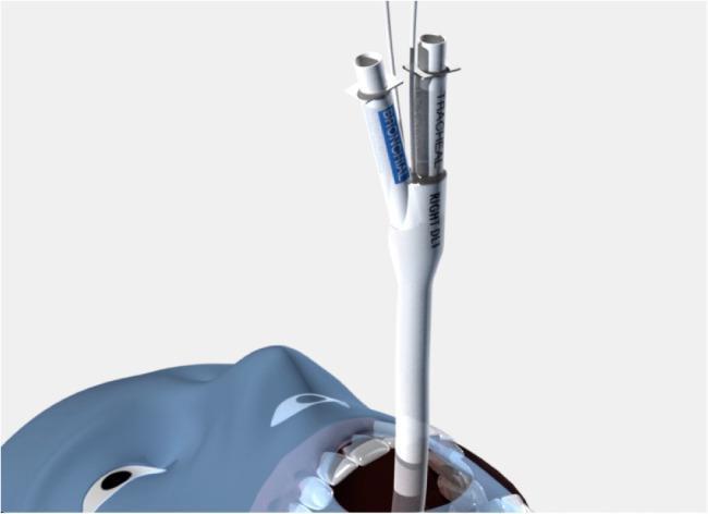
- Two lumens: a longer endobronchial lumen (enters a main bronchus) and a shorter endotracheal lumen (terminates in the lower trachea)
- Two cuffs: an endobronchial cuff (bronchial seal) and an endotracheal cuff (tracheal seal)
- A preformed curve that allows preferential entry into the desired bronchus
- Available sizes: 35F, 37F, 39F, and 41F
- Designed for either the right or left bronchus (right-sided tubes have a proximal portal/modified cuff for ventilation of the right upper lobe)
Indications
Absolute indications:
- Protection against spillage of blood, pus, or lavage fluid from one lung to the other
- Bronchopleural fistula - to prevent loss of ventilation
- Tracheobronchial disruption
- Large lung cyst or bulla (risk of rupture)
- Severe hypoxemia due to unilateral lung disease
- Bronchoalveolar lavage (whole lung)
Relative (procedure-related) indications:
- Pneumonectomy, lobectomy, segmental resection
- Thoracoscopy / VATS (video-assisted thoracoscopic surgery)
- Repair of thoracic aortic aneurysm
- Esophageal surgery
- Single-lung transplantation
- Anterior approach to thoracic spine
Left vs Right-Sided DLT
A left-sided DLT is preferred for most procedures (both left and right-sided surgery) because the left mainstem bronchus is longer (~5 cm from carina to bifurcation) leaving a wider margin of safety for positioning.
A right-sided DLT is specifically indicated when:
- Distorted anatomy of the left main bronchus (e.g., endobronchial or extrabronchial mass)
- Compression of the left main bronchus by a descending aortic aneurysm
- Left-sided pneumonectomy
- Left-sided single lung transplantation
- Left-sided sleeve resection
Size Selection
| Patient | Size |
|---|---|
| Women < 1.6 m | 35 Fr |
| Women > 1.6 m | 37 Fr |
| Men < 1.7 m | 39 Fr |
| Men > 1.7 m | 41 Fr |
The largest DLT that safely fits gives less resistance, requires less cuff pressure, and is less prone to migration.
Placement Technique
- Use a curved (MacIntosh) blade laryngoscope (provides more room for the large tube)
- Pass the tube with the distal curvature concave anteriorly through the vocal cords
- Rotate 90° (toward the side of the bronchus to be intubated) after the tip passes the cords
- Advance until resistance is felt (average depth ~29 cm at the teeth)
- Confirm position with flexible fiberoptic bronchoscopy - this is the most reliable method
- Auscultation and chest ultrasound are alternatives if bronchoscopy is unavailable
Advantages
- Ease of placement relative to bronchial blockers
- Ability to ventilate one or both lungs independently
- Ability to suction either lung
- Allows CPAP to be applied to the non-ventilated lung during OLV
Complications / Problems of Malposition
- Too deep: endobronchial lumen enters too far - upper lobe obstruction
- Too shallow: inadequate lung isolation
- Cuff herniation: bronchial cuff herniates over carina - airway obstruction
- Hypoxemia during OLV
- Tracheobronchial trauma (especially with oversized tubes)
During OLV, the dependent lung should be ventilated with a tidal volume giving plateau pressure < 25 cmH₂O, with rate adjusted to maintain PaCO₂ of 35 ± 3 mmHg.
Sources: Morgan and Mikhail's Clinical Anesthesiology, 7e; Barash, Cullen, and Stoelting's Clinical Anesthesia, 9e; Miller's Anesthesia, 10e
This is a shared conversation. Sign in to Orris to start your own chat.