i have a presentation to present, what are the things that are missing? basics are not kept here, but i want to understand the basics in very detail, help me learn and understand that and explain each and every slide in detail and what to say in the images as well

Reading File
Reading File
Running Command
Finding Sources
Running Command
Reading File
Reading File
Running Command
Reading File
Running Command
Reading File
Running Command
Finding Sources
Loading Image
Loading Image
Loading Image
I now have everything I need. Let me write the comprehensive slide-by-slide guide.

ARMD Presentation - Complete Guide: What's Missing + Slide-by-Slide Explanation

I've read all 71 slides of your deck. It is extremely well-researched and covers advanced content beautifully. But you're right - several basics are either assumed or skipped, and as a resident presenting to a likely mixed audience (faculty + peers), you'll be asked foundational questions. Below is a full guide.

WHAT IS MISSING FROM YOUR PRESENTATION

1. Basic Eye Anatomy (No Introductory Slide)

Your Slide 4 jumps directly into macula anatomy assuming the audience knows where the macula sits in the overall eye. You have no diagram of the globe, retinal layers, or foveal anatomy. You should be able to verbally explain: the eyeball → posterior segment → retina → macula → fovea → photoreceptors hierarchy.

2. The Visual Cycle / Retinoid Cycle (Not Explained)

Your Slide 11 mentions A2E and lipofuscin as byproducts of the visual cycle, but you never explain the visual cycle itself. Since A2E is a key mechanism slide, you'll be asked "how does lipofuscin form?" You need to know this.

3. What Exactly Is the RPE? (Mentioned But Not Explained)

The RPE is mentioned dozens of times but its 11 functions are only listed in your speaker notes (Slide 4). You need to be able to verbally explain why the RPE is the most important cell in AMD - it's the housekeeper of the photoreceptors.

4. Bruch's Membrane - Its 5 Layers

Your Slide 4 says "5-layered; collagen + elastin + basement membranes" but no examiner will accept that. You need to know the 5 layers in order.

5. What Is the Macula Clinically?

Your Slide 2 is blank ("Learning Objective" with no content visible in the text extraction). You have no learning objectives written. You should add these.

6. The Amsler Grid - How to Use It

Your Slide 22 mentions Amsler grid as a clinical sign, and Slide 68 explains home monitoring, but you never show/explain what a positive Amsler grid looks like and why it happens (metamorphopsia from photoreceptor displacement).

7. How Anti-VEGF Injections Work Procedurally

You cover the drugs in detail but never explain the basic intravitreal injection procedure - this is a fundamental question in any exam.

8. No Slide on Normal Fundoscopy

Before showing drusen and RPE changes, a normal fundus photo would anchor the audience.

THE BASICS YOU MUST KNOW COLD

THE EYE - Rapid Overview to Know Verbally

The eye has three layers: the outer fibrous coat (cornea + sclera), the middle uveal coat (choroid + ciliary body + iris), and the inner neural layer (retina). The posterior segment is what matters in AMD. The retina is a 10-layered structure. From outside to inside: RPE → photoreceptors (rods & cones) → outer nuclear layer → outer plexiform layer → inner nuclear layer → inner plexiform layer → ganglion cell layer → nerve fiber layer → inner limiting membrane.

THE MACULA - What It Is

The macula is the central 5.5 mm of the retina responsible for all your detailed, color, and reading vision. Inside it: the fovea (1.5 mm, only cones, no rods), and inside the fovea, the foveola (0.35 mm, purest cone-only zone, highest acuity). This is why AMD causes central vision loss but not peripheral blindness - the macula is damaged but the peripheral retina is fine.

THE RPE - Why It's the Star of AMD

The Retinal Pigment Epithelium is a single layer of hexagonal pigmented cells sitting between the photoreceptors above and Bruch's membrane below. Think of it as the "life support system" for photoreceptors. Its key functions:
  1. Phagocytosis - eats 11-15% of shed photoreceptor outer segments every day (rods shed at dawn, cones throughout the day)
  2. Visual cycle - recycles vitamin A (retinal) to regenerate rhodopsin so you can keep seeing
  3. Outer blood-retinal barrier - tight junctions prevent leakage from choroid into retina
  4. Fluid transport - pumps fluid from subretinal space into choroid
  5. VEGF secretion (basolateral) - keeps the choriocapillaris healthy; in AMD this goes wrong
  6. Melanin - absorbs scattered light, reducing glare
  7. Vitamin A storage
When the RPE fails → photoreceptors die → vision is lost. That is AMD in one sentence.

BRUCH'S MEMBRANE - The 5 Layers (Must Know)

Bruch's membrane is a 5-layered acellular sheet sandwiched between the RPE and the choriocapillaris. Layers from RPE side outward:
  1. RPE basement membrane
  2. Inner collagenous zone
  3. Elastic layer (middle)
  4. Outer collagenous zone
  5. Choriocapillaris basement membrane
With age, lipid and debris accumulate in this membrane → it thickens and becomes less permeable → waste cannot be cleared → drusen form. In wet AMD, new vessels break through this membrane.

THE VISUAL CYCLE (Why Lipofuscin Forms)

Light → photoreceptor bleaches rhodopsin (11-cis retinal → all-trans retinal) → all-trans retinal is toxic → RPE converts it back to 11-cis retinal → sent back to photoreceptors. When this cycle runs imperfectly, a toxic byproduct called A2E (a bis-retinoid) accumulates in RPE lysosomes. This is lipofuscin. A2E:
  • Inhibits lysosomal enzymes (RPE can no longer digest shed outer segments)
  • Activates complement (C3, MAC)
  • Induces RPE apoptosis
  • Fluoresces on fundus autofluorescence (FAF) imaging - this is why FAF is used to track AMD

SLIDE-BY-SLIDE WHAT TO SAY


Slide 1 - Title Slide

"Good morning/afternoon. I am Dr. Anisha Thapa, third year resident at BEH, NAMS. Today I will be presenting on Age-Related Macular Degeneration, covering its pathogenesis, clinical features, imaging, and the latest treatment advances including FDA approvals from 2023."

Slide 2 - Learning Objectives (BLANK - needs content)

What's missing: This slide is blank. You should say your objectives verbally or add them. Suggested objectives:
  • Describe the anatomy and physiology of the macula and RPE
  • Explain the pathogenesis of dry and wet AMD including genetic and complement mechanisms
  • Classify AMD using AREDS/Beckman criteria
  • Apply a multimodal imaging approach
  • Summarize management including AREDS2 supplements, anti-VEGF agents, and the 2023 FDA-approved GA treatments

Slide 3 - Definition & Overview

"AMD is a neurodegenerative disease of the central retina affecting people over 50. The word 'age-related' is key - it does not occur in young people. It is the number one cause of legal blindness in the developed world for those over 65. Globally WHO ranks it third after cataract and glaucoma. There are two late forms - Geographic Atrophy, which is dry AMD, and Neovascular AMD, which is wet AMD. Despite wet AMD being less common in total number of patients, it causes 90% of severe vision loss, which is why the bulk of our treatment efforts go there."
Key numbers to have ready: 3.5 million USA, projected 5-9 million by 2050. ~8.5% of global blindness.

Slide 4 - Anatomy of the Macula

Here explain the key structures. Say:
"The macula is the central 5.5 mm of the retina responsible for all of our fine, detailed vision - reading, recognizing faces, threading a needle. Within it is the fovea at 1.5 mm, and at the very center, the foveola at just 0.35 mm which contains only cones - no rods at all. This is why AMD patients lose central detail vision but can still walk down a street - their peripheral retina is intact."
"The RPE is arguably the most important cell in AMD. It is a single layer of hexagonal, pigmented cells that serves as the caretaker of the photoreceptors above it. It phagocytoses shed outer segments - about 11-15% per day - regenerates visual pigment, maintains the outer blood-retinal barrier, and secretes VEGF basally to keep the choriocapillaris healthy."
"Below the RPE sits Bruch's membrane - a 5-layered structure of basement membranes and collagen. Think of it as a filter. With aging, waste accumulates here and it becomes clogged - this is where drusen form. Below Bruch's is the choriocapillaris, the fenestrated capillary bed that is the primary nutrient source for the RPE and photoreceptors."

Slide 5 - Epidemiology & Prevalence

"Prevalence doubles with every decade after age 50. Whites are most affected, followed by Asians, then Hispanics and African Americans. An important recent development is that the incidence of new neovascular AMD cases is actually declining in the anti-VEGF era because we treat early. Geographic Atrophy had no approved treatment until 2023 - a historic milestone I'll return to."

Slide 6 - Risk Factors: Non-Modifiable

"Age is by far the strongest risk factor. Family history gives a 3-4 times increased risk. Regarding hyperopia - for every additional +1 diopter, risk increases about 13%. The association with light iris color is modest and the mechanism is thought to be reduced uveal pigment protection."

Slide 7 - Risk Factors: Modifiable

"Smoking is the strongest modifiable risk factor - 2-4 times increased risk, and about 20% of AMD cases in women are directly attributable to it. This is important for patient counselling. The omega-3 link is why the Mediterranean diet is recommended. The genetic interaction is clinically relevant - obese patients with CFH or ARMS2 mutations have compounded risk."

Slide 8 - Genetics of AMD

Before presenting the table, explain the concept:
"AMD is polygenic - no single gene causes it. Genome-wide association studies have identified over 40 susceptibility loci. The two most important are CFH on chromosome 1 and ARMS2/HTRA1 on chromosome 10, together accounting for roughly 50% of the genetic risk."
"CFH - complement factor H - normally inhibits the alternative complement pathway. The Y402H polymorphism reduces this inhibition, allowing complement to run unchecked on the RPE. This is why AMD is now understood as partly a complement-driven inflammatory disease."
"ARMS2 - its exact function is still debated, but it is a major risk allele for both dry and wet forms."

Slide 9 - Pathogenesis Flowchart

This is your most important conceptual slide. Walk through it step by step:
"The sequence begins with genetic susceptibility - particularly CFH and ARMS2 - combined with environmental hits like smoking and diet. This causes RPE dysfunction: the RPE accumulates lipofuscin (a toxic waste product called A2E) and its ability to phagocytose shed outer segments declines."
"This dysfunction leads to extracellular deposits in Bruch's membrane - first basal laminar deposits, then the drusen we see clinically. Drusen then trigger two processes: complement activation and oxidative stress, both of which cause inflammation through CFH dysfunction."
"From this point, there are two possible late-stage outcomes: Geographic Atrophy - which is slow, progressive RPE cell death leading to bare patches of retina - or Neovascular AMD, where the hypoxic RPE produces VEGF which drives new, leaky blood vessels through Bruch's membrane."

Slide 10 - Complement Cascade in AMD

"Understanding the complement system is essential because our newest dry AMD treatments target it directly."
"The complement system has three activation pathways - classical, lectin, and alternative - all converging on C3. Normally, CFH dampens the alternative pathway by cleaving C3b. In AMD, mutant CFH cannot do this. The result: C3 convertase generates C3a (an anaphylatoxin that attracts macrophages) and C3b, which drives C5 convertase. C5 convertase generates C5a and the membrane attack complex (MAC, C5b-9). MAC deposits directly on RPE cells and choriocapillaris endothelium, causing lysis and photoreceptor death."
"The composition of drusen tells us this is an inflammatory process - vitronectin, C5b-9, C3d, CFH, and immunoglobulins have all been found in drusen by proteomics."
"This cascade is why pegcetacoplan (a C3 inhibitor) and avacincaptad pegol (a C5 inhibitor) are the two FDA-approved treatments for geographic atrophy in 2023."

Slide 11 - Lipofuscin, A2E & RPE Toxicity

"Lipofuscin is a yellow-brown pigment that accumulates in RPE lysosomes over a lifetime. The principal toxic component is A2E, which stands for N-retinylidene-N-retinylethanolamine. It forms when two molecules of retinaldehyde condense with phosphatidylethanolamine - a byproduct of an imperfect visual cycle."
"A2E is harmful in multiple ways: it inhibits lysosomal enzymes so the RPE can't digest shed outer segments, it directly activates complement (C3, MAC), and it induces RPE apoptosis. Clinically, we detect it using fundus autofluorescence - areas of bright autofluorescence represent stressed RPE loaded with lipofuscin."
"Therapeutically, ALK-001 - a deuterated vitamin A - slows A2E formation by making the retinal molecule more resistant to the condensation reaction. It is in Phase 2/3 trials (GO-STAR)."

Slide 12 - VEGF Signalling Pathway

"In neovascular AMD, the key mediator is VEGF-A. When the RPE becomes hypoxic - from aging, drusen compression, or choriocapillaris loss - HIF-1 alpha (hypoxia-inducible factor) is activated, which turns on VEGF-A gene transcription. The dominant isoform is VEGF-A165."
"VEGF-A binds two receptors: VEGFR-1 (Flt-1), which acts as a decoy receptor and has weak signalling; and VEGFR-2 (KDR/Flk-1), the main signalling receptor. VEGFR-2 activation drives the PI3K/Akt and RAS/RAF/ERK pathways leading to endothelial cell proliferation, survival, migration - and ultimately neovascularisation."
"Each anti-VEGF drug blocks at a different point. Pegaptanib only blocks VEGF-A165. Ranibizumab and bevacizumab block all VEGF-A isoforms. Aflibercept acts as a 'VEGF trap' - it is a decoy receptor that also binds VEGF-B and PlGF. Faricimab is unique: it blocks VEGF-A AND angiopoietin-2 simultaneously."

Slide 13 - Drusen (Introduction)

Looking at the fundus photos above - the first image shows fine yellow-white drusen scattered throughout the macular area:
Dry AMD with fine drusen
"Drusen are the clinical hallmark of AMD. The word comes from German - 'geode' - a nodular stone with crystals inside. They appear as small yellow-white deposits on fundoscopy, located at the interface between the RPE and Bruch's membrane. They represent accumulated debris - lipids, complement proteins, oxidized proteins - that the RPE could not clear."
"Historically, everyone thought drusen were just aging deposits. Now we know they are biologically active - they trigger complement activation, inflammasome activity, and serve as the launching pad for both GA and CNV."

Slide 14 - Drusen Types & Clinical Significance

"Three types matter clinically: hard, soft, and subretinal drusenoid deposits (SDD)."
"Hard drusen are small (under 63 microns), have sharp edges, and carry low-to-moderate AMD risk. Think of them as relatively inert."
"Soft drusen are larger (over 63 microns, with large being over 125 microns), have indistinct borders, and carry high AMD risk. Confluent soft drusen - where multiple soft drusen have merged - carry very high risk of progression. The contents are membranous debris, lipids, and cholesterol."
"SDD (also called reticular pseudodrusen) are different - they sit ABOVE the RPE in the subretinal space, not below it. They are best seen on near-infrared reflectance imaging. They are linked specifically to Type 3 MNV (RAP) and geographic atrophy progression."
The second fundus photo (with large confluent soft drusen and central hemorrhage) shows advanced wet AMD:
Advanced wet AMD with soft drusen and subretinal hemorrhage

Slide 15 - Types of Macular Neovascularization (MNV)

"The new classification abandoned the old 'classic' and 'occult' terminology and replaced it with anatomical types based on where the neovascular membrane sits."
"Type 1 MNV: vessels grow from the choroid but stay under the RPE. The old name was 'occult CNV.' On FA it shows late, ill-defined leakage. It is the most common type in AMD. Because it is contained under the RPE, it tends to be more chronic and less acutely destructive."
"Type 2 MNV: vessels break through the RPE and grow into the subretinal space, between the RPE and photoreceptors. Old name: 'classic CNV.' On FA you see early, well-defined, bright hyperfluorescence. More acutely destructive because vessels are directly next to photoreceptors."
"Type 3 MNV: Retinal Angiomatous Proliferation (RAP). Vessels originate from the retinal capillaries, grow intraretinally, then downward toward the RPE. Associated with SDD, bilateral involvement, and poor prognosis."

Slide 16-17 - SDD vs Soft Drusen

"The key teaching point here is location. Soft drusen are SUB-RPE - below the RPE. SDD are SUPRA-RPE - above the RPE, in the subretinal space. This single anatomical difference has major consequences. On OCT, soft drusen produce a pigment epithelial detachment (PED) pattern - the RPE is pushed up. SDD appear as hyperreflective granular material between the photoreceptors and the RPE apex. On FA, SDD are actually hypofluorescent - the overlying photoreceptors block the signal. The best imaging modality for SDD is near-infrared reflectance."

Slide 18-19 - Polypoidal Choroidal Vasculopathy (PCV)

"PCV is now classified within the pachychoroid spectrum - a group of conditions associated with a thick choroid (pachychoroid). It is characterized by a branching inner-choroidal vascular network with terminal aneurysmal, polyp-like dilations. These polyps can rupture causing massive subretinal hemorrhage."
"It is critical to recognize PCV separately from typical nAMD because the treatment response differs. Anti-VEGF alone gives partial response for the polyps. The EVEREST II trial showed combined ranibizumab + PDT gave 69% polyp regression versus 35% with ranibizumab alone. The gold standard investigation is ICGA (indocyanine green angiography) - FA does not show the branching network."
"In Nepal/Asia, PCV accounts for 22-54% of what we call 'wet AMD' - so when you see wet AMD in an Asian patient, especially with serosanguineous PED and no significant drusen, think PCV and get an ICGA."

Slide 20-21 - Classification (AREDS/Beckman)

"The most clinically used classification today is the Beckman Initiative (2013): No AMD → Early AMD (small/medium drusen without RPE changes) → Intermediate AMD (large drusen ≥125 microns, or extensive medium drusen, or RPE abnormalities) → Late AMD (either GA or nAMD). This classification determines who gets AREDS2 supplements."
"The AREDS Simplified Severity Scale gives you a prognostic score (0-4) based on two simple features per eye: presence of large drusen and/or advanced AMD in that eye. A score of 4 carries a 50% five-year risk of progressing to advanced AMD - which is why those patients need supplements and close monitoring."

Slide 22 - Clinical Features: Dry AMD

"Dry AMD is a slowly progressive disease. Patients often do not notice it until late. The earliest symptom is difficulty with dark adaptation - they find it hard to adjust when entering a dark room. This precedes even visible drusen by months to years (measurable with AdaptDx instrument)."
"The hallmark sign on fundoscopy is drusen. As it progresses, you see RPE pigmentary changes - areas of hyperpigmentation (clumping) and hypopigmentation (atrophy). Late-stage dry AMD is Geographic Atrophy (GA): well-demarcated, circular/oval areas of RPE loss where you can see the underlying choroidal vessels. GA typically spares the fovea initially - growing around it in a 'horseshoe' pattern - causing severe peripheral and reading difficulty before final foveal loss."
"The Amsler grid is a simple 10×10 cm grid held at 30 cm, tested one eye at a time. A normal Amsler grid looks like graph paper. If lines appear wavy (metamorphopsia), missing, or the central square disappears, that suggests macular disease - the photoreceptors are being displaced by fluid or membrane growth under them."

Slide 23 - Clinical Features: Wet AMD

"Unlike dry AMD which evolves over years, wet AMD can cause severe central vision loss within days to weeks. The classic symptom is metamorphopsia - distortion of straight lines (a door frame appears wavy). Patients also describe a central gray or dark spot (scotoma) and sudden blurring."
"On OCT you see the hallmarks of exudation: SRF (subretinal fluid - between photoreceptors and RPE), IRF (intraretinal fluid - within retinal layers appearing as cystoid spaces), and PED (pigment epithelial detachment - RPE lifted off Bruch's membrane by fluid or fibrovascular tissue). Subretinal hemorrhage appears as a dark red elevated lesion on fundoscopy."
"The fellow eye risk is about 10-12% per year of developing CNV - so both eyes must always be monitored. Risk is highest when the fellow eye already has multiple confluent soft drusen and RPE clumping."

Slide 24-25 - Massive Subretinal Hemorrhage

"Massive subretinal hemorrhage is a retinal emergency. Blood is directly toxic to photoreceptors - hemoglobin breaks down to iron, which generates free radicals, and photoreceptor damage becomes irreversible within 24-72 hours. The window for treatment is 7-14 days."
"Treatment is based on clot size and location. Small extrafoveal clots: intravitreal anti-VEGF alone. Large subfoveal clots: either (A) intravitreal tPA (tissue plasminogen activator at 100 micrograms) + C3F8 gas injection to displace the clot inferiorly away from the fovea, followed by positioning the patient face-down; or (B) vitrectomy + subretinal tPA delivery if vitreous hemorrhage is also present."

Slide 26 - RPE Tear

"RPE tears are a specific complication of large fibrovascular PEDs. The mechanism: a large, tense PED under anti-VEGF treatment contracts. The RPE sheet cannot accommodate this contraction, so it splits - one portion scrolls toward the vascular stalk (hyporeflective scroll on OCT), leaving the other portion as denuded Bruch's membrane (hyperfluorescent on FA due to window defect). If this tear involves the fovea, central vision is severely and permanently lost."
"The counterintuitive management: you do NOT stop anti-VEGF after an RPE tear. CNV activity continues on the bare Bruch's membrane underneath - stopping treatment will allow it to grow. You continue injections while watching for any photoreceptor recovery over months."

Slide 27 - Disciform Scar

"A disciform scar is the end stage of untreated or poorly controlled wet AMD. The CNV regresses but is replaced by fibrocellular tissue - a permanent scar composed of fibrovascular tissue, macrophages, RPE remnants, and fibroblasts. On OCT this appears as a hyperreflective dome-shaped subretinal mass with overlying retinal thinning and loss of the ellipsoid zone (the photoreceptor layer signal)."
"Final VA is typically 6/60 or worse centrally, but peripheral vision is preserved - patients are NOT totally blind. Management for active fibrovascular scars: continue anti-VEGF. For quiescent fibrocellular scars: consider implantable miniature telescope (IMT) or low-vision rehabilitation."

Slide 28 - Investigations & Imaging

Explain each modality simply before presenting the table:
"Fundus photography is our baseline documentation tool - every AMD patient gets color fundus photos. Fluorescein angiography (FFA) is our standard for characterizing CNV - the dye leaks from abnormal vessels, telling us the type and extent of CNV. We use sodium fluorescein IV; the dye is not iodine-based so allergy is less common than contrast CT."
"ICG angiography uses indocyanine green, which binds plasma proteins and stays in choroidal vessels longer, making it ideal for seeing through the RPE at the choroidal circulation. This is essential for PCV and Type 1 (occult) CNV."
"SD-OCT (spectral domain OCT) is mandatory for every AMD patient at every visit. It gives us a cross-sectional view of the retinal layers, showing fluid compartments (SRF, IRF, PED), drusen morphology, and ellipsoid zone integrity. The ellipsoid zone is the bright line on OCT representing the inner/outer segment junction of photoreceptors - its disruption predicts visual loss."
"OCT-A is non-invasive, dye-free flow imaging. It detects non-exudative CNV (Type 1 MNV that is 'dry' but active) and maps the choriocapillaris flow deficits."
"FAF (fundus autofluorescence) uses the natural fluorescence of lipofuscin in the RPE. GA appears as dark (hypo-AF) areas because the RPE is gone. The pattern around the GA margin predicts progression rate - a banded or diffuse hyper-AF pattern means fast progression."

Slide 29 - FAF Patterns in GA

"There are 5 FAF junctional zone patterns described by Schmitz-Valckenberg in 2008. 'None' pattern means no increased AF at the GA margin - this progresses slowest, about 5 times slower than the diffuse pattern. 'Diffuse' means widespread increased AF well beyond the GA margin - fastest progression. In clinical practice, this helps you predict how often to monitor and counsel patients about prognosis."

Slide 30 - Dark Adaptation

"Dark adaptation testing with the AdaptDx instrument (MacuLogix) measures the Rod Intercept (RI) - how many minutes it takes for rod-mediated vision to recover after a bright light bleach. Normal is under 6.5 minutes. In AMD, rhodopsin regeneration is delayed because the visual cycle is impaired in the stressed RPE."
"This is the earliest functional biomarker in AMD - Owsley et al. showed it delays preceding visible drusen on fundoscopy by months to years. The FARM study showed eyes with delayed RI at baseline were twice as likely to develop AMD at 2 years. Clinically, this is used to screen patients over 50 and to monitor AREDS2 supplement response."

Slides 31-32 - Advanced Functional Tests & Imaging Algorithm

"Microperimetry maps macular sensitivity point by point in decibels and tracks fixation stability. In GA, it maps the scotoma accurately and guides eccentric viewing rehabilitation. MAIA is the standard device."
"For your imaging algorithm slide: SD-OCT is mandatory at every AMD visit. For dry AMD/drusen only, add FAF for GA monitoring and NIR for SDD. For wet AMD with fluid on OCT, add FFA for CNV typing and ICGA if PCV is suspected."

Slide 33 - Differential Diagnosis

"The classic differentials you must know: Stargardt disease mimics dry AMD but occurs in patients under 50, has an ABCA4 mutation, and shows characteristic fleck pattern on FA. Pattern dystrophy causes butterfly-shaped RPE deposits. Best disease has the 'egg-yolk' vitelliform lesion with reduced electro-oculogram (EOG). CSCR (central serous chorioretinopathy) is in men under 50, stress-related, with serous detachment but no drusen. Ocular histoplasmosis is distinguished by the geographic context (endemic area), peripapillary atrophy, and absence of drusen."

Slide 34-35 - AREDS Supplements

"AREDS (2001) was a pivotal multicenter RCT showing that high-dose antioxidants + zinc reduced progression to advanced AMD by 25% in patients with intermediate AMD or advanced AMD in one eye. The original formula: Vitamin C 500mg + Vitamin E 400 IU + Beta-carotene 15mg + Zinc 80mg + Copper 2mg (copper added to prevent zinc-induced copper deficiency)."
"AREDS2 (2013) improved the formula by replacing beta-carotene with lutein 10mg + zeaxanthin 2mg. Why? Beta-carotene increased lung cancer risk in smokers. Lutein/zeaxanthin are the carotenoids naturally found in the macula, are safe in smokers, and performed at least as well as beta-carotene. There was NO additional benefit from omega-3 (DHA/EPA) in AREDS2."
"Critical prescribing point: AREDS2 supplements are indicated only for intermediate AMD (in one or both eyes) or advanced/late AMD in one eye. They are NOT indicated for early AMD or healthy eyes - no benefit was shown in those groups."

Slides 36-37 - Anti-VEGF Therapy

This is the heart of AMD management. Here is what to say for each drug:
"Pegaptanib (Macugen, 2004) was the first approved anti-VEGF. It is an RNA aptamer that only blocks VEGF-A165, the dominant isoform. It was largely superseded because it doesn't block all isoforms. Rarely used today."
"Bevacizumab (Avastin): A full-length IgG1 antibody designed for colorectal cancer. Used off-label in the eye at 1.25 mg. CATT trial showed it was non-inferior to ranibizumab with monthly dosing at a fraction of the cost. Widely used globally, especially in resource-limited settings including Nepal."
"Ranibizumab (Lucentis, 2006): A Fab fragment (48 kDa) - specifically designed for intravitreal use, smaller molecule, theoretically better penetration through retinal layers. MARINA and ANCHOR trials showed it gained +7-11 letters at 1 year vs vision loss with sham/PDT."
"Aflibercept (Eylea, 2011): A fusion protein with domains from VEGFR-1 and VEGFR-2 fused to an IgG Fc. Acts as a VEGF trap. Broader target: VEGF-A, VEGF-B, AND PlGF. Higher binding affinity than ranibizumab. VIEW 1&2 showed non-inferiority to monthly ranibizumab with a q8w dosing schedule - reducing injection burden."
"Brolucizumab (Beovu, 2019): Single-chain antibody (scFv), smallest anti-VEGF at 26 kDa. Highest molar concentration per injection. HAWK/HARRIER showed superior fluid resolution and 56% of patients maintained q12w dosing. Caution: post-marketing reports of intraocular inflammation/occlusive vasculitis led to prescribing precautions."
"Faricimab (Vabysmo, 2022): The newest and most innovative - a bispecific antibody targeting BOTH VEGF-A AND Angiopoietin-2. I'll explain the Ang-2 mechanism on the dedicated slide. YOSEMITE/LUCERNE trials showed 53% of patients on q16w dosing at 2 years - the longest approved interval."

Slide 38 - Historical (Pre-Anti-VEGF) Treatments

"Before anti-VEGF, we had laser. Thermal laser photocoagulation destroyed CNV but also destroyed the overlying retina - only useful for extrafoveal CNV. Photodynamic therapy (PDT) with verteporfin was a step forward: the drug is activated by non-thermal laser, selectively destroying CNV vessels without direct thermal retinal damage. TAP study (1999) showed PDT slowed vision loss in predominantly classic CNV. But neither treatment improved vision - they only slowed loss. PDT is still used today for PCV and myopic CNV."

Slides 39-40 - Dosing Strategies & Real-World Gap

"The three main dosing strategies: Fixed monthly (best outcomes but high burden), PRN as-needed (fewer injections but undertreatment risk), and Treat & Extend (T&E) - start monthly, extend by 2 weeks when dry, shorten when fluid returns. T&E is preferred in most practices as it personalizes treatment."
"The real-world gap is a major problem. In clinical trials with monthly dosing, patients gained 7-10 ETDRS letters at 1 year. In the real world (LUMINOUS registry, IRIS registry), the gain was only 1-3 letters. The gap is mainly undertreatment - missed appointments, logistic barriers, patients stopping early. The SEVEN-UP study followed patients for 7 years and found a mean loss of 8.6 letters from the peak. This drives the development of extended-interval agents and sustained-delivery systems."

Slides 41-42 - Treatment Flowchart & Complications

For your flowchart slide, walk through it step by step as the clinical algorithm.
"Complications of intravitreal injection: The most feared is endophthalmitis at 0.05% per injection. This means for a patient receiving 8 injections per year, the 10-year risk is about 4% - clinically significant. Patients must know to call immediately if pain, redness, or vision loss occurs after injection. Macular atrophy at 5 years affects up to 40% - this is both natural progression and possibly accelerated by discontinuous treatment."
"Brolucizumab-specific: 1-6% intraocular inflammation rate; rare but serious occlusive vasculitis has been reported. Monitor carefully in first 4 injections."
"IOP spike (pressure rise) occurs transiently in 30% right after injection - significant for glaucoma patients; always check IOP before injecting these patients."

Slide 43 - Treatment Summary Table

"This table is your quick reference. For dry AMD: lifestyle changes and AREDS2 supplements are Level A evidence for intermediate/advanced AMD. For geographic atrophy: we now have two FDA-approved treatments from 2023 - first time in history. For wet AMD: anti-VEGF is Level A, the gold standard. The most recent extended-interval options are faricimab q16w and Eylea HD q12w. For end-stage bilateral disease: low vision rehabilitation with IMT and visual aids."

Slides 44-45 - Tachyphylaxis & Combination Therapy

"Tachyphylaxis means the drug stops working even though disease is active. It affects about 10-14% of patients on ranibizumab or bevacizumab. The mechanism is thought to involve upregulation of alternative angiogenic pathways like PDGF and angiopoietin-2 - which is why faricimab (blocking Ang-2) is attractive in tachyphylaxis. Switching agents improves outcomes in 50-60% of refractory cases in the GEFAL/BRAMD trials."

Slide 46 - Systemic Safety & Special Populations

"Two common clinical questions: Should we stop warfarin before intravitreal injection? NO - the CATT data shows no significant increase in subretinal hemorrhage. The risk of a systemic thromboembolic event from stopping anticoagulation exceeds the minimal surgical risk. Should patients with cardiovascular disease worry about anti-VEGF systemically? Systemic levels after intravitreal injection are less than 0.1% of therapeutic serum levels in oncology, and MARINA/ANCHOR/CATT showed no significant increase in arterial thromboembolism."

Slides 47-50 - Clinical Trials (AREDS, MARINA, ANCHOR, CATT, VIEW, HAWK, YOSEMITE)

Know these headlines:
  • MARINA (2006): Ranibizumab monthly → +7.2 letters vs -10.4 sham at 12 months. First trial showing vision GAIN.
  • ANCHOR (2006): Ranibizumab vs PDT → +11.3 letters vs -9.5 PDT. Confirmed anti-VEGF superior to PDT.
  • CATT (2011): Bevacizumab = ranibizumab monthly; PRN slightly less effective but non-inferior at 2 years.
  • VIEW 1&2 (2012): Aflibercept q8w = monthly ranibizumab. Proven extended dosing.
  • HAWK/HARRIER (2020): Brolucizumab non-inferior to aflibercept; 56% on q12w. Post-marketing vasculitis concern.
  • YOSEMITE/LUCERNE (2022): Faricimab q16w non-inferior at 1 year; 53% maintained q16w at 2 years.

Slides 51-52 - Faricimab & Port Delivery System (PDS)

"Faricimab is ophthalmology's first dual-mechanism biologic. Ang-2 is elevated in nAMD - it destabilizes vessels by antagonizing the Tie2 receptor on endothelium. By blocking both Ang-2 AND VEGF-A, faricimab restores vascular stability more completely than blocking VEGF alone. This is why it achieves better anatomical dryness and longer intervals."
"The Port Delivery System (Susvimo) is an implantable titanium reservoir placed through a pars plana incision, sitting just under the conjunctiva. It continuously releases ranibizumab at 100 mg/mL (10× normal concentration) through a semi-permeable membrane. It is refilled in-office with a needle every ~6 months. For patients receiving monthly injections, this is transformative."

Slides 53-54 - Geographic Atrophy Treatments (2023 Historic Milestone)

"2023 was historic for dry AMD. For the first time in history, two drugs were approved to slow GA growth.
Pegcetacoplan (Syfovre, FDA February 2023): C3 inhibitor - targets the complement cascade at the C3 level, blocking all three pathways simultaneously. The OAKS/DERBY Phase 3 trials showed ~22-26% reduction in GA growth rate vs sham. Given as monthly intravitreal injection.
Avacincaptad pegol/Izervay (FDA August 2023): C5 inhibitor - targets further downstream in the cascade. GATHER1 showed 35% reduction in GA growth at 12 months, GATHER2 showed 17.7%. Monthly IVT.
Important caveat: Both drugs slow growth, they do not restore lost vision. Also, there is a concern that complement inhibition may increase conversion to wet AMD (neovascularization) - both trials showed this numerically, though it was managed with anti-VEGF. Patients must be counselled accordingly.
Lampalizumab (targeting Factor D) failed Phase 3 trials - showing that not all complement targets are equal."

Slides 55-56 - Gene Therapy & Emerging Treatments

"The concept of gene therapy in nAMD is elegant: a single injection delivers a viral vector carrying the gene for an anti-VEGF protein, which the retinal cells then produce continuously - potentially eliminating the need for repeat injections for years. The vector used is rAAV (recombinant adeno-associated virus), which is non-integrating and safe for the eye."
"RGX-314 delivers an anti-VEGF Fab subretinally via AAV8. Phase II/III (ATMOSPHERE/LIBERTY) trials are ongoing. ADVM-022 uses an intravitreal route with aflibercept gene construct. The appeal is a functional 'cure' - one injection → 5+ years of anti-VEGF. Challenges remain: dose titration, immune responses, and the need for very long follow-up."

Slide 57 - Biosimilars

"Biosimilars are coming. In 2025, three aflibercept biosimilars (Opuviz, Pavblu, Ahzantive) are FDA-approved. They are 40-60% cheaper than the originator drug. For a country like Nepal where anti-VEGF access is limited by cost, biosimilars represent a major public health opportunity. Non-inferiority to originators has been demonstrated in Phase 3 RCTs."

Slide 58 - Angiopoietin-Tie Pathway

"The Tie2 receptor on endothelial cells normally receives stabilizing signals from Ang-1 (released by pericytes). In AMD, hypoxia and VEGF upregulate Ang-2, which competes with Ang-1 at the Tie2 receptor and destabilizes vessels - increasing leakage and inflammation by upregulating ICAM-1 and VCAM-1. Faricimab blocks Ang-2, allowing Ang-1 to activate Tie2 again, restoring vascular stability. This is why the combination of VEGF-A + Ang-2 blockade is mechanistically superior to VEGF-A blockade alone."

Slides 59-62 - AI, Recent Advances, Eylea HD

"AI in AMD is already clinical reality. The Google DeepMind/Moorfields study showed AI matching retinal specialists in referring AMD patients appropriately from OCT scans. Home monitoring with ForeseeHome (a preferential hyperacuity perimetry device) detects early conversion to wet AMD at home, alerting the clinic before a crisis. Home OCT devices are now becoming available."
"Eylea HD (high-dose aflibercept 8mg, FDA August 2023): same drug, same 50 µL injection volume, but 4 times the molar dose. PULSAR trial: 78% of patients maintained q12w dosing after loading - a real advance in reducing injection frequency. The higher dose suppresses VEGF longer between injections."

Slide 63 - Photobiomodulation (PBM)

"PBM uses low-level red/near-infrared light to stimulate mitochondrial cytochrome C oxidase in RPE cells, increasing ATP production and reducing oxidative stress. The LumiThera Valeda device delivers 670/790/880 nm light. LIGHTSITE III (Phase 3) showed a statistically significant improvement of +4.1 letters vs +1.0 sham at 13 months. This is the first non-pharmacological treatment showing benefit in intermediate dry AMD."

Slides 64-65 - Stem Cell Therapy & Prognosis

"Stem cell therapy aims to replace lost RPE in geographic atrophy with a patch of laboratory-grown RPE derived from human embryonic stem cells or iPSCs. Early human trials show safety and stabilization of vision but we are still far from Phase 3. The challenges are surgical delivery, immune rejection, and ensuring the transplanted cells actually integrate and function."
"For prognosis: dry AMD - most patients maintain reading vision for years if the fovea is spared. GA grows at ~1.78 mm²/year on average but ranges widely. Untreated wet AMD: 50% lose 3 lines of vision at 2 years, 90% by 5 years. With anti-VEGF: 30-40% GAIN 3 lines at 1 year. AMD rarely causes total blindness - peripheral vision is always preserved."

Slides 66-68 - Prevention, Low Vision Rehab, Patient Education

"Smoking cessation is the single most impactful thing we can tell patients. Mediterranean diet (oily fish, leafy greens, nuts) reduces risk. Annual dilated fundoscopy from age 55."
"Amsler grid monitoring at home: each eye separately, at 30 cm. Report any new distortion or missing areas within 24 hours as this may signal conversion to wet AMD. Important limitation: Amsler grid misses up to 50% of early wet AMD conversions. ForeseeHome is more sensitive (83%) but not universally available."
"For end-stage patients: optical magnifiers, electronic CCTV magnifiers, eccentric viewing training (using peripheral vision near the scotoma), and the implantable miniature telescope (IMT, approved for end-stage bilateral disease). Depression is common - about 40% of patients - always screen and refer."

Slide 69 - Clinical Approach Algorithm

"Walk through this as your clinical summary: Patient ≥50 with central vision symptoms → history + dilated exam + Amsler + Snellen VA → imaging with SD-OCT mandatory → classify as dry or wet → treat accordingly. For dry intermediate/advanced: AREDS2 supplements ± GA treatment if applicable. For wet: 3 loading anti-VEGF injections then T&E. Both require home monitoring, fellow eye surveillance, and patient education."

Slide 70 - Key Take-Home Points

"End with this table as your summary. The key messages are: AMD is the #1 cause of legal blindness in developed world over 65. The disease is driven by complement + VEGF. Two historic first approvals in 2023 for dry AMD - pegcetacoplan and avacincaptad. Faricimab and Eylea HD represent the extended-interval frontier for wet AMD. Gene therapy Phase 3 trials are running now and results are expected in 2025-2026."

Slide 71 - References

"My primary references are the AAO BCSC Retina and Vitreous 2024-25, Kanski's Clinical Ophthalmology 10th edition, Yanoff & Duker Ophthalmology 4th edition, and Ryan's Retina."

LIKELY EXAMINER QUESTIONS - BE READY

  1. "What is the visual cycle and how does it relate to AMD?" - Explain rhodopsin bleaching, all-trans retinal toxicity, RPE-mediated recycling, and A2E formation as the byproduct.
  2. "Why does AMD cause central vision loss but not blindness?" - Macula is central 5.5mm; peripheral retina intact; patients can walk but can't read faces.
  3. "What are the 5 layers of Bruch's membrane?" - RPE BM / Inner collagenous / Elastic / Outer collagenous / Choriocapillaris BM.
  4. "Why do you not stop anticoagulation before intravitreal injection?" - Risk of systemic thromboembolism from stopping > risk of ocular hemorrhage; CATT data supports continuing.
  5. "What is the difference between SRF, IRF, and PED on OCT?" - SRF: between photoreceptors and RPE (dark space); IRF: within retinal layers (cystoid spaces); PED: RPE lifted off Bruch's membrane.
  6. "Why is ICGA better than FFA for PCV?" - ICG binds plasma proteins, stays in choroidal vessels longer, penetrates through RPE; FA cannot show the branching choroidal network or distinguish from occult CNV.
  7. "Name the two FDA-approved GA treatments from 2023." - Pegcetacoplan (Syfovre) February 2023 - C3 inhibitor; Avacincaptad pegol (Izervay) August 2023 - C5 inhibitor.
  8. "What is the mechanism of faricimab that makes it different?" - Bispecific antibody blocking both VEGF-A AND Angiopoietin-2; Ang-2 blockade restores Tie2-mediated vascular stability.

The presentation content itself is excellent - very thorough and up to date through 2025. The main gaps are the basics (eye anatomy, RPE physiology, visual cycle, Bruch's membrane layers) and the learning objectives on Slide 2. You will be asked foundational questions about these, so prepare to explain them verbally even though they are not on the slides. Good luck with your presentation tomorrow!

i have not added the epidemiology in nepal, help me add the data of nepal

Searching PubMed

age-related macular degeneration Nepal epidemiology prevalence

Searching the Web

AMD ARMD epidemiology prevalence Nepal data statistics

Reading Article
Reading Article
Reading Web Page

AMD prevalence Nepal Bhaktapur retina study statistics data

Searching PubMed

macular degeneration Nepal Bhaktapur retina study prevalence

Reading Article
Reading Article
Reading Article
Searching the Web

Nepal AMD blindness burden 2023 2024 global disease burden ARMD retinal disease

I now have all the Nepal-specific data I need. Here is a comprehensive breakdown:

AMD Epidemiology in Nepal - Complete Data for Your Presentation

This is actually a remarkable finding that will make your presentation stand out: Nepal has the highest age-standardized prevalence rate (ASPR) for AMD-related low vision and blindness among all 204 countries in the world (GBD 2021 data). That is a powerful opening line for your Nepal slide.

SLIDE CONTENT TO ADD

Here is the ready-to-present slide with all data, fully referenced:

Suggested Slide Title: AMD in Nepal – Epidemiology & Burden


GLOBAL CONTEXT:
  • Nepal has the highest ASPR for AMD-related low vision and blindness among 204 countries globally (GBD 2021)
  • ASPR increased from 308.55 per 100,000 (1990) to 399.22 per 100,000 (2021); EAPC = +0.56
  • AMD contributes to 8.7% of total blindness in Nepal (national population-based data)
  • Retinal disease is the 2nd most common cause of blindness in Nepal; AMD is the leading cause within retinal disease

BHAKTAPUR RETINA STUDY (2017) — Largest Population-Based Study in Nepal: (Thapa R et al., Clin Ophthalmol 2017; PMID 28579747) (n = 1,860 subjects, age ≥60 years, 30 clusters, Bhaktapur district)
AMD TypePrevalence (95% CI)
Any AMD35.43% (33.25–37.65)
Mild Dry AMD26.02% (24.04–28.08)
Intermediate Dry AMD7.69% (6.52–8.99)
Geographic Atrophy1.02% (0.61–1.59)
Wet AMD0.70% (0.37–1.19)
Early AMD (overall)33.71%
Late AMD (overall)1.72%
Bilateral involvement63.13% of all AMD subjects
Bottom line: 1 in 3 Nepalis aged ≥60 years has some form of AMD.

VISUAL IMPACT:
  • Among dry AMD patients: 36.53% had visual impairment; 2.78% were blind
  • Among wet AMD patients: 46.1% had visual impairment; 23.08% were blind
  • In the KMCTH hospital study (Pokharel et al., 2009): 52.5% of AMD patients had VA 6/24–6/60; 15% had VA <3/60

RISK FACTORS IN THE NEPALI POPULATION (unique/important contrasts with global data):
Risk FactorNepal-Specific Finding
SmokingStrongest significant risk factor (OR 1.02 per cigarette/day; P=0.007); 69.9% of hospital AMD patients had smoking history (Thapa 2011)
Pseudophakia (post-cataract surgery)OR 1.45 (95% CI: 1.12–1.87; P=0.005) — unique to Nepal; prolonged UV exposure during surgery or pre-op AMD
Hypertension45.4% of AMD patients had hypertension (KUMJ 2011); OR 1.21 in multivariate (Bhaktapur Retina Study)
Agriculture occupation42.5% of AMD patients; prolonged outdoor sunlight/UV exposure
Illiteracy75.6% of AMD patients illiterate; barrier to early diagnosis
AgePrevalence 19.3% at >71 years (KMCTH study)

PATTERN IN NEPAL (Tilganga Institute Study, Thapa et al., KUMJ 2011; n=141 patients):
  • Mean age at presentation: 69.5 years
  • Male predominance: 58.9% male (contrast with global female predominance)
  • Dry AMD: 62.4% | Wet AMD: 37.6%
  • Bilateral involvement: 88.7% of patients
  • Presenting VA <6/18 in 71.4% of AMD eyes — reflects late presentation
  • Most common pattern: One eye dry + fellow eye wet = 44% of patients

AWARENESS — A CRITICAL PUBLIC HEALTH GAP: (Bhaktapur Retina Study awareness sub-study, BMC Ophthalmol 2015; PMID 26714483)
  • Only 7.6% of the Bhaktapur population was aware of AMD
  • Only 7.5% knew that smoking worsens AMD
  • Only 7.4% knew sunlight exposure is a risk
  • Awareness was lower in: older age groups, females, illiterates, farmers, and those with poor visual acuity — the exact groups most at risk

RETINAL DISEASE BURDEN OVERALL (Bhaktapur Retina Study 2020, PMID 32801619):
  • Prevalence of any retinal disorder: 52.37% in those ≥60 years
  • AMD was the most common retinal disease (35.43%)
  • Followed by: hypertensive retinopathy (4.35%), ERM (3.66%), BRVO (2.90%), diabetic retinopathy (2.15%)

KEY MESSAGES FOR VERBAL PRESENTATION

"Nepal carries a disproportionately high burden of AMD. The GBD 2021 data identified Nepal as having the highest age-standardized prevalence rate for AMD-related blindness and low vision among all 204 countries globally. The Bhaktapur Retina Study - our best population-based evidence from Nepal - found that one in three people over the age of 60 has some form of AMD. Despite this, only 7.6% of the population is even aware the disease exists. Smoking and post-cataract surgery pseudophakia were the two statistically significant risk factors in our population - the smoking association aligns with global data, but the pseudophakia link reflects both the high cataract burden and possible pre-existing AMD that goes undetected before surgery. The late presentation pattern is alarming - over 70% of eyes presenting to Tilganga had vision below 6/18 at first visit - suggesting that by the time patients reach us, significant irreversible damage has already occurred. This makes awareness campaigns, community screening, and primary eye care integration absolutely critical in the Nepali context."

WHERE TO INSERT IN YOUR PRESENTATION

Best placement: After Slide 5 (Global Epidemiology) - insert as a new Slide 6 titled "AMD in Nepal" before moving to risk factors. This keeps the logical flow: global epidemiology → Nepal-specific epidemiology → risk factors.

REFERENCES TO CITE ON THE SLIDE

  1. Thapa R et al. Prevalence of and risk factors for AMD in Nepal: the Bhaktapur Retina Study. Clin Ophthalmol 2017; PMID 28579747
  2. Thapa R et al. Prevalence, Pattern and Risk Factors of Retinal Diseases in Nepal: the Bhaktapur Retina Study. Clin Ophthalmol 2020; PMID 32801619
  3. Thapa R et al. Age-related macular degeneration in Nepal. Kathmandu Univ Med J 2011; PMID 22609500
  4. Thapa R et al. Population awareness of AMD in Nepal. BMC Ophthalmol 2015; PMID 26714483
  5. Pokharel S et al. Pattern of AMD. JNMA 2009; PMID 20795460
  6. BMC Public Health 2024 - Global burden of AMD 1990–2021 (GBD 2021 data - Nepal highest ASPR globally)

All data above is directly sourced from published peer-reviewed papers. The "Nepal = highest ASPR globally" finding is from the GBD 2021 analysis published in BMC Public Health 2024 - this is your strongest headline statistic and will definitely generate discussion in your presentation.

i have data of our hospital BEH from jan 2025-dec 2025, total opd patient: 1791779, AMD: 1402, male: 812, female: 590

Running Command
Here is everything calculated and ready for your slide:

BEH Hospital Data - Calculated Statistics

Raw Numbers

ParameterValue
Total OPD (Jan–Dec 2025)1,791,779
Total AMD patients1,402
AMD as % of total OPD0.08% (0.78 per 1,000 OPD visits)
Monthly average~117 AMD cases/month

Sex Distribution

n%
Male81257.9%
Female59042.1%
M:F ratio1.38 : 1

Suggested Slide Content (add to your Nepal Epidemiology slide)

BEH NAMS Hospital Data (Jan–Dec 2025) — Unpublished
"At our institution, 1,402 AMD cases were recorded over 12 months out of 1,791,779 total OPD attendees — a prevalence of 0.08% among all OPD patients (~0.78 per 1,000 visits). Monthly average: ~117 new AMD cases per month."
Male predominance: 57.9% male vs 42.1% female (M:F = 1.38:1) This is consistent with the Tilganga hospital data (Thapa et al., 2011: 58.9% male) and stands in contrast to the global trend of slight female predominance — possibly reflecting healthcare-seeking behaviour patterns in Nepal where males access tertiary care more readily.

Key Talking Points for Presentation

What to say about the sex ratio: "Our BEH data shows male predominance of 1.38:1 — mirroring the Tilganga study. This likely reflects a healthcare access bias in Nepal rather than true biological sex predisposition, since globally females have slightly higher AMD prevalence. Women in Nepal, particularly in rural areas, face barriers to accessing tertiary eye care — a public health concern in itself."
What to say about the 0.08% figure: "This is an OPD-based hospital figure, so it is a referred, symptomatic population — it is NOT a true prevalence figure. The actual community prevalence, as shown by the Bhaktapur Retina Study, is as high as 35% in those over 60. Our hospital data reflects only those who made it to a tertiary centre, meaning the real burden is vastly larger and mostly undiagnosed at community level."
What to say about the monthly average: "With ~117 AMD patients per month, BEH is one of the highest-volume AMD referral centres in Nepal. This translates to a significant anti-VEGF injection workload and highlights the need for sustained drug supply, trained personnel, and ideally an AMD-dedicated clinic."

How to Frame the Full Nepal Slide (Combined)

A clean structure for your slide would be:
Section A — Global Context: Nepal = highest ASPR globally (GBD 2021) Section B — Community Data: Bhaktapur Retina Study — 35.43% in ≥60 yrs Section C — Our Institution (BEH): 1,402 AMD patients in 2025 | M:F = 1.38:1 | ~117/month Section D — Awareness Gap: Only 7.6% of population aware of AMD (Bhaktapur study)
This four-part structure goes from global → national → institutional → public health gap, which is a very professional presentation format and will impress the examiners.

i am not able to understand anything, explain me from the very basics

Asking for Preferences
Finding Sources
Generating Image
Finding Sources
Generating Image
Finding Sources
Generating Image
Loading Image
Loading Image
Loading Image
Good. Let's go from the very beginning. Read everything carefully - by the end of this, you will understand AMD fully.

PART 1 — THE EYE: How It Works

Step 1: Think of the Eye Like a Camera

Your eye works exactly like a camera:
Camera partEye equivalent
Camera body (outer shell)Sclera (white wall)
LensCornea + Lens (focuses light)
Dark interiorVitreous (gel filling the eye)
Film / sensorRetina (catches the image)
That's it. Light enters → gets focused → hits the back wall (retina) → signal goes to the brain → you see.

Step 2: The Eye in Cross-Section

Cross-section of the human eye
Look at this diagram. Starting from the front and moving to the back:
FRONT OF THE EYE (Anterior Segment):
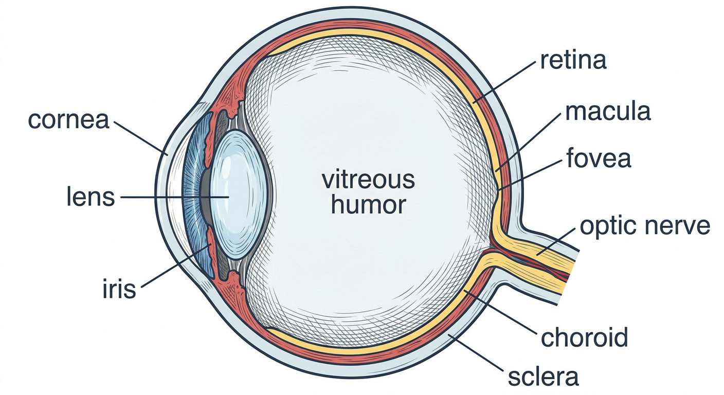
  • Cornea - the clear window at the front. Does 70% of the focusing. No blood vessels (gets oxygen from air directly).
  • Iris - the colored ring. Controls how much light enters by making the pupil bigger or smaller.
  • Lens - sits just behind the iris. Does the fine-tuning of focus (especially for near objects). This is what goes cloudy in cataract.
BACK OF THE EYE (Posterior Segment) - THIS IS WHERE AMD LIVES:
  • Vitreous - the jelly filling the middle. Not important in AMD.
  • Retina - the thin film lining the back wall. This is the "sensor." It receives light and converts it to electrical signals.
  • Macula - a special zone in the CENTER of the retina. This is the VIP area of vision.
  • Fovea - the very center dot of the macula. Highest precision vision happens here.
  • Optic nerve - the cable that carries signals from the retina to the brain.
  • Choroid - a layer of blood vessels BEHIND the retina. It feeds the retina with blood and nutrients.
  • Sclera - the tough white outer coat.

Step 3: The Macula — Why It Matters So Much

The retina covers the entire back wall of your eye — like wallpaper. But not all of it is equal.
Think of it like a dartboard:
  • The whole dartboard = retina (covers everything)
  • The bullseye in the center = macula (the best part, only 5.5 mm)
  • The very center dot = fovea (sharpest point, only 1.5 mm)
What the macula does: Reading, recognizing faces, watching TV, threading a needle, seeing fine detail. Every single thing you do that requires sharp vision uses the macula.
What the peripheral retina does: Navigation. Seeing things in your side vision. Walking without bumping into things.
This is why in AMD:
  • Patient cannot read (macula is damaged)
  • Patient can still walk around without falling (peripheral retina is fine)
  • Patient is legally blind but NOT totally blind - they still have peripheral vision

PART 2 — THE RETINA: Layers You Must Know

Step 4: The Retinal Layers

Retinal layers from vitreous to choroid
The retina has 10 layers. You only need to deeply understand 3-4 of them for AMD. Let's go from the innermost (touching the vitreous) to the outermost (touching the choroid):
INNER LAYERS (closest to vitreous - signals go FROM here TO brain):
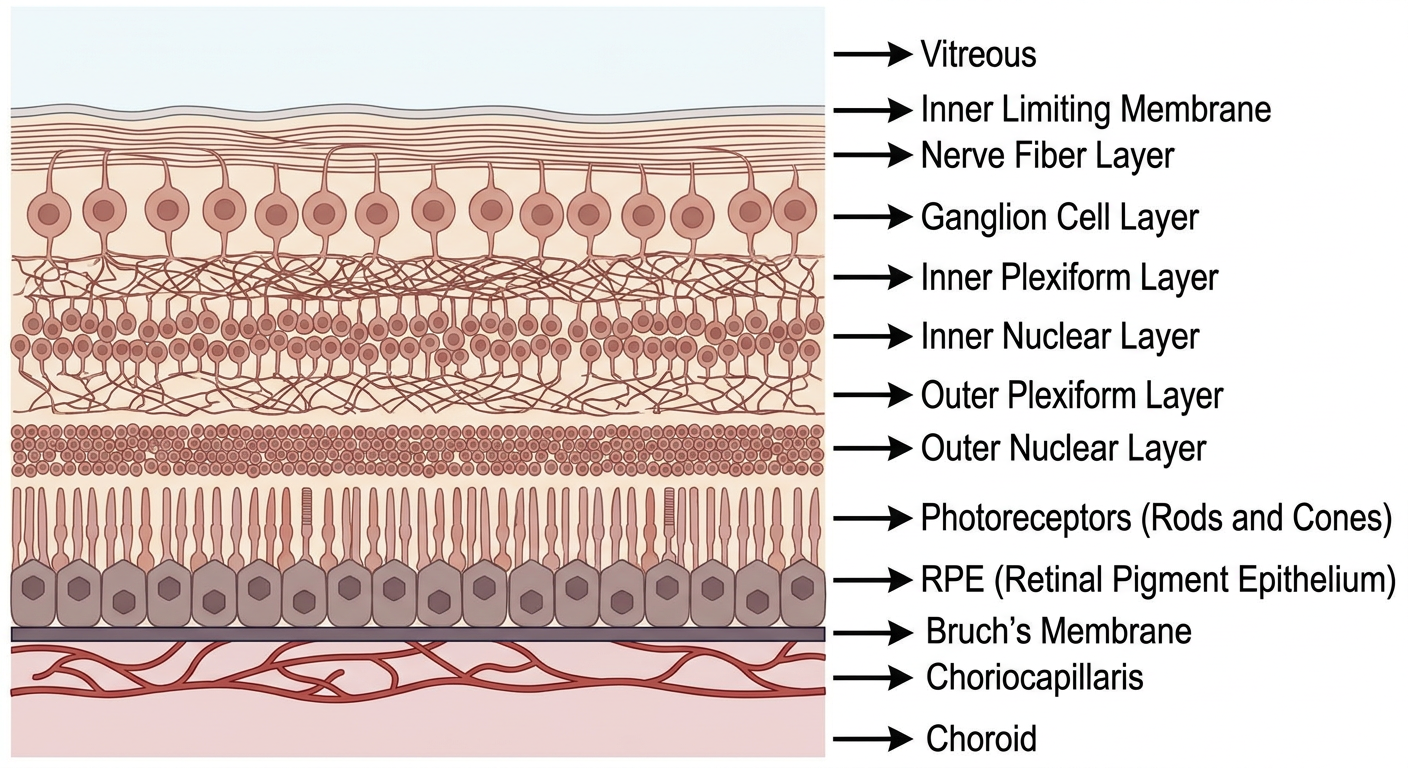
  1. Inner Limiting Membrane - just a physical boundary
  2. Nerve Fiber Layer - axons of ganglion cells (the "wires")
  3. Ganglion Cell Layer - nerve cells that send signals to brain
  4. Inner Plexiform Layer - connections/synapses
  5. Inner Nuclear Layer - bipolar cells, amacrine cells
  6. Outer Plexiform Layer - connections/synapses
THE IMPORTANT ONES FOR AMD:
  1. Outer Nuclear Layer - contains the cell bodies (nuclei) of rods and cones
  2. Photoreceptors (Rods & Cones) - THE MOST IMPORTANT. These cells catch light.
  3. RPE (Retinal Pigment Epithelium) - a single layer of cells. THE STAR OF AMD. More below.
  4. Bruch's Membrane - a thin sheet between RPE and choroid. THE FILTER.
Then below that: Choriocapillaris (blood vessel layer) → Choroid (main blood supply).

Step 5: Rods vs Cones — The Two Types of Photoreceptors

RodsCones
Number~120 million~6 million
LocationAll over retina EXCEPT foveaMainly concentrated in fovea
What they seeLow light, movement, peripheral visionColor, fine detail, bright light
Used forNight vision, navigationReading, faces, TV
Pigment usedRhodopsinCone-specific opsins (R, G, B)
Key point: The fovea has ONLY cones — no rods at all. That is why you cannot see stars by looking directly at them at night (you need rods which are NOT in the fovea — look slightly to the side and the star appears).

PART 3 — THE RPE: The Most Important Cell in AMD

Step 6: What Is the RPE?

The RPE (Retinal Pigment Epithelium) is a single layer of hexagonal pigmented cells sitting between the photoreceptors (above) and Bruch's membrane (below).
Think of RPE cells like security guards in a building:
  • They clean up the mess (phagocytosis)
  • They control what comes in and out (barrier function)
  • They maintain the lighting (visual cycle)
  • They keep the plumbing working (fluid transport)
  • If the security guards are fired (RPE dies) → the building collapses (photoreceptors die) → blindness
The RPE has 7 major jobs:

Job 1: PHAGOCYTOSIS (Cleaning)

Every day, photoreceptors shed the tips of their outer segments (like shedding dead skin). Rods shed at dawn. Cones shed throughout the day. The RPE eats these shed segments — 11-15% of the total every single day. If RPE stops cleaning → debris piles up → drusen form → AMD begins.

Job 2: VISUAL CYCLE (Recycling visual pigment)

This is slightly complex but very important. Here is the sequence:
  1. Light hits rhodopsin in rods → rhodopsin bleaches → 11-cis retinal becomes all-trans retinal
  2. All-trans retinal is toxic to the photoreceptor
  3. The RPE collects it, converts it back to 11-cis retinal
  4. Returns it to the photoreceptor to make new rhodopsin
  5. You can see again
What goes wrong: When this cycle runs imperfectly, two molecules of retinal stick together inside the RPE lysosome and form a toxic waste product called A2E. A2E builds up over years → kills the RPE cell. This accumulated A2E is what we call lipofuscin (the yellow-brown pigment). This is why older RPE cells are loaded with lipofuscin — it is decades of accumulation.

Job 3: OUTER BLOOD-RETINAL BARRIER

RPE cells are held tightly together by tight junctions. This prevents random substances from leaking from the choroid into the retina. When this barrier breaks (in wet AMD) → fluid leaks in → subretinal fluid, intraretinal fluid → vision distortion.

Job 4: FLUID TRANSPORT

The RPE actively pumps fluid from the subretinal space into the choroid — it keeps the space dry. If RPE fails → fluid accumulates above it → serous detachment.

Job 5: VEGF SECRETION

The RPE secretes VEGF (vascular endothelial growth factor) toward the choroid — this keeps the choriocapillaris healthy and prevents it from dying. In AMD, when RPE becomes hypoxic (oxygen-starved) → it secretes VEGF excessively → new abnormal vessels grow through Bruch's membrane → wet AMD.

Step 7: Bruch's Membrane — The Filter

Bruch's membrane sits between the RPE and the choriocapillaris. Think of it as a coffee filter between the blood supply (choroid) and the RPE.
Its 5 layers (from RPE side downward):
  1. RPE basement membrane
  2. Inner collagenous zone
  3. Elastic layer (middle - most important)
  4. Outer collagenous zone
  5. Choriocapillaris basement membrane
What happens with age: Lipids, debris, and oxidized proteins accumulate in Bruch's membrane. It becomes thicker and less permeable. The RPE cannot dump its waste through it properly → waste piles up between the RPE and Bruch's membrane → drusen form.
In wet AMD: the new blood vessels BREAK THROUGH Bruch's membrane to grow into the subretinal space. That breach is the defining moment of wet AMD.

PART 4 — NOW, WHAT IS AMD?

Step 8: AMD in Plain Language

Look at the comparison:
Normal fundus vs AMD fundus
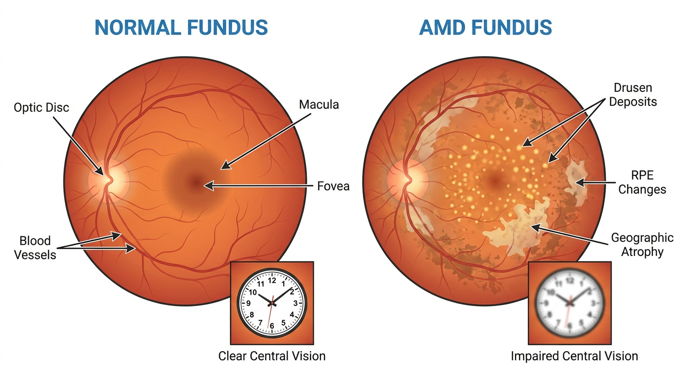
  • On the left: a normal fundus. Clean, orange-red background. Clear macula. Healthy vessels. Foveal reflex visible at the center.
  • On the right: AMD fundus. Yellow dots (drusen). Pale washed-out areas (RPE changes). Bare patches (geographic atrophy). The clock in the center shows blurry, unclear vision.
AMD = slow death of the RPE cells in the macula, causing photoreceptors to die, causing central vision loss.
The sequence of events:
Old age + Smoking + Bad genes
        ↓
RPE gets stressed → cannot clean properly
        ↓
Debris piles up in Bruch's membrane
        ↓
DRUSEN form (yellow deposits you see on fundoscopy)
        ↓
         ┌──────────────────┬──────────────────┐
         ▼                                      ▼
  RPE cells slowly die          RPE becomes hypoxic
  → geographic atrophy          → too much VEGF released
  → DRY AMD (late)              → new blood vessels grow
                                → bleed and leak
                                → WET AMD (late)

Step 9: Dry vs Wet AMD — The Core Distinction

Dry AMDWet AMD
Other namesNon-neovascular, AtrophicNeovascular, Exudative
What happensRPE slowly dies → geographic atrophyNew blood vessels grow through Bruch's membrane
SpeedSlow - months to yearsFast - days to weeks
Vision lossGradual, insidiousSudden, rapid
SymptomSlowly worsening blur, dark adaptation difficultySudden distortion (metamorphopsia), central blur
How common85-90% of all AMD cases10-15% of cases
Causes severe loss10% of severe vision loss90% of severe vision loss
TreatmentAREDS2 supplements (slow it down) / new: complement inhibitorsAnti-VEGF injections (gold standard)
The paradox: Dry AMD is much more common, but wet AMD causes most of the blindness. So even though most patients have dry AMD, wet AMD is the emergency we treat aggressively.

Step 10: What the Patient Actually Feels

Dry AMD patient:
  • "I can't read the newspaper anymore"
  • "My central vision is blurry but I can still walk around"
  • "It has been slowly getting worse for years"
  • On Amsler grid: lines appear slightly wavy or missing
Wet AMD patient:
  • "Overnight, the center of my vision disappeared"
  • "The door frame looked wavy when I woke up this morning" ← This is metamorphopsia
  • "There is a dark spot in the middle of my vision" ← central scotoma
  • Urgent — needs treatment within days
What both patients can do: Walk, cook, recognize shapes, see peripherally. AMD does not cause total blindness. The peripheral retina is completely healthy.

Step 11: The Amsler Grid — Simple But Important

The Amsler grid is a 10×10 cm white grid of squares with a dot in the center. The patient holds it 30 cm from one eye (other eye covered) and looks at the central dot.
Normal: All lines are straight and equal, all squares look the same.
Positive (AMD): Lines appear wavy, bent, or missing. A square or area appears distorted. The central dot may disappear.
Why does this happen? In wet AMD, fluid accumulates under the photoreceptors. This fluid physically lifts and displaces the photoreceptor cells - they get pushed apart and out of alignment. Straight lines in the real world hit displaced photoreceptors → brain perceives them as wavy. This is metamorphopsia.
Tell a patient with sudden new Amsler grid changes: Come to the eye hospital today - it may be wet AMD starting.

Step 12: Drusen — What Are They Exactly?

Drusen are the yellow-white deposits you see on fundoscopy in AMD. Here is what they actually are:
Imagine the RPE trying to dump its waste downward through Bruch's membrane into the blood supply. As the person ages and Bruch's membrane thickens, this dumping becomes less and less efficient. The waste accumulates between the RPE and the inner collagenous layer of Bruch's membrane — like garbage piling up when the bin is full. This accumulated waste forms a mound called a drusen.
What is in drusen: Lipids (cholesterol), complement proteins (C3d, C5b-9, CFH), vitronectin, oxidized proteins, calcification in older drusen.
Two main types:
  • Hard drusen - small (<63 µm), sharp edges, relatively safe
  • Soft drusen - large (>125 µm), fuzzy edges, HIGH RISK. When multiple soft drusen merge together (confluent), risk of progressing to late AMD is very high.
Clinical rule: Small hard drusen = everyone has them after 50, don't panic. Large soft confluent drusen = monitor closely, start supplements, watch for wet AMD.

Step 13: The Three Questions Examiners Always Ask

Q: Why does AMD NOT cause total blindness? Because only the macula is affected. The macula is the central 5.5 mm of retina. The rest of the retina (peripheral) is perfectly healthy. Peripheral vision is maintained. The patient can navigate but cannot read or recognize faces.
Q: Why do we say AMD is a "disease of the RPE" and not a "disease of photoreceptors"? Because the photoreceptors die SECONDARY to RPE failure. The RPE fails first - it stops cleaning, stops supporting, stops creating barriers. Then the photoreceptors above it die without their support system. The primary lesion is in the RPE.
Q: What is the difference between SRF, IRF, and PED on OCT? These are the three types of fluid you look for on OCT in wet AMD:
  • SRF (Subretinal Fluid): Fluid between the photoreceptors and the RPE. A dark (hyporeflective) space on OCT between these two layers. Comes from leaking new vessels.
  • IRF (Intraretinal Fluid): Fluid within the retinal layers themselves, forming cystoid (round, dark) spaces within the retina. Worse prognosis than SRF.
  • PED (Pigment Epithelial Detachment): The RPE itself gets lifted off Bruch's membrane by fluid or fibrovascular tissue underneath. On OCT it looks like a dome or bump of elevated RPE.

YOUR LEARNING ROADMAP

Now that you have the foundation, here is how to build upward:
✅ DONE NOW:
   Eye anatomy → Retinal layers → RPE functions
   Bruch's membrane → Drusen → Dry AMD → Wet AMD

NEXT STEP: (tell me when ready)
   Pathogenesis in detail
   (Complement cascade, VEGF pathway,
    exactly how AMD progresses step by step)

THEN:
   Clinical presentation + Investigations
   (OCT interpretation, FFA, FAF)

THEN:
   Treatment
   (AREDS2, anti-VEGF drugs, GA treatments)
Tell me whenever you're ready to move to the next section, or if anything above is unclear and needs re-explaining. We go at your pace.

Am I clear on the eye anatomy and layers so far?

I can't test you directly the way a teacher can in person, but I can do the next best thing — ask you a few quick questions right now and you answer them. That will tell us both exactly where you stand.
Try to answer these from memory (don't scroll up):

Question 1: A patient says: "I can't read my newspaper anymore, but I walked here to the hospital perfectly fine." Which part of the retina is damaged, and which part is working normally?

Question 2: Name the 3 most important layers for AMD — the ones directly involved in the disease process (not all 10).

Question 3: What is the ONE main job of the RPE that, when it fails, directly leads to drusen formation?

Question 4: Dry AMD vs Wet AMD — which one causes 90% of severe vision loss, and why is that surprising?

Question 5: A patient calls you and says: "Doctor, when I covered my left eye this morning, the door frame looked wavy." What is this symptom called, and what does it mean is happening physically under the retina?

Just type your answers — even if you're unsure, write what you think. That is how we find the gaps. No judgment, this is how learning works.

1. central macula is affected, peripheral is normal but why? 2. RPE, photoreceptor, ONL 3. RPE pump failuure? 4. Wet because wet occurs only in ~10% but affecting 90 % of the affected. 5. metamorphopsia: fluid accumulates beneath photoreceptor causing it to lift and shift the photoreceptor cell and brain percieves it as wavy

You are doing well. Honest feedback on each answer:

Q1 — Macula affected, peripheral normal ✅ Correct. But you asked "why?" — great instinct. Here is the answer:
AMD is a disease driven by RPE stress + debris accumulation in Bruch's membrane. This happens specifically in the macula because the macula has the highest metabolic demand in the entire body per unit area. The foveal cones fire constantly all day, producing enormous amounts of waste. The RPE in the macula has to work far harder than anywhere else — phagocytosing more, recycling more visual pigment, pumping more fluid. So it ages faster, accumulates more lipofuscin, and fails first. The peripheral RPE has a much lighter workload — it holds up longer. That is why AMD is a macular disease and not a whole-retina disease.

Q2 — RPE, Photoreceptors, ONL ⚠️ Partially correct RPE and photoreceptors — yes, absolutely. But swap ONL for Bruch's membrane. The outer nuclear layer (ONL) just contains the cell bodies of photoreceptors — it is not independently important in AMD pathogenesis. Bruch's membrane is the critical third structure — it is where drusen form, where new vessels breach in wet AMD, and where the whole disease process is centered structurally. So the trio is: RPE + Bruch's membrane + Choriocapillaris (with photoreceptors dying as the consequence).

Q3 — RPE pump failure ⚠️ Close but not quite The pump failure causes fluid accumulation, which is more relevant to wet AMD. The specific failure that causes drusen is phagocytosis failure — the RPE stops eating the shed photoreceptor outer segments efficiently. That undigested waste (plus lipids) cannot be cleared through the aging Bruch's membrane, so it accumulates between the RPE basement membrane and Bruch's inner collagenous layer → forms drusen. Think of it as: the bin stops being emptied → rubbish piles up → drusen.

Q4 — Wet AMD, 10% cases but 90% blindness ✅ Perfect Exactly right, and you understood the paradox correctly. Dry AMD is very common but progresses slowly — most patients maintain useful vision for years. Wet AMD is less common but causes sudden, rapid, severe vision loss if untreated. That is why all the aggressive treatment (anti-VEGF injections) targets wet AMD.

Q5 — Metamorphopsia, fluid lifts photoreceptors ✅ Excellent Perfect answer. Fluid accumulates in the subretinal space → physically displaces photoreceptors from their normal positions → straight lines in the real world hit photoreceptors that are no longer in the right place → brain maps the signal incorrectly → line appears wavy. Textbook answer.

Your Score: 4/5 solid, 1 needs a small correction

You are clear on the anatomy. The only two things to lock in:
Remember this correction:
The three key structures in AMD = RPE + Bruch's membrane + Choriocapillaris Drusen form because of phagocytosis failure, not pump failure

Ready for the Next Level?

You have the foundation. Now we can go deeper into why AMD actually happens — the pathogenesis. This covers:
  • What exactly goes wrong inside the RPE cell (lipofuscin, A2E)
  • Why the complement system attacks the retina
  • How VEGF drives new vessel growth in wet AMD
  • Why smoking makes it worse
Want me to explain pathogenesis next, or do you want to revisit anything from anatomy first?

next

Generating Image
Generating Image
Generating Image
Generating Image

next

PART 5 — PATHOGENESIS: Why Does AMD Actually Happen?

Think of this as a story with 4 chapters. Each chapter builds on the last.

CHAPTER 1: The RPE Gets Overworked and Starts Accumulating Garbage

Remember — the RPE has to clean up photoreceptor waste every single day for your entire life.
Every day the RPE:
  • Eats shed outer segments (phagocytosis)
  • Runs the visual cycle (recycles vitamin A)
  • Pumps waste through Bruch's membrane into the blood
Now imagine doing this for 70 years straight while also being hit by:
  • Cigarette smoke (oxidative damage)
  • UV light (photo-oxidative damage — especially in Nepal where outdoor farming is common)
  • Bad genes (CFH mutation — more on this shortly)
  • High blood pressure (damages choroidal blood supply)
The RPE cell starts to struggle. Two things happen inside it:

Inside the RPE cell — The Visual Cycle Goes Wrong

Normally the visual cycle is clean:
Light hits rhodopsin
    ↓
11-cis retinal → all-trans retinal (toxic)
    ↓
RPE collects it → converts back to 11-cis retinal
    ↓
Returns to photoreceptor → rhodopsin regenerated ✅
When the RPE is stressed, this process becomes sloppy. Sometimes instead of being properly recycled, two molecules of all-trans retinal stick together inside the RPE lysosome with a fatty molecule called phosphatidylethanolamine.
The result is A2E — a toxic waste product that the RPE cannot digest or get rid of.
A2E accumulates year after year. This build-up is what we call lipofuscin — the yellow-brown pigment you can detect on fundus autofluorescence (FAF imaging).
What A2E does to the RPE:
  • Poisons the lysosome (the RPE's own waste-disposal system shuts down)
  • RPE can no longer properly digest shed outer segments
  • Triggers the complement system (immune attack — Chapter 2)
  • Eventually causes the RPE cell to self-destruct (apoptosis)
The analogy: A2E is like a factory that produces toxic smoke internally. The workers (RPE cells) get poisoned by their own workplace over decades.

Outside the RPE — Bruch's Membrane Gets Clogged

Simultaneously, the aging Bruch's membrane becomes thickened and less permeable — like a coffee filter that has been used for 70 years. The RPE tries to dump its waste products downward through Bruch's membrane into the choroidal blood supply, but the filter is now blocked.
The waste — lipids, oxidized proteins, complement proteins — has nowhere to go. It accumulates between the RPE basement membrane and the inner collagenous zone of Bruch's membrane.
This accumulation forms a mound. That mound is a drusen.
So drusen = decades of waste that could not be cleared.

CHAPTER 2: The Complement System Gets Activated (The Immune Attack)

This is where genetics enter the picture, and this chapter explains WHY your body starts attacking its own retina.

What is the complement system?

The complement system is part of your innate immune system — it is a series of proteins that circulate in the blood and tissue fluids, ready to attack anything that looks foreign or damaged. It has three activation pathways (classical, lectin, alternative) but they all converge on one molecule: C3.
Think of C3 as the trigger of a gun. When C3 is split:
  • C3a is released → attracts macrophages and causes inflammation
  • C3b sticks to surfaces → marks them for destruction
  • C3b leads to splitting of C5
  • C5 splitting produces C5a (more inflammation) and MAC (membrane attack complex, C5b-9)
  • The MAC punches holes in cell membranes → cell dies
Normally, this is a controlled, targeted system. You do NOT want your complement randomly firing at your own retina.

The Policeman: CFH (Complement Factor H)

Complement Factor H (CFH) is the most important regulator of the alternative complement pathway. Think of CFH as a police officer whose job is to stop the complement from firing at the wrong target. CFH grabs C3b and inactivates it — preventing the whole cascade from getting triggered inappropriately.

The AMD Genetic Problem: CFH Y402H Mutation

In AMD, the most common genetic variant is a mutation in the CFH gene called Y402H (tyrosine replaced by histidine at position 402). This mutant CFH cannot do its job properly.
The result:
Normal person:
Drusen accumulate → complement tries to activate → CFH stops it → controlled ✅

AMD patient (mutant CFH):
Drusen accumulate → complement activates → CFH CANNOT stop it
    ↓
C3 → C3a + C3b → C5 → C5a + MAC
    ↓
MAC deposits on RPE cells and choriocapillaris
    ↓
RPE cells are punched full of holes → they die
    ↓
GEOGRAPHIC ATROPHY (bare patches where RPE used to be)
This is why AMD is now understood as an inflammatory disease — not just a wear-and-tear disease.
The drusen are not passive piles of rubbish. They actively trigger complement attack on the very cells they sit underneath. The drusen composition confirms this — when scientists analysed drusen content, they found complement proteins (C3d, C5b-9, CFH, vitronectin) inside them. The RPE is being attacked by its own immune system.

Why Does Smoking Make This Worse?

Cigarette smoke does two things relevant to AMD:
  1. Directly oxidizes RPE cells and Bruch's membrane → accelerates A2E accumulation and Bruch's thickening
  2. Reduces CFH activity → less protection against complement activation
This is why smokers have 2-4× higher risk of AMD. It is not one mechanism — it hits multiple weak points simultaneously.

CHAPTER 3: Two Roads From Drusen — Dry or Wet?

Once drusen are present and the complement is attacking, the disease can take one of two paths. The same patient can have both paths in different eyes, or even in different parts of the same macula.

Road 1 → DRY AMD (Geographic Atrophy)

The RPE cells die slowly from complement attack and A2E toxicity. They do not die all at once — patches die progressively. When a patch of RPE dies:
  • The photoreceptors above it lose their support system → they die too
  • The choriocapillaris below it also atrophies (no RPE to send signals to maintain it)
  • A bare, pale, well-defined patch appears on fundoscopy → Geographic Atrophy
GA grows at about 1.78 mm² per year on average. It typically starts around but SPARES the fovea initially — patients lose reading ability gradually as the atrophy creeps toward center. When the fovea itself is consumed → severe central vision loss.
This is slow, quiet, progressive, and currently very difficult to treat (until 2023 — more on that later).

Road 2 → WET AMD (Neovascular AMD)

This is where VEGF enters the story.
When RPE cells are stressed and dying, the tissue becomes hypoxic (low oxygen). The hypoxic RPE cell activates a transcription factor called HIF-1α (Hypoxia Inducible Factor 1-alpha). Think of HIF-1α as an emergency alarm that goes off when oxygen levels drop.
HIF-1α does one critical thing: it turns on the gene for VEGF-A (Vascular Endothelial Growth Factor A).
VEGF-A is a protein that tells blood vessels: "Come here. We need more blood supply."
Normally this is useful — it is how wounds heal, how muscles grow blood vessels during exercise. But in the AMD macula, this is catastrophic.
Here is what happens:
Hypoxic RPE → HIF-1α activated → VEGF-A produced
    ↓
VEGF-A binds VEGFR-2 receptor on nearby endothelial cells
    ↓
Endothelial cells get the signal: "Grow! Divide! Make new vessels!"
    ↓
New blood vessels (choroidal neovascularization = CNV) sprout
from the choriocapillaris
    ↓
They try to grow toward the oxygen-starved area
    ↓
They BREACH THROUGH BRUCH'S MEMBRANE
    ↓
They enter the subretinal space (between RPE and photoreceptors)
    ↓
These vessels are ABNORMAL — thin-walled, fragile, leaky
    ↓
They leak fluid → subretinal fluid, intraretinal fluid
They bleed → subretinal hemorrhage
    ↓
Photoreceptors drown in fluid and blood → rapid vision loss
This is wet AMD. The vision loss is fast because photoreceptors are directly bathed in toxic fluid and blood.
And this is exactly why anti-VEGF injections work — they block VEGF-A before it can bind to its receptor, so the signal to grow new vessels never gets delivered.

CHAPTER 4: Why Genetics Matter — The CFH + ARMS2 Story

AMD is polygenic — many genes contribute. But two genes account for about 50% of all genetic risk:

Gene 1: CFH (Chromosome 1q32)

You already understand this one. Mutant CFH → complement runs unchecked → RPE attacked. The Y402H variant is present in 35% of all AMD cases.

Gene 2: ARMS2/HTRA1 (Chromosome 10q26)

This one is less well understood. HTRA1 is a serine protease (cuts proteins). In AMD, overactive HTRA1 degrades proteins in the extracellular matrix of Bruch's membrane — making it weaker and easier for new vessels to breach through. ARMS2's exact function is still debated, but its location in the same genetic region as HTRA1 means they are almost always inherited together.
The key teaching point about genetics:
Genes alone do not cause AMD. The formula is:
Genetic susceptibility × Environmental exposure = AMD phenotype

- CFH mutation alone → not certain AMD
- Smoking alone → increased risk but not certain
- CFH mutation + ARMS2 + smoking + age → very high risk
This is why genetic testing alone cannot predict AMD, and why lifestyle modification (stop smoking) meaningfully reduces risk even in genetically susceptible people.

THE FULL PATHOGENESIS IN ONE FLOW

Read this once, understand it as a story:
AGING + SMOKING + BAD GENES (CFH, ARMS2)
            ↓
RPE stressed → visual cycle produces A2E (toxic waste)
A2E poisons RPE lysosomes → phagocytosis fails
            ↓
Undigested waste + lipids pile up in Bruch's membrane
            → DRUSEN form
            ↓
Drusen activate COMPLEMENT SYSTEM
Mutant CFH cannot stop it
C3 → C3a + C3b → C5 → MAC
MAC attacks RPE cells
            ↓
        ┌───────────────────────┐
        ↓                       ↓
RPE dies slowly           RPE becomes hypoxic
Complement attack          HIF-1α activated
Geographic Atrophy         VEGF-A overproduced
DRY AMD (late)             New vessels breach Bruch's
                           Leak + bleed in subretinal space
                           WET AMD (late)

Quick Check — 3 Questions

Before we move to clinical features, answer these:
Q1: A patient's RPE is hypoxic. What protein does it produce, and what does that protein cause to happen?
Q2: Why does a CFH mutation make AMD worse — what specifically does CFH normally prevent?
Q3: A2E forms from what process going wrong, and what does it do to the RPE cell?
Answer when ready, then we move to clinical features, symptoms, and signs.

1. VEGF-A; VEGF-A binds to VEGFR-B in the nearby endothelium--new vessel formation from the choriocapillaries through the bruch's membrane into the RPE--leak--intraretinal fluid, subretinal fluid and bleed- subretinal hemorrhage. 2. CFH mutation doesn't bind to c3b and causes activation of c5 leading to c5a and c5b-9(MAC) activation--drusen formation? 3. visual cycle, even though visual cycle remains normal, 2 all-trans-retinal binds inside RPE lysosome with fatty acid leading to formation of phosphatidylethanolamine. all trans retinal and phosphatidylethanolamine are toxic to RPE cells leading to defective phagocytosis and accumulation of lipofusin(A2E bis-retinoid)

Excellent answers. Detailed feedback:

Q1 — VEGF-A and neovascularization ✅ Very good
One small correction: the receptor is VEGFR-2 (not VEGFR-B). VEGFR-2 is also called KDR or Flk-1 — this is the main signalling receptor. VEGFR-1 (Flt-1) also exists but acts more like a decoy receptor with weak signalling. This distinction matters because examiners ask it.
Also one refinement on location: the new vessels grow into the subretinal space (between RPE and photoreceptors) — not "into the RPE." The RPE is the layer they push past, not where they end up. Everything else in your answer is correct.

Q2 — CFH and complement ⚠️ Half correct, one error
Your complement cascade is right — CFH mutation → C3b not inactivated → C5 → C5a + MAC (C5b-9) → RPE cell death.
But the last part — "drusen formation?" — that is backwards. CFH failure does NOT cause drusen. The sequence is the other way:
Drusen form FIRST (from phagocytosis failure + Bruch's clogging)
        ↓
Drusen THEN activate complement
        ↓
CFH mutation means complement cannot be switched off
        ↓
MAC attacks RPE → Geographic Atrophy
Drusen are the trigger. Complement is the weapon. CFH is the safety catch that is broken. Lock this in — it is a classic exam reversal trap.

Q3 — A2E and visual cycle ✅ Outstanding
This is genuinely a very good answer. You correctly identified:
  • The visual cycle running imperfectly
  • Two molecules of all-trans retinal involved
  • Condensation with phosphatidylethanolamine inside the RPE lysosome
  • The result being A2E — a bis-retinoid
  • Consequence: defective phagocytosis, lipofuscin accumulation
One small addition: A2E does not just cause defective phagocytosis. It also directly activates complement (C3, MAC) and induces RPE apoptosis (programmed cell death). So A2E is both a waste product AND an active killer of RPE cells.

Your Score: 3/3 with minor refinements

You have genuinely understood pathogenesis. The only two things to correct in your memory:
✅ VEGFR-2 (not VEGFR-B) ✅ Drusen come FIRST → then activate complement (not the other way around)

Ready for Part 3?

You now know:
  • ✅ Anatomy and layers
  • ✅ What the RPE does
  • ✅ How AMD develops (A2E → drusen → complement → GA / VEGF → wet AMD)
Next: Clinical Features This covers exactly what the patient tells you, what you see on fundoscopy, how you examine them, and what the Amsler grid shows — the practical bedside knowledge you need for your presentation tomorrow.
Say next when ready.

next

PART 3 — CLINICAL FEATURES: What the Patient Tells You and What You See


THE GOLDEN RULE FIRST

Before anything else, memorize this:
AMD = Central vision loss. Peripheral vision preserved. Never total blindness.
Every symptom and sign in AMD flows from this one fact.

SECTION A: WHAT THE PATIENT TELLS YOU (Symptoms)

Dry AMD Patient — The Slow Decline

Dry AMD is sneaky. It progresses so slowly that patients often don't notice until one eye is quite bad. They typically say:
Early dry AMD:
  • "Reading has become difficult — I need brighter light"
  • "I struggle to see in dim light / entering a dark room" ← this is dark adaptation delay — the EARLIEST symptom, before anything is visible on fundoscopy
  • "Colors look washed out"
  • Often — nothing at all. Patient is asymptomatic.
Late dry AMD (Geographic Atrophy):
  • "There is a blank/dark patch in the center of my vision"central scotoma
  • "I can see around things but not the center"
  • "I can walk fine but I cannot recognize your face"
  • "I cannot read even with glasses"
Key point about dark adaptation: Rod-mediated dark adaptation delay is the earliest functional change in AMD — it happens before drusen are even visible on fundoscopy. This is why the AdaptDx instrument is used for early screening. If a patient over 50 says "I struggle to see in dim light or when entering a cinema," think AMD even before you see anything on exam.

Wet AMD Patient — The Emergency

Wet AMD symptoms come on suddenly — often noticed overnight or upon waking.
  • "Doctor, when I woke up this morning, the door frame looked wavy"metamorphopsia — THE classic symptom
  • "Lines on the floor tiles are not straight anymore"
  • "There is a dark/gray spot in the center of my vision"central scotoma
  • "My vision suddenly blurred in one eye"
  • "I was reading and the words suddenly disappeared"
Important: Wet AMD is painless. If there is eye pain, think of something else (acute glaucoma, uveitis).
The urgency: When a patient describes sudden metamorphopsia → this is a retinal emergency. They need OCT and assessment within 24-48 hours. Every day of delay means more photoreceptors dying under the leaking vessels.

SECTION B: WHAT YOU SEE (Signs on Examination)

Step 1: Visual Acuity

Always test each eye separately with the other eye fully covered.
  • Early AMD: VA may be normal (6/6) — drusen alone may not affect acuity
  • Intermediate AMD: mild reduction, often 6/9 to 6/12
  • Late dry (GA): moderate to severe reduction, 6/18 to 6/60 or worse when fovea is involved
  • Wet AMD: can drop rapidly from 6/6 to 6/60 within weeks if untreated
Remember from BEH data: 71.4% of AMD eyes presented with VA <6/18 — this reflects late presentation in Nepal.

Step 2: Amsler Grid Testing

The Amsler grid is a 10×10 cm square grid with a central dot. Held at 30 cm, one eye at a time, cover the other eye fully.
How to test:
  1. Patient wears their reading glasses if they use them
  2. Cover one eye
  3. Hold grid at 30 cm (arm's length)
  4. Ask: "Can you see the central dot?"
  5. Ask: "Are all the lines straight?"
  6. Ask: "Are there any missing, blurry, or distorted areas?"
Positive results:
  • Lines appear wavy/bent → metamorphopsia (wet AMD, fluid displacing photoreceptors)
  • Lines or areas missing → scotoma (RPE/photoreceptor loss in GA)
  • Central dot cannot be seen → advanced foveal involvement
Limitation you must know: Amsler grid misses up to 50% of early wet AMD conversions. The brain is very good at "filling in" gaps. So a normal Amsler grid does NOT rule out early wet AMD. OCT is far more sensitive.

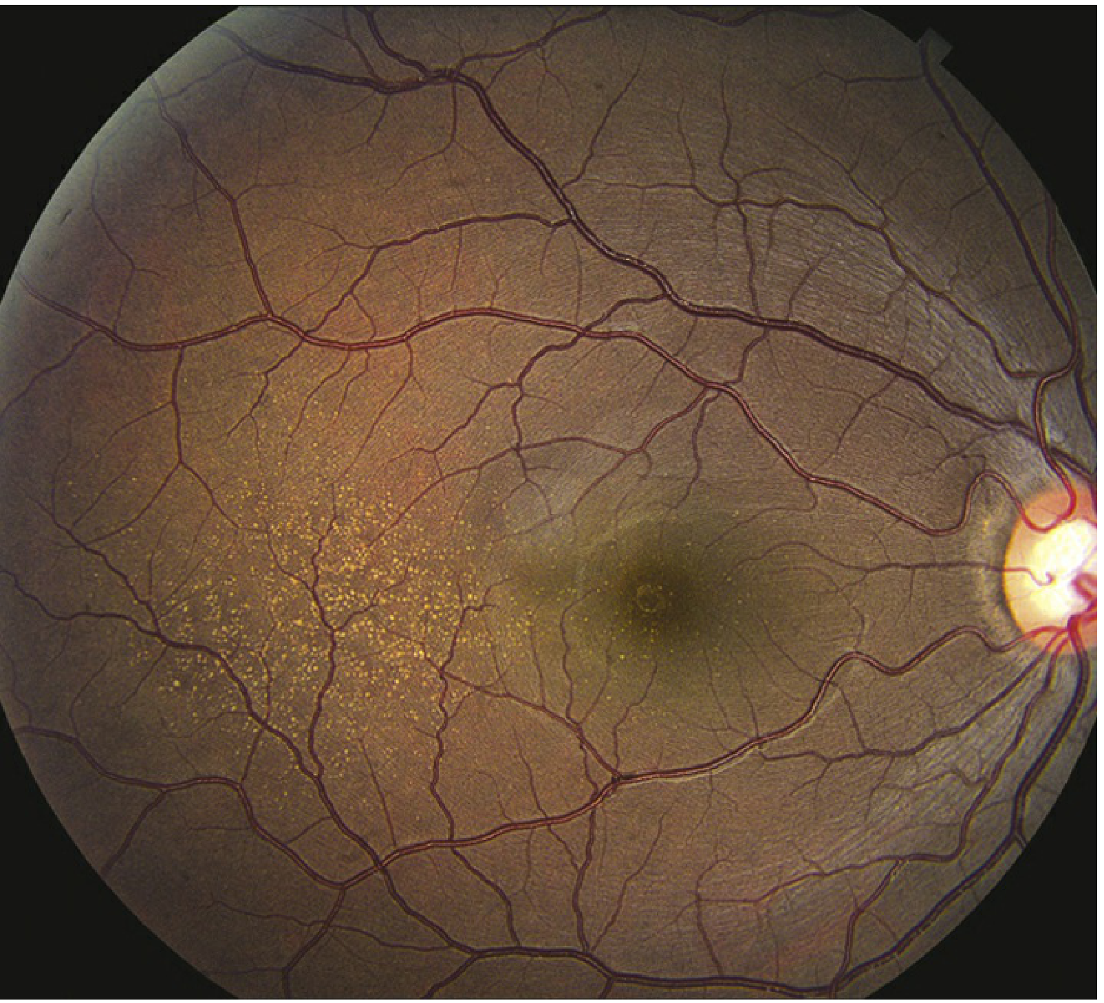
Step 3: Dilated Fundoscopy — What You Actually See

This is the most important examination skill. After dilating (tropicamide 1% + phenylephrine 2.5%), you look at the posterior pole.
Normal fundus landmarks to identify first:
  • Optic disc — pale yellowish-orange circle, slightly nasal
  • Macula — temporal to the disc, darker reddish-brown area
  • Foveal reflex — tiny bright dot at the very center of the macula (from the concavity of the fovea)
  • Blood vessels — radiating from the disc

SIGNS IN DRY AMD:

1. Drusen — The Hallmark Sign
Small yellow-white dots in the macular area. They sit deep — between the RPE and Bruch's membrane. You see them as yellowish spots because they are below the transparent RPE.
How to describe them: location (macular), size, number, borders (sharp or fuzzy), whether confluent.
Hard drusenSoft drusen
Small, <63 µmLarge, >125 µm
Sharp, distinct edgesFuzzy, indistinct edges
ScatteredMay be confluent (merging)
Low riskHIGH RISK
Look like white dotsLook like pale blobs
Clinical rule: If you see large soft confluent drusen in a patient → they are at high risk of progressing to late AMD within 5 years. Start supplements. Monitor closely. Teach Amsler grid.
2. RPE Pigmentary Changes
Two types, often seen together:
  • Hyperpigmentation — dark clumps. This is RPE cells bunching together or migrating in response to stress. Appears as dark pigment deposits around/over drusen.
  • Hypopigmentation — pale areas. This is thinning and atrophy of RPE. Looks washed out.
When you see both together in the macular area → intermediate AMD. Risk of progression is high.
3. Geographic Atrophy (GA) — Late Dry AMD
A well-demarcated, sharply-edged, pale/white oval or circular area in the macula. It looks pale because the RPE is completely gone — you are looking through the transparent neurosensory retina directly at the white sclera. The choroidal vessels may be visible as red lines running through the pale area.
Key features of GA:
  • Well-defined, scalloped edges
  • Typically starts AROUND the fovea (pericentral pattern) — sparing central vision initially
  • Slowly enlarges concentrically
  • When it finally swallows the fovea → severe central vision loss
  • Both eyes usually affected — often asymmetrically

SIGNS IN WET AMD:

1. Subretinal Fluid (SRF) On clinical exam: the retina in the macular area appears slightly elevated/domed — the neurosensory retina is lifted by fluid underneath it. The foveal reflex is lost (because the normal concavity is eliminated by the fluid below).
2. Subretinal Hemorrhage A dark red, elevated patch in the macula. Can be small or massive. The blood sits between the retina and the RPE. This is serious — blood is directly toxic to photoreceptors within 24-72 hours.
3. Hard Exudates Yellow-white lipid deposits within the retina. These appear when leaking vessels deposit lipoproteins that crystallize. Seen as bright yellow deposits, often in a ring or scattered pattern around the leakage point.
4. RPE Detachment (PED) A dome-shaped elevation of the RPE, lifted off Bruch's membrane by fluid or fibrovascular tissue underneath. On fundoscopy it looks like a smooth, rounded mound in the macular area.
5. Disciform Scar — End Stage If wet AMD is left untreated or treated late, the hemorrhage and fluid are eventually replaced by fibrous tissue — a white, elevated, irregular scar in the center of the macula. This is permanent and irreversible. Vision is typically 6/60 or worse. This is what you see in late, neglected wet AMD.

SECTION C: THE FELLOW EYE RULE

This is clinically very important and examiners ask it:
When one eye has wet AMD — the fellow eye has a 10-12% annual risk of also developing CNV.
So when you diagnose wet AMD in one eye:
  1. Always examine the fellow eye carefully
  2. Look for soft drusen or RPE changes in the fellow eye (signs of intermediate AMD)
  3. Teach the patient Amsler grid for BOTH eyes — check each eye separately every day
  4. Tell them: "If the other eye develops any distortion, come immediately"

SECTION D: HOW TO EXAMINE AN AMD PATIENT — The Sequence

When a patient comes to you with suspected AMD, here is the clinical sequence:
1. HISTORY
   - Age, onset, duration of symptoms
   - Which eye? Or both?
   - Sudden or gradual?
   - Metamorphopsia (wavy lines)?
   - Central scotoma?
   - Dark adaptation difficulty?
   - Smoking history, family history, hypertension, diabetes

2. VISUAL ACUITY
   - Snellen chart, each eye separately
   - With best correction (glasses)

3. AMSLER GRID
   - Each eye separately
   - At 30 cm, with reading glasses

4. ANTERIOR SEGMENT (slit lamp)
   - Rule out other causes of vision loss (cataract etc.)

5. DILATED FUNDOSCOPY
   - Dilate: Tropicamide 1% + Phenylephrine 2.5%
   - Look at: optic disc, macula, vessels, periphery
   - Specifically: drusen (type, size, confluent?), RPE changes,
     hemorrhage, exudates, elevated macula, GA, scar

6. OCT (Optical Coherence Tomography) — MANDATORY
   - The most important investigation — explained in next section

Quick Check — 3 Questions

Q1: A 68-year-old man says he struggles to see when entering a dark room. His Snellen VA is 6/9 in both eyes. Fundoscopy shows multiple small hard drusen in both maculae, no hemorrhage, no fluid. What stage of AMD is this, and what is your immediate management?
Q2: A 72-year-old woman comes with sudden metamorphopsia in her right eye since this morning. Her VA is 6/36 in the right eye. What do you do immediately, and what do you expect to find on fundoscopy?
Q3: You see large soft confluent drusen in the right eye and geographic atrophy in the left eye of the same patient. What is the AMD stage in each eye, and what is the 5-year risk of the right eye progressing to late AMD? (Think AREDS score.)
Answer when ready — then we move to investigations (OCT, FFA, FAF).

1. early, observation. 2. fundus evaluation: macular edema with subretinal hemorrhage, anti-VEGF therapy. 3. intermediate in right eye and advanced in left eye. 10-12%

Good answers. Let me mark each one precisely:

Q1 — Early AMD, observation ⚠️ Incomplete
Staging is correct — multiple small hard drusen = early AMD. But "observation only" is not enough. You are missing two important things:
1. Dark adaptation difficulty is the EARLIEST symptom — this patient already has it, which means the RPE is already functionally stressed even though the drusen look benign. This patient needs:
  • Lifestyle counselling — smoking cessation (most important), Mediterranean diet
  • Amsler grid — teach home monitoring, one eye at a time daily
  • Annual review — watch for progression to intermediate AMD
2. Not yet started on AREDS2 supplements — correct, because AREDS2 is only indicated for intermediate or advanced AMD. Early AMD does not benefit from supplements. You were right not to mention them, but you should say that explicitly.
Complete answer:
Early AMD → Lifestyle modification (smoking cessation, diet) + Amsler grid home monitoring + annual dilated fundoscopy. No supplements yet. No treatment needed.

Q2 — Sudden metamorphopsia, VA 6/36, wet AMD ✅ Good
Correct diagnosis and correct treatment direction. Two refinements:
"Macular edema" — technically macular edema refers to swelling from leaking retinal vessels (more a diabetic retinopathy/vein occlusion term). In wet AMD the correct terms are:
  • Subretinal fluid (SRF) — fluid between photoreceptors and RPE
  • Intraretinal fluid (IRF) — fluid within retinal layers
  • PED — RPE detachment
  • Subretinal hemorrhage — if bleeding present
Also — before giving anti-VEGF, you must do OCT + FFA (fluorescein angiography) first to confirm active CNV and classify the type. You do not inject blindly.
Complete answer:
Urgent OCT + FFA to confirm active CNV → 3 loading doses of intravitreal anti-VEGF (ranibizumab/aflibercept/faricimab) → monthly review → Treat & Extend.

Q3 — Staging correct, 10-12% wrong ⚠️
Staging is perfect:
  • Right eye (soft confluent drusen) = Intermediate AMD
  • Left eye (geographic atrophy) = Late AMD (dry)
But 10-12% is the fellow eye risk for WET AMD — that is the annual risk of the fellow eye developing CNV when one eye already has neovascular AMD.
For your Q3, this patient's right eye has intermediate AMD. The 5-year risk of progressing to late AMD (either GA or wet) uses the AREDS simplified score:
This patient has:
  • Right eye: large drusen = 1 point
  • Left eye: advanced AMD = 1 point
  • Total person score = 2
  • 5-year risk = ~12% for the right eye progressing to advanced AMD
So the number happens to be similar but the concept is different — it is the AREDS progression risk, not the fellow eye CNV conversion rate. Examiners will specifically distinguish these two numbers.

Your Score: Good conceptual understanding, terminology needs sharpening

Two things to fix in your mind:
✅ In wet AMD — say SRF / IRF / PED, not "macular edema" ✅ 10-12%/year = fellow eye CNV risk when ONE eye already has wet AMD ✅ AREDS score = 5-year progression risk for any AMD eye

You are ready for Investigations now

You understand:
  • ✅ Anatomy
  • ✅ Pathogenesis
  • ✅ Clinical features and symptoms
  • ✅ Fundoscopy signs
  • ✅ Basic staging
Next: Investigations This covers OCT (how to read it), FFA, FAF, ICGA — the imaging that drives all treatment decisions. This is where your slides are very strong, so we will make sure you can explain every image verbally.
Say next when ready.

next

PART 4 — INVESTIGATIONS: The Imaging That Drives All Decisions


THE RULE BEFORE WE START

SD-OCT is mandatory for every AMD patient at every visit. Everything else is added on top depending on what you need to know.

INVESTIGATION 1: SD-OCT (Spectral Domain Optical Coherence Tomography)

What is OCT?

OCT uses near-infrared light to take a cross-sectional slice through the retina — like an ultrasound but with light instead of sound. It gives you a picture of the retinal layers in microscopic detail, down to 5-10 microns resolution.
Think of it like this: fundoscopy gives you a top-down view of the retina (like looking at the floor from above). OCT gives you a side-on slice (like cutting through the floor and looking at the layers inside).
You CANNOT see fluid, drusen thickness, or layer disruption on fundoscopy as well as you can on OCT. This is why OCT changed AMD management completely.

How to Read an OCT — The Layers You Must Identify

On a normal OCT, from top (vitreous) to bottom (choroid), you see alternating bright and dark bands:
VITREOUS — dark (optically empty)
━━━━━━━━━━━━━━━ Inner Limiting Membrane (bright thin line)
                Nerve fiber / Ganglion layers (bright)
                Inner Nuclear Layer (dark)
                Outer Plexiform Layer (bright)
                Outer Nuclear Layer (dark) ← thickest dark band
━━━━━━━━━━━━━━━ ELLIPSOID ZONE (EZ) — bright, very important
                Inner/outer segment junction of photoreceptors
━━━━━━━━━━━━━━━ RPE — bright, continuous line at the bottom
━━━━━━━━━━━━━━━ Bruch's Membrane (just below RPE, hard to separate)
                Choriocapillaris / Choroid — dark vessels
The Ellipsoid Zone (EZ) is the single most important line on OCT in AMD. It represents the junction between the inner and outer segments of photoreceptors — think of it as the "health indicator" of your photoreceptors. When EZ is intact and continuous → photoreceptors are alive. When EZ is disrupted, thinned, or absent → photoreceptors are dead or dying → vision loss is irreversible.

The 3 Types of Fluid on OCT — MUST KNOW

This is the most common exam question about OCT in AMD.
1. SRF — Subretinal Fluid
Location: Between the photoreceptors and the RPE
On OCT: A dark (hyporeflective) space separating the photoreceptor layer from the RPE line. The RPE line is pushed downward, the photoreceptors are pushed upward. There is a clear gap between them filled with fluid.
Clinical meaning: Leaking from CNV underneath the RPE. The fluid is lifting the photoreceptors off their RPE support. This is what causes metamorphopsia — the photoreceptors are physically displaced by the fluid underneath.
2. IRF — Intraretinal Fluid
Location: Within the retinal layers themselves
On OCT: Round or oval dark (hyporeflective) spaces — cysts — within the retinal layers, particularly in the outer nuclear layer or inner nuclear layer. They look like bubbles or holes inside the retina.
Clinical meaning: More severe leakage — fluid has entered the retinal layers themselves. IRF is associated with worse visual outcomes than SRF alone. If you see large IRF cysts near the fovea → expect significant vision loss.
3. PED — Pigment Epithelial Detachment
Location: The RPE itself is lifted off Bruch's membrane
On OCT: The bright RPE line is no longer flat — it is domed upward, forming a tent or bump. There is a dark space between the RPE and Bruch's membrane (the flat line below it).
Three types of PED:
  • Serous PED — pure fluid underneath, very dark space, smooth dome → often in CSCR
  • Fibrovascular PED — heterogeneous (mixed bright and dark) material underneath RPE → this is wet AMD, CNV pushing RPE up
  • Drusenoid PED — multiple soft drusen merging and pushing RPE up → dry AMD, high risk
The key clinical question at every AMD visit: Do you see SRF? IRF? PED? Are they new, same, or resolved compared to last visit? This determines whether you inject or extend the interval.

OCT Signs in Dry AMD

  • Drusen: Small bumps under the RPE line — the RPE line undulates up and down with each drusen
  • RPE atrophy: The RPE line becomes thin, patchy, or disappears completely
  • Geographic Atrophy on OCT: Complete absence of the RPE line + loss of the ellipsoid zone above it + increased signal penetrating through to the choroid (because the RPE that normally blocks light penetration is gone — this is called enhanced transmission signal or "choroidal hypertransmission")
  • EZ disruption: Loss of the bright ellipsoid zone band → dead photoreceptors → irreversible vision loss

OCT Signs in Wet AMD

  • SRF — dark space between photoreceptors and RPE
  • IRF — dark cysts within retinal layers
  • Fibrovascular PED — domed RPE with heterogeneous material below
  • CNV membrane — can sometimes be seen as a hyperreflective (bright) lesion below the RPE or in the subretinal space
  • Hyperreflective foci — small bright dots in the outer retina — represent migrating RPE cells or macrophages — marker of active disease
  • Subretinal hyperreflective material (SHRM) — bright material in the subretinal space — blood, fibrin, or early fibrosis

INVESTIGATION 2: FFA (Fundus Fluorescein Angiography)

What is FFA?

You inject sodium fluorescein dye intravenously (antecubital vein). The dye travels to the retinal and choroidal circulation. You photograph the eye with a blue light that makes fluorescein glow green. You take sequential photos as the dye passes through, showing how vessels fill and whether they leak.
Timing of phases:
  • Choroidal flush (0-1 sec) — choroid fills first, background flush
  • Arterial phase (1-3 sec) — retinal arteries fill
  • Arteriovenous / venous phase (3-25 sec) — veins fill
  • Late phase (5-10 min) — dye washes out from normal vessels; leaking vessels retain dye
In AMD: FFA shows whether CNV is present and what type it is.

FFA in AMD — The Two Classic CNV Patterns

Classic CNV (Type 2 MNV):
  • Vessels are ABOVE the RPE (in the subretinal space)
  • FFA: Early, well-defined, bright hyperfluorescence in the arterial phase
  • Late phase: Leakage expands beyond the original lesion margins (dye spills out)
  • Looks like a lacy or wheel-spoke pattern of vessels early on
Occult CNV (Type 1 MNV):
  • Vessels are BELOW the RPE (sub-RPE space)
  • FFA: Late, ill-defined, stippled hyperfluorescence — the RPE partially blocks the early signal
  • Also called "fibrovascular PED" pattern or "late leakage of undetermined source"
  • More common than classic CNV
Clinical importance of FFA: Previously used to guide thermal laser and PDT treatment. Now mainly used to:
  • Classify CNV type (classic vs occult vs mixed)
  • Confirm active leakage before starting anti-VEGF
  • Trial eligibility (some trials required FFA-confirmed CNV)
  • Detect late-stage fibrosis or atrophy

INVESTIGATION 3: ICGA (Indocyanine Green Angiography)

Why ICGA when we have FFA?

Fluorescein is a small molecule that leaks out of the fenestrated choriocapillaris quickly — so it cannot give you a clean picture of the choroidal circulation. Also, the RPE blocks some of the fluorescein signal.
ICG dye (indocyanine green) binds tightly to plasma proteins — it stays inside choroidal vessels much longer. It also uses near-infrared light (835 nm) which penetrates through the RPE and subretinal hemorrhage that would block visible light.
Result: ICGA shows the choroidal circulation clearly — something FFA cannot do.
When do you need ICGA in AMD?
1. PCV (Polypoidal Choroidal Vasculopathy):
  • ICGA gold standard — shows the branching inner-choroidal vascular network and the polypoidal (aneurysmal) dilations
  • FFA cannot show these polyps clearly
  • This distinction matters because PCV treatment (PDT + anti-VEGF) differs from typical wet AMD
2. Occult CNV / Type 1 MNV:
  • ICGA shows the "hot spot" — the plaque of vessels under the RPE that FFA cannot characterize properly
3. RAP (Retinal Angiomatous Proliferation / Type 3 MNV):
  • ICGA shows the characteristic "hot spot" adjacent to a retinal vessel and the anastomosis
Remember: In Nepal, PCV is more common than in Western populations (22-54% of "wet AMD" in Asian populations). So ICGA is more frequently needed in your practice than the global literature suggests.

INVESTIGATION 4: FAF (Fundus Autofluorescence)

What is FAF?

No dye injection needed. The camera uses a specific wavelength of blue light (488 nm) to excite the natural fluorescence of lipofuscin in RPE cells. RPE cells loaded with lipofuscin (A2E) glow brightly — hyper-autofluorescent. Dead RPE cells with no lipofuscin appear dark — hypo-autofluorescent.
In AMD:
  • Geographic Atrophy = dark (hypo-AF) area — RPE is gone, no lipofuscin to fluoresce
  • Stressed RPE with excess lipofuscin = bright (hyper-AF) areas — RPE is overloaded but still alive
  • The edge (junctional zone) of GA — this is where FAF is most useful

FAF Junctional Zone Patterns — Predict Progression Rate

The pattern of autofluorescence at the EDGE of a GA lesion predicts how fast that GA will grow:
PatternWhat it looks likeGrowth rate
NoneNo increased AF at the marginSlowest
Focal1-2 small bright spots at edgeSlow-moderate
BandedContinuous bright ring around GAFast
PatchyScattered bright areas beyond GAFast
DiffuseWidespread bright AF beyond GAFastest
Clinical use: A patient with diffuse pattern needs more frequent monitoring and earlier intervention. A patient with none/focal pattern can have longer intervals between visits.

INVESTIGATION 5: OCTA (OCT Angiography)

What is OCTA?

OCTA is the newest modality. It uses motion contrast — it detects moving red blood cells within vessels by comparing sequential OCT scans. No dye injection. Non-invasive.
What it shows: A map of blood flow in different layers of the retina and choroid. You can isolate the choriocapillaris layer, the outer retina layer, and see CNV as a flow signal.
Advantages:
  • No dye injection (good for patients with dye allergy or renal impairment)
  • Can detect non-exudative CNV — Type 1 MNV that is not leaking yet (not visible on FFA)
  • Shows choriocapillaris flow deficits (predict GA progression)
Limitations:
  • Cannot show leakage (only flow — no information about whether vessels are active/leaking)
  • Artifacts from eye movement
  • Cannot replace FFA for trial eligibility or leakage mapping

THE IMAGING DECISION ALGORITHM

When a patient comes to you with AMD, use this mental map:
ANY AMD PATIENT
→ SD-OCT mandatory (every visit, always)

DRY AMD / DRUSEN ONLY:
→ Add FAF (monitor GA progression, junctional zone pattern)
→ Add NIR reflectance (detect SDD — subretinal drusenoid deposits)
→ Colour fundus photo (baseline documentation)

WET AMD / FLUID ON OCT:
→ Add FFA (confirm CNV type, leakage, trial eligibility)
→ Add ICGA (if PCV suspected — Asian patient, serosanguineous PED, no drusen)
→ Add OCTA (non-invasive CNV map, choriocapillaris assessment)

Quick Check — 3 Questions

Q1: On OCT, you see a dark space between the photoreceptor layer and the RPE line. What is this called, and what is causing it?
Q2: A 65-year-old Nepali woman has wet AMD. FFA shows late, ill-defined, stippled hyperfluorescence. ICGA shows a branching vascular network with bright polypoidal nodules. What is the diagnosis, and why is ICGA better than FFA here?
Q3: You see geographic atrophy on FAF as a dark area. The junctional zone around it shows a continuous bright ring. What does this pattern mean, and what do you tell the patient?
Answer when ready.

1. subretinal fluid, causing photoreceptor cells to shift causing metamorphopsia. 2. RAP or type 3 MNV, ICGA binds to proteins much longer than fluorescein which are moved faster by the choriocapillaries. 3. diffuse, frequent follow up; rapid progression

Good effort. Precise feedback:

Q1 — Subretinal fluid ✅ Correct
SRF is right. The mechanism (photoreceptors displaced → metamorphopsia) is also correct — well done for connecting the imaging finding back to the symptom.
One small addition: SRF alone does not always cause metamorphopsia. It causes metamorphopsia specifically when the fluid is subfoveal — displacing the foveal cones. If SRF is extrafoveal, vision may be relatively preserved. Always note where the fluid is relative to the fovea on OCT.

Q2 — Wrong diagnosis ⚠️ Important correction
The findings described are PCV (Polypoidal Choroidal Vasculopathy) — NOT RAP/Type 3 MNV.
Here is how to distinguish them:
FeaturePCVRAP (Type 3 MNV)
ICGA findingBranching network + polypoidal nodulesHot spot adjacent to retinal vessel
OriginInner choroidal vesselsRetinal capillaries growing downward
PatientAsian/African, often femaleOlder, bilateral, often with SDD
PED typeSerosanguineous, largeSmall PED with retinal-choroidal anastomosis
The branching inner-choroidal network + polypoidal dilations on ICGA = PCV by definition. This is the gold standard diagnostic finding for PCV.
Your ICGA explanation was partially right — ICG does bind to plasma proteins and stays in choroidal vessels longer. But the key additional point is that ICG uses near-infrared light (835 nm) which penetrates through the RPE and even through hemorrhage — this is what makes it see the choroidal network that fluorescein (which uses visible light blocked by RPE) cannot show.

Q3 — Banded pattern, not diffuse ⚠️ Small but important error
A continuous bright ring around the GA margin = Banded pattern — not diffuse.
Diffuse means widespread increased autofluorescence extending well beyond the GA margin in multiple directions — not just a ring.
The distinction matters:
PatternDescription
BandedContinuous ring AROUND GA margin
DiffuseWidespread AF extending far BEYOND GA, not just at margin
Your management advice was correct — both banded and diffuse indicate fast progression and need frequent follow-up. But the pattern name was wrong for the description given.

Your Score: 1/3 fully correct, 2/3 with fixable errors

Three things to lock in:
Branching network + polypoidal nodules on ICGA = PCV (not RAP) ✅ ICG penetrates RPE using near-infrared light — this is why it sees choroid ✅ Continuous ring = Banded pattern. Widespread beyond margin = Diffuse pattern

Where You Stand Now

✅ Anatomy — solid ✅ Pathogenesis — solid ✅ Clinical features — solid with some terminology refinements ✅ Investigations — good conceptual understanding, a few classification errors to fix
One section left: Treatment
This covers:
  • AREDS2 supplements — who gets them and why
  • Anti-VEGF drugs — each one, how they work, the key trials
  • GA treatments (the 2023 milestone drugs)
  • Dosing strategies (T&E vs PRN vs fixed)
  • What to say when asked about injections
This is the section most examiners focus on. Say next when ready.

next

PART 5 — TREATMENT: Everything From Supplements to Injections


THE TREATMENT MAP FIRST

Before details, understand the big picture:
EARLY AMD
→ No treatment. Lifestyle only.

INTERMEDIATE AMD
→ AREDS2 supplements + monitoring

LATE DRY AMD (Geographic Atrophy)
→ Pegcetacoplan OR Avacincaptad (FDA 2023 — first ever GA treatments)
→ Low vision rehabilitation

LATE WET AMD (Neovascular)
→ Anti-VEGF intravitreal injections — GOLD STANDARD
→ Loading × 3 → then Treat & Extend

END STAGE (bilateral, scarred)
→ Low vision aids, rehabilitation

TREATMENT 1: LIFESTYLE & PREVENTION

For ALL AMD patients and high-risk individuals:

Smoking cessation — the single most impactful thing you can do. 2-4× risk reduction. In Nepal, 69.9% of AMD patients at Tilganga had a smoking history — this is your most actionable public health message.
Mediterranean diet:
  • Oily fish (omega-3) twice a week
  • Leafy green vegetables daily (kale, spinach — high in lutein and zeaxanthin)
  • Nuts, olive oil
  • Reduces AMD risk independently of supplements
Sun protection — UV-blocking sunglasses, especially relevant for Nepali farmers with prolonged outdoor exposure
Blood pressure control — hypertension damages the choriocapillaris
Annual dilated eye exam from age 55

TREATMENT 2: AREDS2 SUPPLEMENTS

The Background — Why These Supplements?

The idea: if oxidative stress damages the RPE, can antioxidant vitamins protect it? The AREDS trial (2001) tested this in a large RCT of 4,757 patients over 5 years.

AREDS (2001) — The Original Trial

Formula: Vitamin C 500mg + Vitamin E 400 IU + Beta-carotene 15mg + Zinc 80mg + Copper 2mg
Result: 25% reduction in risk of progressing to advanced AMD in patients with intermediate AMD or advanced AMD in one eye.
Problem discovered: Beta-carotene increased lung cancer risk in smokers. Could not be given to smoking patients.

AREDS2 (2013) — The Improved Trial

Change: Replaced beta-carotene with Lutein 10mg + Zeaxanthin 2mg
Why lutein/zeaxanthin? These are the carotenoids naturally concentrated in the macula (the "macular pigment"). They absorb blue light and have antioxidant properties. Safer in smokers. Performed at least as well as beta-carotene.
Also tested: Omega-3 (DHA + EPA) — added no additional benefit in AREDS2.
Current AREDS2 formula:
  • Vitamin C 500mg
  • Vitamin E 400 IU
  • Lutein 10mg
  • Zeaxanthin 2mg
  • Zinc 80mg
  • Copper 2mg

WHO GETS SUPPLEMENTS? — The Critical Prescribing Rule

AMD StageSupplements?
No AMD❌ No benefit
Early AMD❌ No benefit shown
Intermediate AMDYES — start AREDS2
Advanced in ONE eyeYES — protect the other eye
Advanced bilateral✅ Yes (ongoing, though limited benefit at this stage)
Exam trap: A patient with small hard drusen only (early AMD) — do NOT prescribe AREDS2. No evidence of benefit. This is a very common exam mistake.

TREATMENT 3: ANTI-VEGF THERAPY (Wet AMD)

The Concept — Why Anti-VEGF Works

You know from pathogenesis: hypoxic RPE → HIF-1α → VEGF-A → VEGFR-2 on endothelium → new vessel growth.
Anti-VEGF drugs are injected directly into the vitreous (intravitreal injection). They flood the vitreous and diffuse to the subretinal space where they intercept VEGF-A before it can bind to VEGFR-2. No binding = no signal = no new vessel growth = no leakage = vision preserved.

The 6 Anti-VEGF Drugs — Know Each One

1. Pegaptanib (Macugen) — 2004
  • RNA aptamer (not an antibody)
  • Only blocks VEGF-A165 (one isoform)
  • Given every 6 weeks
  • Historically first, now obsolete — only slowed vision loss, didn't improve it
  • Rarely used today
2. Bevacizumab (Avastin) — Off-label
  • Full IgG1 antibody, originally designed for colon cancer
  • Blocks ALL VEGF-A isoforms
  • 1.25 mg dose
  • NOT FDA approved for eye use — used off-label
  • CATT trial (2011): Non-inferior to ranibizumab
  • Cost: cheapest option — very important in Nepal where cost is a major barrier
  • Most widely used globally due to cost
3. Ranibizumab (Lucentis) — 2006
  • Fab fragment (smaller piece of antibody, 48 kDa)
  • Designed specifically for the eye
  • Blocks ALL VEGF-A isoforms
  • 0.5 mg dose
  • MARINA trial: +7.2 letters vs -10.4 sham
  • ANCHOR trial: +11.3 letters vs -9.5 PDT
  • These were the landmark trials that established anti-VEGF as gold standard
4. Aflibercept (Eylea) — 2011
  • Not an antibody — a fusion protein ("VEGF trap")
  • Acts like a decoy receptor: has domains from both VEGFR-1 and VEGFR-2 fused to an IgG Fc
  • Broader target: VEGF-A + VEGF-B + PlGF (placental growth factor)
  • Higher binding affinity than ranibizumab
  • 2 mg dose, given every 8 weeks after loading (fewer injections than monthly ranibizumab)
  • VIEW 1&2 trial: Non-inferior to monthly ranibizumab with q8w dosing
5. Brolucizumab (Beovu) — 2019
  • Single-chain antibody fragment (scFv) — smallest anti-VEGF (26 kDa)
  • Highest molar concentration per injection — more drug molecules per dose
  • 6 mg dose, potentially every 12 weeks
  • HAWK/HARRIER: Non-inferior to aflibercept; superior fluid resolution; 56% maintained q12w
  • Important caution: Post-marketing reports of intraocular inflammation and occlusive vasculitis — can cause severe vision loss. Prescribe carefully, warn patients.
6. Faricimab (Vabysmo) — 2022
  • First bispecific antibody in ophthalmology
  • Blocks TWO targets: VEGF-A AND Angiopoietin-2 (Ang-2)
  • Why add Ang-2 blockade? Ang-2 is elevated in wet AMD and destabilizes vessels by blocking the Tie2 receptor. Blocking both VEGF-A and Ang-2 = more complete vascular stabilization
  • 6 mg dose, up to every 16 weeks (longest approved interval)
  • YOSEMITE/LUCERNE: 53% of patients maintained q16w dosing at 2 years
  • FDA approved January 2022

Memory Aid for the Drugs in Order:

"People Buy Real Apples Before Friday" Pegaptanib → Bevacizumab → Ranibizumab → Aflibercept → Brolucizumab → Faricimab

TREATMENT 4: HOW INJECTIONS ARE GIVEN (The Procedure)

Examiners ask this. Know it cold.
Site: Pars plana, 3.5-4 mm from the limbus (avoids lens anteriorly and retina posteriorly)
Step by step:
  1. Informed consent — explain procedure, risks, need for multiple injections
  2. Topical anaesthesia (proxymetacaine drops) or subconjunctival lignocaine
  3. Povidone iodine 5% — applied to conjunctival sac for 3 minutes (most important step to prevent endophthalmitis)
  4. Sterile drape, lid speculum
  5. Measure 3.5 mm (pseudophakic) or 4 mm (phakic) from limbus, superior or inferotemporal quadrant
  6. 30-gauge needle, 0.05 mL (50 µL) injection
  7. Check optic nerve perfusion after injection (light perception or fundoscopy)
  8. Post-injection: topical antibiotic drops (controversial — some centres omit)
  9. Warn patient: mild redness and discomfort normal. Report immediately if: severe pain, redness, vision loss → endophthalmitis
Complications:
  • Endophthalmitis: 0.05% per injection — most serious
  • Retinal detachment: 0.01-0.04%
  • RPE tear: 2-5% (with large fibrovascular PED)
  • IOP spike: 30% transient — check in glaucoma patients
  • Traumatic cataract: 0.01%

TREATMENT 5: DOSING STRATEGIES

Loading Phase — Always First

3 monthly injections for all anti-VEGF drugs (4 monthly for faricimab) Goal: Rapidly suppress CNV activity and dry the retina

Then — Three Options for Maintenance:

1. Fixed Monthly (PRN — As Needed)
  • 3 loading → monthly review → inject only if fluid returns on OCT
  • Fewer injections but undertreatment risk
  • Real-world studies show PRN leads to worse outcomes than T&E
  • Used mainly where resources/follow-up are difficult
2. Treat & Extend (T&E) — Preferred in Most Centres
  • 3 loading → monthly review
  • If retina is dry (no SRF/IRF): extend next interval by 2 weeks
  • If retina is active (fluid present): shorten interval by 2 weeks or maintain
  • Maximum extension varies by drug: q12w (ranibizumab), q16w (faricimab)
  • Personalised for each patient — some patients need monthly, others can go 3-4 months
  • Fewer visits than fixed monthly, better outcomes than PRN
3. Fixed Interval (Aflibercept q8w)
  • After loading, inject every 8 weeks regardless
  • Simple, predictable
  • May over-treat some and under-treat others

The Real-World Problem in Nepal:

Clinical trials: patients gained +7-10 letters/year with monthly dosing. Real world (registries): patients gained only +1-3 letters/year.
Why the gap? Missed appointments. Cost. Long travel distances. Patient stopping early when vision improves. This is a major problem in Nepal where:
  • Patients come from distant hilly regions
  • Cost of repeated injections is prohibitive
  • Only a few centres offer intravitreal injections

TREATMENT 6: GA TREATMENTS (2023 — Historic)

For decades, geographic atrophy had NO treatment. In 2023, two drugs were approved for the first time in history.

Drug 1: Pegcetacoplan (Syfovre) — FDA February 2023

Target: C3 (complement component 3) Mechanism: Pegaptanib-conjugated complement inhibitor that blocks C3 from being activated. Blocking C3 interrupts ALL THREE complement pathways simultaneously — classical, lectin, AND alternative. This is the broadest possible complement blockade. Trial: OAKS/DERBY Phase 3 — ~22-26% reduction in GA growth rate vs sham Dose: 15 mg intravitreal injection monthly or every other month Concern: Small increased risk of conversion to wet AMD (CNV) — patients need monitoring

Drug 2: Avacincaptad Pegol (Izervay) — FDA August 2023

Target: C5 (complement component 5) Mechanism: Blocks C5 from being split into C5a and MAC. More downstream than pegcetacoplan. Trial: GATHER1 Phase 3 — 35% reduction in GA growth rate at 12 months Dose: 2 mg intravitreal injection monthly Concern: Also increased rate of wet AMD conversion

Important Honest Point:

Neither drug reverses or cures GA. They only slow the rate of growth. Vision already lost does not return. These drugs slow the progression so patients retain useful vision longer. This must be clearly communicated to patients.

THE TREATMENT SUMMARY TABLE

StageTreatmentKey Trial/Evidence
Early AMDLifestyle only
Intermediate AMDAREDS2 supplementsAREDS2 (2013)
Late Dry — GAPegcetacoplan or AvacincaptadOAKS/DERBY, GATHER1 (2023)
Wet AMDAnti-VEGF (ranibizumab/aflibercept/faricimab)MARINA, ANCHOR, VIEW, YOSEMITE
Wet AMD extendedBrolucizumab q12w or Faricimab q16wHAWK, YOSEMITE/LUCERNE
End-stage bilateralLow vision aids, IMT, rehabilitation

FINAL CHECK — 5 Questions

Q1: A patient with intermediate AMD asks you: "Doctor, what tablets should I take?" Name the AREDS2 formula components.
Q2: You start a new wet AMD patient on intravitreal aflibercept. How many loading injections, at what interval, and what is the maintenance schedule?
Q3: A patient on ranibizumab for 18 months initially responded well but now has persistent fluid despite 3 consecutive monthly injections. What is this phenomenon called, and what do you do?
Q4: Faricimab blocks two targets. Name both and explain why blocking both is better than blocking VEGF alone.
Q5: A patient with GA asks: "Doctor, will the new injection stop my vision loss?" What do you tell them — honestly and correctly?
These are all questions your examiner will ask tomorrow. Answer when ready.
This is a shared conversation. Sign in to Orris to start your own chat.